
Abstract
Abstract
Collecting blood from laboratory animals is necessary for a wide variety of scientific studies, but the small size of the zebrafish makes this common procedure challenging. We developed a novel, minimally invasive method to collect repeated blood samples from adult zebrafish. This method minimizes trauma to the zebrafish and yields a low mortality rate of 2.3%. The maximum volume of blood that can be collected using this technique is approximately 2% of body weight. To avoid blood loss anemia and hemorrhagic death, we recommend that the total blood sample volume collected over repeat bleeds should be ≤0.4% of body weight per week, and ≤1% of body weight per 2 weeks. Additionally, we applied this method to the study of zebrafish glycolipid metabolism by measuring blood glucose and plasma triacylglyceride levels weekly over a 5-week period in both control and overfed zebrafish. The overfed fish developed significantly increased fasting blood glucose levels compared with normally fed fish. This new method of blood collection is essential for zebrafish or other small aquarium fish research requiring repeated blood samples, and increases the utility of the zebrafish as a model animal in hematological studies of human diseases.
Introduction
The small size of the zebrafish makes blood collection relatively difficult. Several methods have been developed to collect blood from adult zebrafish. Eames et al. performed decapitation with scissors and collected the blood by holding a heparinized microcapillary tube adjacent to the severed heart. 12 Another lethal blood sampling method is tail ablation. 14 Jagadeeswaran et al. harvested blood from a lateral incision (∼0.3 cm in length) in the region of the dorsal aorta using a micropipette tip. 8 Subsequently, a similar technique was reported that used a diagonal incision between the anal fin and the caudal fin. 15 Cardiac puncture on live fish is a potential alternative that would allow repeated measurements from the same individual.16–18 However, since the heart size of adult zebrafish is very small (∼1–2 mm), this technique requires a very high level of accuracy. One group has successfully performed repeated blood sampling by cardiac puncture, 11 but the volume of the blood samples obtained was approximately 50 nL, which is a very small amount and would restrict the analyses that could be performed. Thus, although these methods are effective for a single blood collection, even the nonlethal methods have serious drawbacks that include large incisional wounds and damage to the heart, both of which may potentially result in fish death.
In this study we developed a novel, minimally invasive method for obtaining blood from adult zebrafish, which enables repeated blood sampling from the same fish and results in a low mortality rate. We then utilized this method to perform repeated blood glucose and plasma triacylglyceride (TG) measurements during feeding experiments. In previous studies, zebrafish with diet-induced obesity have exhibited dyslipidemia, liver steatosis, and visceral adiposity19,20; however, diabetes has not been detected. The ability to take repeated samples from individual zebrafish using our blood collection method will clarify the relationship between overnutrition and glucose intolerance in this species. Understanding this relationship is necessary to determine if the zebrafish is a suitable model for glycolipid metabolism in obese humans.
Materials and Methods
Animals and maintenance
Zebrafish (AB strain; the Zebrafish International Research Center, Eugene, OR) were maintained under standard laboratory conditions at 28°C with a light:dark cycle of 14:10 h. 21 Fish were fed twice daily with commercial dry food and once daily with live Artemia nauplia. The zebrafish used in this study were 6-month-old males and females that were bred in our facility.
Minimally invasive blood sample collection
We developed the technique described below to allow multiple blood collections from the same zebrafish with minimal trauma to the fish.
Needle preparation
Glass microcapillary needles were prepared by pulling a 1.0-mm-outer-diameter glass capillary (GD-1; Narishige, Tokyo, Japan) with a needle puller (PC-10; Narishige). The tips of the needles were cut obliquely using fine scissors to result in a tip diameter of approximately 100–300 μm (Fig. 1A). Note that if the tip diameter is too narrow, blood will not enter the needle and the collection will be unsuccessful. Heparin (Wako Pure Chemical Industries, Osaka, Japan) was dissolved in saline to a concentration of 5 mg/mL. The precut needles were then heparinized using an aspirator tube assembly (Drummond, New Bethlehem, PA) or a bulb dispenser (Drummond) (Fig. 1B). The aspirator tube assembly is a rubber hose that has a mouthpiece on one end and a rubber nosepiece to hold the capillary tube on the other. When the needle tip is immersed in the heparin solution, the liquid flows into the needle via capillary action, and is then expelled by blowing through the mouthpiece end of the aspirator tube assembly (the needles can also be heparinized by mouth suction to save time). The heparinized needles were stored on adhesive tape in a Petri dish and allowed to dry naturally before use. Using this technique, a large number of needles can be prepared for later use.
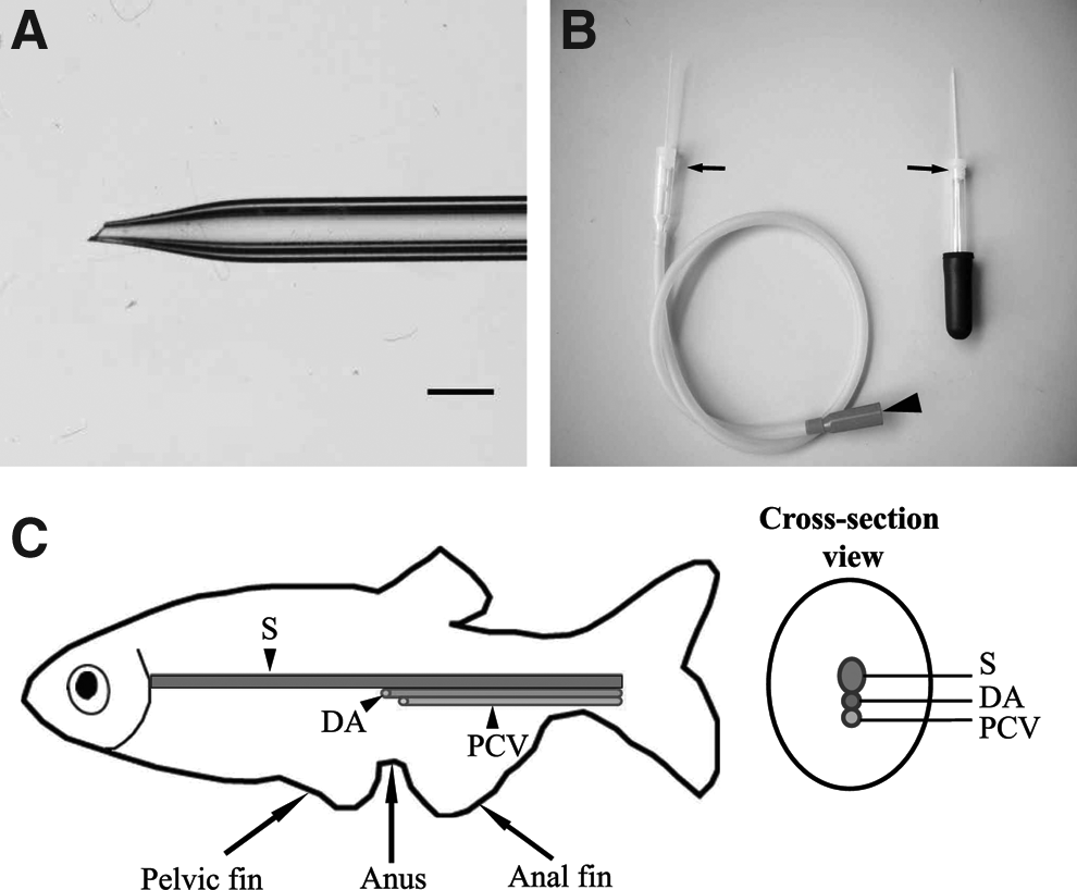
Equipment and anatomic reference diagram for blood collection.
Anesthesia and preparation
Adult zebrafish were anesthetized by placing them in a tank containing 500 ppm of 2-phenoxyethanol (2-PE; Wako Pure Chemical Industries) for approximately 1 min, or until the fish stopped swimming. 2-PE is one of the most commonly used anesthetics in aquaculture. 22 In our experience, 2-PE has a more rapid effect, lower breathing toxicity, and lower mortality than another frequently used anesthetic, tricaine methanesulfonate (MS-222). Using a skimmer, the anesthetized fish was then lifted from the 2-PE bath and gently placed on a paper towel soaked in the 2-PE solution, and its head was gently covered with soft tissue paper (Kimwipe; Nippon Paper Crecia, Tokyo, Japan) also saturated with the anesthetic solution. Another Kimwipe was used to gently dry off the body surface before blood collection. As shown in Figure 1C, the anus is located between the pelvic fin and anal fin, and the dorsal aorta and the posterior cardinal vein are just ventral to the spine. The needle insertion site is along the body axis and posterior to the anus in the region of the dorsal aorta (Fig. 2A). Once the fish has been anesthetized, it is important to perform the blood collection step quickly and carefully.

Procedure for blood collection from adult zebrafish.
Blood collection
Before sample collection a heparinized needle was placed in the nosepiece end of the aspirator tube assembly and the mouthpiece was held in the mouth of the sample collector. The nosepiece end of the aspirator tube and the needle were grasped carefully and the needle was inserted at a 30–45° angle into the blood collection site (Fig. 2B). Care was taken to avoid dulling the needle by contact with the scales, and to avoid puncture of the gastrointestinal tract. Once the needle was felt to touch the spine, suction was applied to the mouthpiece end of the aspiration tube to collect the blood. If blood did not flow, then the needle tip was moved subtly by hand to encourage blood flow. Note that once blood is rising into the needle, it is important to stop shaking and suck gently. In general, the blood will rise slowly into the needle in a pulsatile manner without suction. This is likely because of arterial blood pressure, leading us to conclude that the blood collected was arterial blood from the dorsal aorta. Once the appropriate amount (1–25 μL) of blood was collected, suction was stopped. The needle was removed from the fish and pressure was applied to the puncture site using a Kimwipe to stop any bleeding (bleeding stops after approximately 10–20 s of finger pressure). The blood sample was expelled from the needle onto a clean area of a piece of parafilm for blood glucose measurement (Fig. 2C). Accurate quantities of blood for other analyses can be obtained at this stage using a pipette set to the desired volume. After bleeding from the puncture site stopped, the fish were immediately transferred to a clean water tank.
Sampling volume and frequency
The maximum possible sample volume that can be collected from an individual zebrafish using the technique described above was determined by performing a single maximal blood collection in 34 male and 15 female adult zebrafish. Body weight and length were measured to assess the influence of these factors on the blood sample volume obtained. To discover the effect of repeated blood collections on hemoglobin levels over a 1-week period, five adult male zebrafish weighing approximately 0.5 g each were assigned to each of four blood collection groups. These fish underwent a 2 μL blood collection daily, every second day, or weekly, or a 5 μL blood collection weekly, depending upon their group assignment. Hemoglobin levels were measured at each blood collection using a DRI-CHEM3500V (Fujifilm, Tokyo, Japan).
Overfeeding experiment
Six-month-old male zebrafish were assigned to either an overfeeding or a control group. All fish were housed in groups of three in 2 L tanks. The zebrafish in the overfeeding group were fed 100 mg/fish/day of a commercially available fish food (OTOHIME B-2; Marubeni Nisshin Feed, Tokyo, Japan) divided over five daily feedings. Zebrafish in the control group were fed 20 mg/fish/day of OTOHIME B-2 once daily. OTOHIME B-2 contains a minimum of 11% crude fat, 51% crude protein, 2.3% crude calcium, and 1.5% phosphorous, and a maximum of 15% ash, 3% crude fiber, and 6.5% moisture. The leftover food was removed once daily by vacuuming to avoid water pollution, as described in The Zebrafish Book. 21
Weight and length measurement
The body weights of the zebrafish were measured weekly throughout the study just before blood sample collection. Fish were anesthetized as described above and their body weight (g) was measured after their body surface was dried with a Kimwipe. Fish length (cm) was measured from the anteriormost point of the mouth, to the posteriormost point of the caudal fin. Body–mass index (BMI) was expressed as body weight (g) divided by the square of the body length (cm).
Blood biochemical analysis
The zebrafish were fasted for 14 h before blood collection. Group sizes for the fasting blood glucose measurements were 20 and 22 for the control and overfeeding groups, respectively. The group size for the TG assay was 10 per group. Blood glucose was measured using Glutest Neo Super (Sanwa Kagaku Kenkyusho, Nagoya, Japan), which is a handheld glucometer with a glucose dehydrogenase–flavin adenine dinucleotide electrode that requires a sample volume of 0.6 μL. The manufacturer-provided error range of this glucometer is ±10%. For determination of plasma TG concentrations, blood samples were centrifuged for 3 min at 3500 rpm at room temperature and the plasma was harvested. TG was measured using a Wako L-type TG kit (Wako Pure Chemical Industries) according to the manufacturer's protocol. Blood glucose and plasma TG analyses were performed once a week throughout the overfeeding study.
Individual blood glucose tracking in overfed zebrafish
Six-month-old male zebrafish were housed in groups of three in 2 L tanks and were overfed as described above. Fish in the same tank were individually identified by a right-side puncture site, left-side puncture site, or caudal fin cut in half. The blood glucose analyses were performed once weekly throughout the overfeeding study.
Statistical analysis
The results are reported as means and standard errors. All t-tests were two-tailed, and were unpaired unless stated otherwise. Correlation coefficients (r) were calculated comparing the volume of blood collected from a single, maximal blood draw and BMI. A value of p<0.05 was considered statistically significant.
Results
Collection of blood from adult zebrafish
We developed a minimally invasive method for blood collection from the dorsal aorta of adult zebrafish (Figs. 1 and 2). Because the injury to zebrafish is minimal (a <1 mm puncture; Fig. 2D), most fish survive the blood collection; this allows repeated sample collections from the same fish, which is essential for time-course studies involving blood biochemical parameters.
This method can be used in experiments requiring large quantities of blood. The mean maximal blood volumes collected from 34 male and 15 female zebrafish were approximately 9 and 17 μL, respectively (Table 1). The largest volume of blood collected from an individual fish was 25 μL. We found that the maximal blood volume collected was positively correlated with BMI (r=0.898). Thus, we concluded that the maximum volume of blood collected depended on the size of the fish. In addition, there was no difference in the maximum blood volume collected from male (2.22%±0.06%) and female (2.1%±0.1%) zebrafish when expressed as a percentage of body weight.
Values are mean±standard error.
BMI, body mass index.
Next, we examined the effects of repeated blood sampling over a 1-week period on the blood hemoglobin level (Fig. 3A). Repeated blood sampling (2 μL per sample) of the same fish once daily for 7 days resulted in a rapid decrease in hemoglobin levels from 10.82±0.78 g/dL on day 1 to 2.38±0.8 g/dL on day 7. Two of five fish (40%) were dead on day 5, probably because of severe phlebotomy-induced anemia. Removal of 2 μL blood every 2 days decreased hemoglobin levels from 11±1.29 g/dL to 4.95±1.78 g/dL. One week after a single collection of a 2 or 5 μL blood sample, hemoglobin levels were slightly below normal (from 8.47±0.96 g/dL and 9.72±0.73 g/dL to 6.93±0.93 g/dL and 7.7±0.37 g/dL, respectively). The ratio of hemoglobin on day 6/7:hemoglobin on day 1 was 22%, 49%, 87%, and 76% for fish that had 2 μL samples taken daily, every 2 days, or once weekly, or 5 μL samples taken once weekly, respectively (Fig. 3B). Finally, two weeks after a single collection of a 2 or 5 μL blood sample, the hemoglobin levels had returned completely to normal in all fish. This demonstrates that repeated collection of 2 μL blood per week and 2–5 μL per 2 weeks from individual fish (≥0.5 g in body weight) is safe.
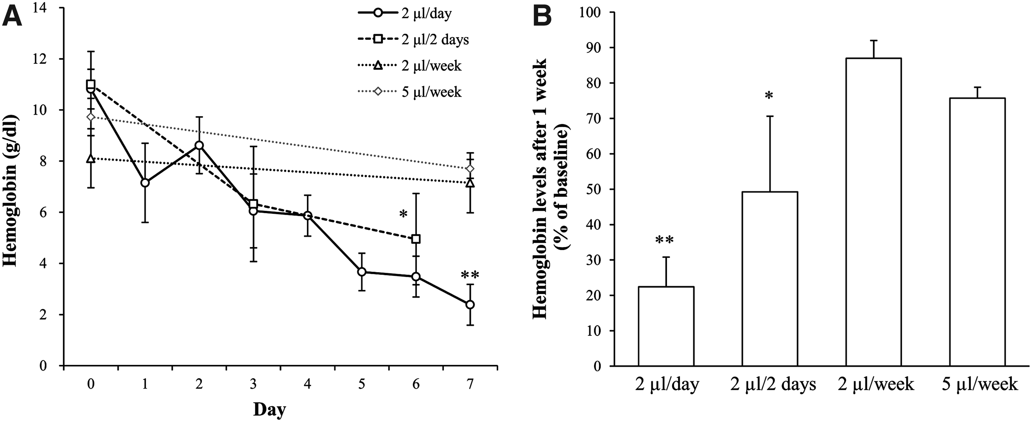
Changes in hemoglobin levels of male zebrafish over a 1-week period with repeated blood sampling.
Monitoring blood glucose and plasma TG during overfeeding
Zebrafish in the overfeeding group exhibited a significant increase (p<0.01) in body weight compared with normally fed fish (Fig. 4A). The average body weight of fish in the overfeeding group was 26% greater than that of fish in the control group at week 5. Correspondingly, the fasting blood glucose levels in the overfeeding group increased markedly from week 1 (p<0.01) onward, and this hyperglycemia was maintained throughout the 5-week study period (Fig. 4B). At week 1 the fasting blood glucose of the fish in the overfeeding group was 55% greater than that of fish in the control group. The rate of increase slowed after week 1, and at week 5 fish in the overfeeding group had a mean blood glucose concentration that was 59% greater than that of the control fish. The plasma TG levels also increased in the overfeeding group (p<0.01) in comparison to the control group at week 1. This trend was sustained throughout the study, and the overfeeding group had a mean plasma TG concentration that was 103% greater than that of the controls at week 4 (Fig. 4C).
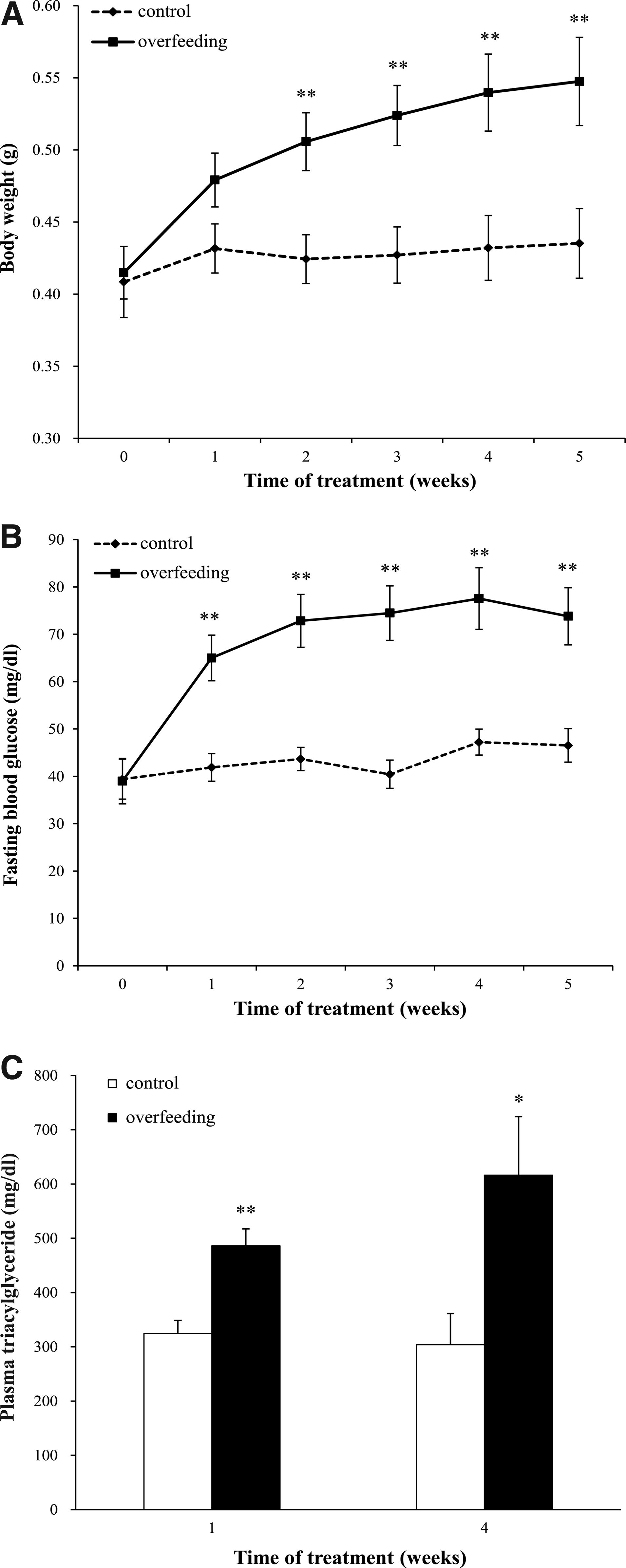
Body weight, fasting blood glucose, and plasma triacylglyceride concentrations in overfed zebrafish. Changes in the body weight
Individual blood glucose changes during overfeeding
To investigate the changes in blood glucose levels of individual fish over time, we performed individual identification of nine fish in the overfeeding group and monitored their blood glucose levels over the 5-week period (Fig. 5). The overfed fish were scatter fed more food than they could eat, and there was no evidence of social dominance limiting the intake of any fish. Fish A, B, C, and D exhibited relatively severe changes in blood glucose, while fish E, F, G, H, and I experienced a milder increase and had much more stable fasting blood glucose levels. This demonstrates that there are varied individual responses to overfeeding.

Changes in the fasting blood glucose concentrations of nine individual overfed male zebrafish over a 5-week period.
Although blood samples were collected repeatedly from the same zebrafish once weekly for a 5-week period, the mean mortality rate was only 2.3%±0.6%. This is an extremely low mortality rate, and demonstrates the usefulness of our blood collection method for studies requiring repeated blood samples from individual fish.
Discussion
Minimally invasive blood collection with a low mortality rate
Blood collection is one of the most common procedures performed in animal research. Unfortunately, there are no established guidelines for blood collection from zebrafish, even though the zebrafish is a commonly used model organism. Although methods for blood collection from zebrafish have been reported, the majority of techniques are lethal. These include decapitation,12,13 tail ablation, 14 and lateral incision.8,15 Here, we developed a new method of blood collection that obtains a sample from the dorsal aorta using a glass capillary needle and an aspirator tube assembly or bulb dispenser. This technique was easy to master and, in our experience, resulted in successful blood collection from 100% of punctures. The injury to the zebrafish was minimized using this technique, and the postsampling mortality was markedly decreased, which enables repeated blood sampling from the same individuals.
We investigated the maximum blood sample volume that could be collected (presented as percentage of body weight) from individual fish using this method (Table 1), and found that approximately 2% of body weight could be collected regardless of sex. This suggests that the total circulating blood volume of the zebrafish is greater than 2% of body weight. Although the maximum volume of blood collected is not equal to the circulating blood volume, this result is consistent with studies of Osteichthyes, the teleost fish, which possess a total blood volume ranging between 1.8% and 3.8% of body weight.23,24 Previous studies have shown that removal of up to 40% of a rat's total blood volume (equal to 2.56% of body weight) results in a 50% mortality rate. 25 A similar mortality rate occurred when we removed a blood volume equal to 2% of body weight from adult zebrafish. However, the predicted circulating blood volume of the zebrafish is smaller than that of the rat (we predict a blood volume of 2%–3.8% of body weight in zebrafish vs. 6.4% in rat 26 ). This suggests that zebrafish may be better able to cope with blood loss than rats.
The main advantage of this method is that it enables repeated blood sampling from the same individual; this is especially useful for toxicokinetic and pharmacokinetic studies. To determine the optimal volume and frequency of blood sampling, we performed periodic blood sampling and measured the changes in hemoglobin levels (Fig. 3). Removal of 2 μL of blood (0.4% of body weight) daily for 1 week resulted in dramatically decreased hemoglobin levels and fish that showed weakness, pale skin, and hypophagia. However, hemoglobin levels had returned to 87% of normal after a recovery period of 1 week (Fig. 3B). In addition, after removal of 5 μL blood (1% of body weight), hemoglobin levels recovered in 2 weeks. These results are consistent with the data in the guidelines for repeated blood sampling of mice and rats,26,27 which state that removal of 7.5% of circulating blood volume (equivalent 0.5% of body weight) requires a 1-week recovery period, and withdrawing 10%–15% of circulating blood volume (equivalent 0.7%–1% body weight) requires 2 weeks for recovery. Accordingly, we recommend that the volume for repeated blood sampling of zebrafish at intervals should be ≤0.4% of body weight per week or ≤1% per 2 weeks.
Blood glucose changes in overfed zebrafish
We applied our new method of blood collection to the study of glycolipid metabolism. It is well known that rodent models develop obesity associated with abnormal glycolipid metabolism when fed a high-fat diet,28–31 but there are no zebrafish studies using repeated glycolipid measurements for obesity modeling. Thus, we monitored the temporal changes in blood glucose that occur in overfed zebrafish. The overfeeding group exhibited significant body weight gain (Fig. 4A) and hypertriglyceridemia (Fig. 4C) compared with the control group, which was consistent with the results of our previous studies.19,20,32 As for glucose metabolism, significantly elevated fasting blood glucose levels were apparent as early as 1 week after beginning the overfeeding treatment, and this trend continued until the end of the study (Fig. 4B). These data suggest that, similar to mammals, overfed zebrafish develop obesity associated with decreased glucose tolerance.33–35 In addition, the rapid increase in fasting blood glucose over week 1 is similar to the changes seen in a previous study of rainbow trout (Oncorhynchus mykiss), which also showed significantly increased circulating glucose levels on day 7 in response to a high-fat diet (containing 20% digestible fat). 36 This may indicate that certain fish species have a tendency for glucose intolerance. We anticipate that the methods presented here will be useful for the study of metabolic disorders such as obesity and type 2 diabetes mellitus.
The changes in fasting blood glucose of each individual overfed fish showed varying patterns. These fish could be classified as those with a relatively large change in blood sugar concentrations and those with comparatively stable blood sugar (Fig. 5). This situation seems similar to that in humans, in which there are interindividual differences in the response to bulimia and obesity. 37 It is well known that genetic factors, including single polymorphisms, are linked to pancreatic beta cell function and correspond to the differences in individual responses to obesity.38–40 The results of our current study provide a good opportunity to figure out the individual differences in the glucose metabolism of zebrafish, thus further confirming its utility as an animal model for human obesity.
In this study, we developed a novel method for repeated blood collection from adult zebrafish. Our method is easy to perform, is minimally invasive, results in a low mortality rate, reduces the number of animals necessary for studies, and allows repeated blood collection, which is difficult using conventional methods. This method will enable zebrafish researchers to perform studies that require repeated blood sampling from individual zebrafish, such as studies of toxicokinetics, pharmacokinetics, serum lipid metabolism, and metabolic disorders. In the future, these data will serve as a reference for performing minimally invasive, ethical blood sampling of adult zebrafish. Furthermore, this collection method can also be useful in medaka (Oryzias latipes), Xiphophorus (Xiphophorus helleri), or other small aquarium fish in biomedical research.
Footnotes
Acknowledgments
We would like to thank Dr. Tomomi Yamada for helpful discussions, and Mrs. Asaka Uechi and Mrs. Eriko Nakanishi for secretarial assistance.
Author Disclosure Statement
No competing financial interests exist.