




Abstract
Abstract
Due to their size and optical clarity, zebrafish embryos have long been appreciated for their usefulness in time-lapse confocal microscopy. Current methods of mounting zebrafish embryos and larvae for imaging consist mainly of mounting in low percentage, low melting temperature agarose in a Petri dish. Whereas imaging methods have advanced greatly over the last two decades, the methods for mounting embryos have not changed significantly. In this article, we describe the development and use of 3D printed plastic molds. These molds can be used to create silicone casts and allow embryos and larvae to be mounted with a consistent and reproducible angle, and position in X, Y, and Z. These molds are made on a 3D printer and can be easily and cheaply reproduced by anyone with access to a 3D printer, making this method accessible to the entire zebrafish community. Molds can be reused to create additional casts, which can be reused after imaging. These casts are compatible with any upright microscope and can be adapted for use on an inverted microscope, taking the working distance of the objective used into account. This technique should prove to be useful to any researcher imaging zebrafish embryos.
Introduction
Z
Classically, zebrafish embryos are prepared for live imaging by mounting in low percentage (0.5%–1%) low melting temperature agarose. 8 Recent work by other groups provides significant improvements over this classical mounting method.9–11 Petzold et al. 11 describe the development of mounting embryos in plastic tubing. This was then further developed by Kaufmann et al., 10 who describe the development of multilayer mounting techniques specifically suited for light sheet microscopy. Imaging multiple embryos using these methods in an automated fashion is cumbersome, although not impossible. The method described by Bishel et al., 9 allows for rapid mounting and imaging of multiple embryos using a microfluidic device. However, this does not allow for pinpoint accuracy in the angular positioning of embryos. Here we developed a universal method that builds on the classical method already used by most zebrafish researchers. We also addressed the previously mentioned issues allowing for more precise and consistent mounting. Several key criteria were taken into consideration; this should be a cheap and relatively simple technique to use, and it should be possible to use this system on already existing microscope platforms without having to make any modification to the platform itself. Furthermore, it should be possible for other research groups to easily deploy this solution and where necessary, modify and improve upon it.
In this article, we provide a description of the development and usage of silicone casts that can be used to consistently and accurately position multiple fish for time-lapse confocal microscopy for a range of developmental stages. Molds to make these casts are produced on a rapid prototype 3D printer. Domestic 3D printers are developing rapidly, making 3D printing available to more researchers then ever before. The 3D models of the molds as well as the 3D models of the zebrafish embryos used to make these molds are freely available for download at www.armi.org.au/Research1/Research_Groups/Currie_Group/Research_projects1/Silicone_Imaging_Casts.aspx. It is strongly encouraged to use and improve upon these 3D models to create molds to suit personal preference and experimental design.
Materials and Methods
Zebrafish husbandry
Zebrafish were maintained on a 14-h light/10-h dark cycle using standard procedures. Both wild-type (WT) and tg(Bactin:GFPcaax) transgenic zebrafish are used. Male and female zebrafish were left to freely breed, eggs were subsequently collected, staged, and maintained on 28°C in an E3 medium. 8 Embryos were specifically staged for 15 somite, 1, 2, and 3 dpf.
Mold design and fabrication
Using the ubiquitously expressing tg(Bactin:GFPcaax) individual ventral sides of embryos at various stages during development were imaged using a confocal microscope (Leica SP5). Imaris (Imaris 7.4; Bitplane) was used to create a surface rendering of the GFP signal and was subsequently exported as a VRML2 file. Meshlab and Solidworks were used to remove residual background noise and fill in the gaps of the 3D rendered zebrafish embryos. Individual lateral sides were then mirrored and stitched together to create a model of whole zebrafish embryos at various developmental stages (15 somite, 1, 2, and 3 dpf).
Taking both potential variation in embryo size and errors in the design process into account, 3D reconstructions were scaled in a range from 100% to 140%. Plastic molds were printed with a 3D printer (objet eden 260v; Stratasys) at a resolution of 50 μm based on the principle of stereolithography using Fullcure720 (Stratasys). Molds can be produced on any other 3D printer with the key requirements of a print resolution of at least 50 μm and the use of a polymer that is harder then silicone.
Cast fabrication and validation
Casts were made using Sylgard184 (Dow Corning), a silicone polymer with a refractive index similar to water. Sylgard184 was mixed to a 10:1 ratio with its curing agent and mixed for 3–4 h at 4°C. For the polymer to harden, the plastic molds were placed in Petri dishes, and treated with stoner rapid release A324 spray (Stoner, Inc.). The preincubated Sylgard184 mixture was poured on top of the mold and left to polymerize for a minimum of 4 days at room temperature. The silicone casts were then removed from the mold and excess silicone was removed using a scalpel blade. To confirm that sizing was in concordance with the final design, WT embryos were placed in the cast and inspected on a stereomicroscope (Zeiss axio imager Z1) for any issues in scaling. A confocal microscope (Leica SP5) was used to assess any issues in the angular positioning of these embryos.
Cast usage
To mount embryos using this cast, the silicone cast is placed in a Petri dish appropriate to the microscope platform. The properties of the silicone cast allow for it to adhere to the Petri dish without the need for any glue or additional supports. Zebrafish embryos are anesthetized using 0.2% tricaine (MS-222) in the E3 medium. Zebrafish embryos are pipetted on to the silicone casts and using fine-tip pipettes any residual air pockets should be removed from the cavities in the cast. Embryos were positioned into the individual pockets and the excess E3 medium is removed. About 0.5% agarose was placed on top of the embryos, excess agarose was removed, and the remaining agarose is left to polymerize. The E3 medium containing 0.2% tricaine (MS-222) is used to flood the Petri dish to keep the embryos anesthetized and hydrated. Embryos are now properly prepared for a wide range of imaging purposes. To clean the silicone casts, the cast is removed from its Petri dish, embryos and agarose are removed, the cast is rinsed using tap water, and left to air dry.
It is important to note that when a cast is made, this should be done on a completely level surface otherwise a slightly slanted cast will result. This can be confirmed using a bullseye spirit level. It is recommended to leave casts to polymerize on a vibration dampening table, thus making sure that an equal thickness throughout the cast is achieved.
Results
To improve the quality and consistency of zebrafish microscopy, casts were developed to hold zebrafish at specific angles. These casts also allow for embryos to be evenly spaced in X and Y, with only minimal variation in the Z plane. Several casts were created to hold embryos at various stages of development, specifically at the 15 somite stage, 1, 2, and 3 dpf. These stages were chosen because they display the most dramatic changes in the embryo size during early development. Later in development, the overall morphology of a larvae does not alter as dramatically (4–6 dpf), and a 3-dpf mold can be easily scaled up to accommodate larger sizes.
In silico 3D reconstruction of zebrafish embryos
To create an in silico reconstruction of an entire zebrafish embryo, the ubiquitously expressing tg(bactin:GFPcaax) was used. At various stages of embryonic development, the entire lateral side was imaged by tile scanning on a Leica SP5 confocal microscope. These tiled confocal stacks were subsequently surface-rendered in Imaris. Surface renderings were transferred to SolidWorks, inspected, and artefacts manually removed. Each lateral side was mirror-imaged and stitched together with the original lateral side to create a 3D reconstruction of an entire zebrafish embryo (Fig. 1A–D).
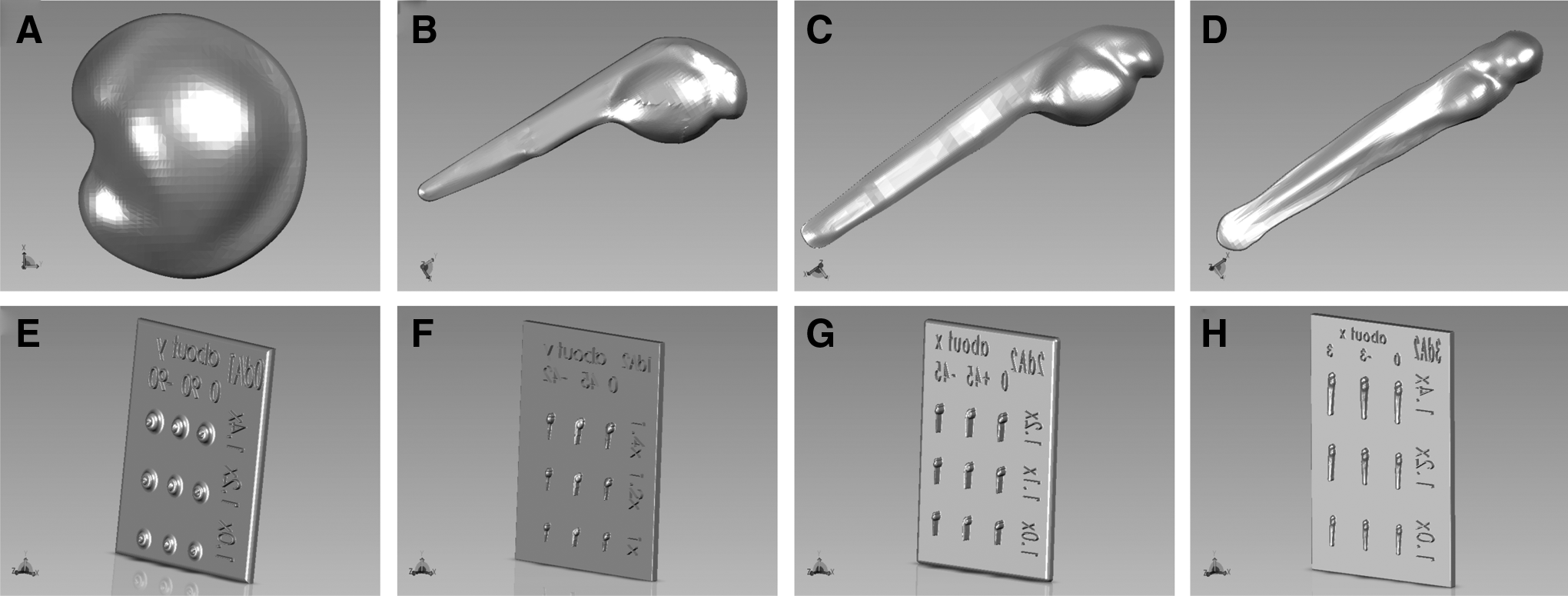
Computer modeling of various zebrafish embryo imprints on a base plate. Various 3D renderings have been made for specific developmental stages, 15 somite
Mold design and fabrication
The 3D in silico models can be used to rapidly test different versions of molds using a 3D printer. To test the validity and to prove the usefulness of this approach, molds were designed for several developmental stages (15 somite, 1, 2, and 3 dpf). To compensate for any potential error in the scaling between the various scanning modeling and printing steps involved, various scaling sizes were chosen ranging from 100% to 140%. Embryo models were positioned at various angles to showcase the consistent and precise mounting capabilities of these molds (Fig. 1E–H). Plates were printed on a rapid prototype 3D printer (object eden 260v; Stratasys) using a Fullcure720 feedstock with a 50-μm resolution, and visually inspected afterward on a dissecting microscope for correct reproduction of the virtual model. The 15 somite mold is designed to mount embryos at 0°, 90°, and −90° from a lateral orientation (rotation around AP axis) (Figs. 1E and 2A). The 1- and 2-dpf molds are designed to mount embryos at 0°, 45°, and −45° from lateral (rotation around AP axis) (Figs. 1F, G and 2B, C). The approach for the 3-dpf mold was slightly different, we opted to precisely control the rotation of the trunk around the DV axis, as such the 3-dpf mold was rotated around the DV axis at 0°, 3°, and −3° (Figs. 1H and 2D). This was done to allow for a single confocal plane to acquire the entire length of the zebrafish embryo. Silicone casts were made using Sylgard184, removed from the molds, and inspected for proper reproduction using a dissecting microscope (Fig. 2A–D).
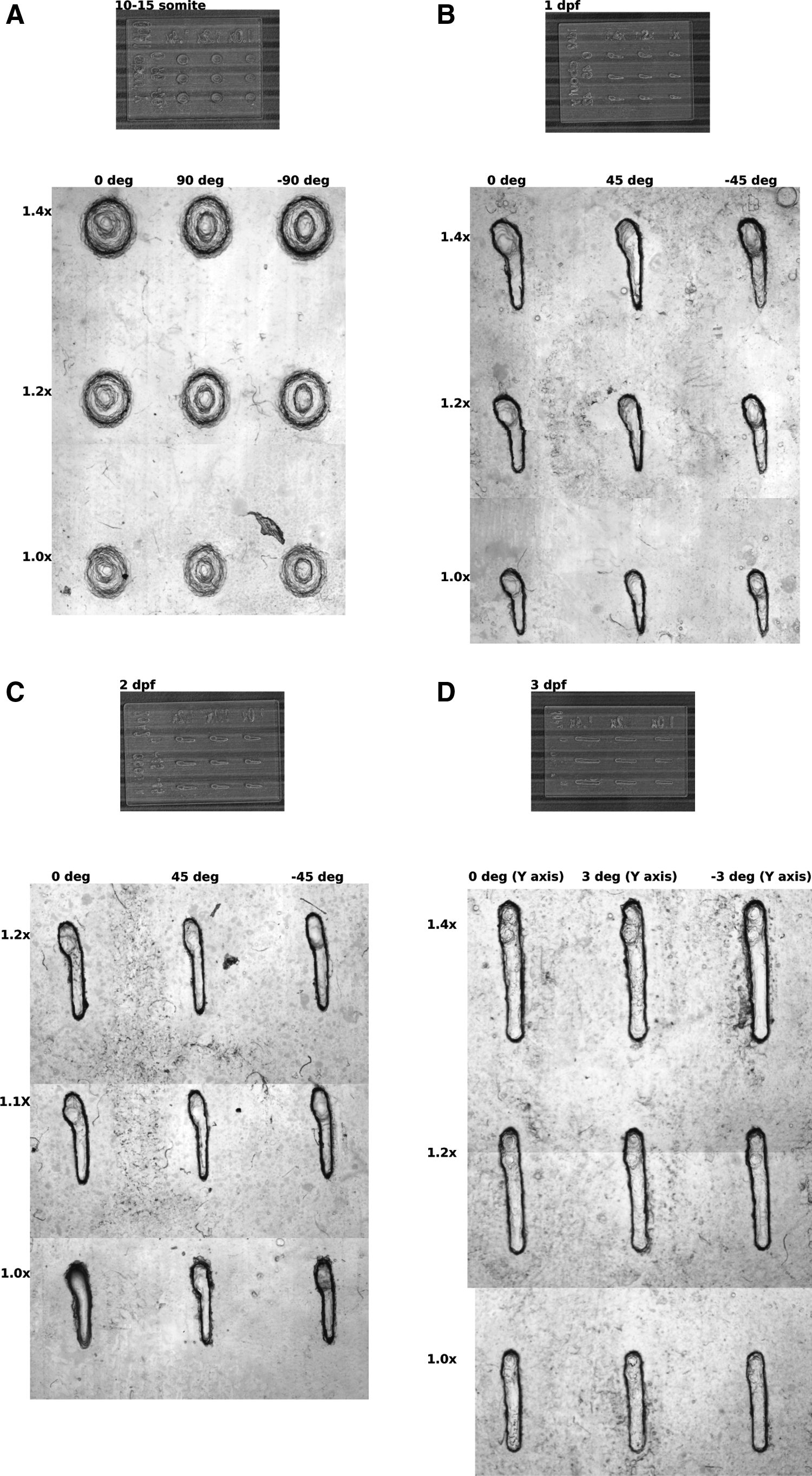
Three-dimensional printed models at various stages, black lines are spaced 5 mm
Validation of silicone casts
To validate correct reproduction of the silicone casts, several parameters were tested. The correct size and angular positioning were crucial parameters to assess. First, any possible error in scaling that could have occurred between the various steps of image acquisition, rendering, digital reconstruction, printing, and cast production was assessed. WT zebrafish embryos were placed in molds and imaged using a stereomicroscope (Zeiss axio imager Z1). Sizes were assessed for all developmental stages, shown here are the 100% and 120% scale sizes for 1 and 2 dpf. Both the 1- and 2-dpf 100% scale are sufficient to properly hold the embryo (Fig. 3A, C). For both developmental stages, the 120% scale is clearly too large (Fig. 3B, D). This showcases the level of accuracy that can be achieved using this production process.
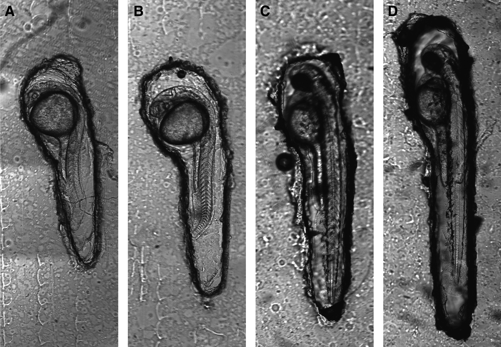
Scale validation of the silicone imaging molds. Embryos are placed into selected molds at 1 dpf
Second, the angular positioning had to be addressed. Angular positioning of the cast on the AP and DV axis should be recapitulated by the orientation of the embryo. This means that if, for example, a cast is made in a way to contain an embryo at 45° from a lateral view, this should be accurately reproduced in the positioning of the embryo. Tg(Bactin:GFPcaax) embryos were placed in several casts designed to position them at different angles, and Z-stacks of individual embryos were acquired. Embryos were observed consistently at the predetermined angles of 0°, +45°, and −45° from lateral (Fig. 4A–C). Creating optical orthogonal slices of each 3D rendered embryo allows for detailed inspection of consistent angular positioning. Three positions along the AP axis are shown (head, yolk, and yolk extension) (Fig. 4). Consistent angular positioning can be observed for fish positioned at 0° (Fig. 4A’–A”’), −45° (Fig. 4B’–B”’), and +45° (Fig. 4C’–C”’) from lateral. The view of embryos positioned at −45° (Fig. 4B–B”’) is largely obstructed by its yolk and as such is difficult to acquire Z-stacks from.
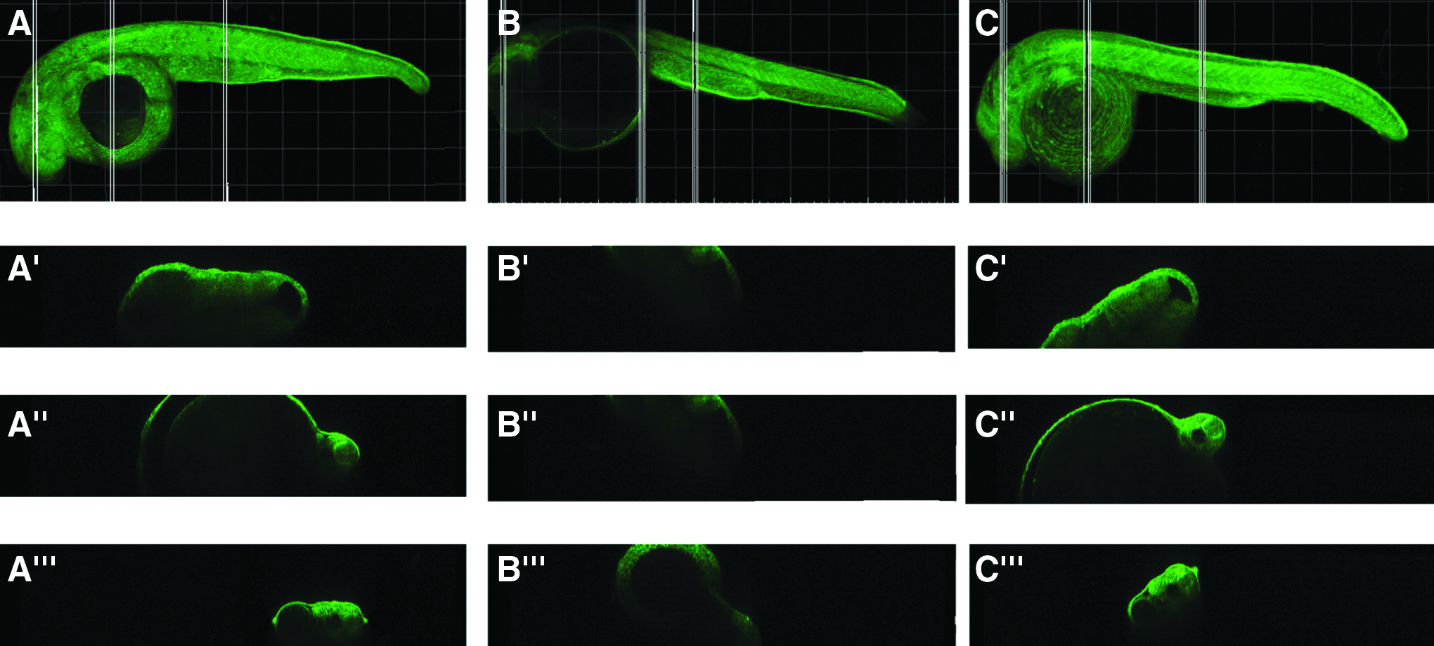
Validation of angular positioning in the silicone imaging molds. Tg(bactin:GFPcaax) embryos at 1 dpf are placed into selected silicone molds and confocal images were acquired. In postprocessing, 20-μm orthogonal sections were taken at three different levels (head, yolk, and yolk extension). One dpf tg(bactin:GFPcaax) embryo, 0° from lateral, maximum projection.
While acquiring these datasets, only minimal variations in focal planes were observed. This showcases the level of precision that can be achieved using these casts.
Discussion and Conclusion
Zebrafish embryos have long been used in combination with confocal microscopes. Although confocal microscopes continue to undergo rapid advances, zebrafish mounting techniques have not altered significantly. With increased demands to collect more data in a shorter amount of time, we developed silicone casts that can be used to consistently position zebrafish embryos at a wide range of angles. These casts can be used in combination with any upright microscope with the caveat that there is sufficient working distance to place a silicone cast. Considering the silicone polymer used has a refractive index close to water, these casts can also be on inverted microscopes. In these cases, the thickness of the silicone mold and working distance of the objectives will be crucial.
We developed molds specific to a variety of developmental stages (10–15 somite, 1, 2, and 3 dpf). Both the complete 3D reconstruction of the individual zebrafish embryos and the completed plates are available for download from www.armi.org.au/Research1/Research_Groups/Currie_Group/Research_projects1/Silicone_Imaging_Casts.aspx. These molds can be easily reproduced using a 3D printer. Multiple casts can be made from a single mold further driving down the total cost per cast.
We confirmed correct size reproduction by inspecting the size difference between the silicone cast and zebrafish embryo. Various casts with different angular positions have been tested and confirmed to accurately position zebrafish on the required angle.
It is recommended to mix the curing agent and Sylgard 184 polymer for 3–4 h at 4°C. In addition, treating the mold with the Stoner rapid release A324 Spray (Stoner, Inc.) is important, this lays down a thin wax-like coating that makes it easier for the polymerized silicone to release from the mold and also increases the level of detail that can be achieved. A second equally important thing to take into account is to leave sufficient time for the silicone to polymerize. In our experience, polymerizing in a temperature controlled room on a true level was important. To do this, we placed the silicone cast on a vibration dampening table in a microscope room. It is recommended leaving the silicone for 4–5 days to completely polymerize. Taking these things into account maximizes the accuracy of reproduction and eliminates the need to remove residual silicone from the plastic mold.
Acquiring confocal time-lapse series on multiple samples is significantly improved by the increased speed offered by galvo-scanners, they do, however, reduce the total travel distance allowed in Z to 500 μm making it crucial to mount embryos in the same Z-plane. This no longer poses a problem using the casts we developed, as all embryos can be easily mounted on the same Z-plane. Not only is multipositioning time-lapse imaging made far easier with this method, mounting zebrafish embryos in these casts allows for imaging at a consistent angle. These two key factors combined will allow for the design of high-throughput experiments with high reproducibility.
Footnotes
Acknowledgments
The authors would like to acknowledge Monash Micro Imaging (MMI) for the use of confocal microscopes and expert advice. Also, the authors acknowledge FishCore for zebrafish husbandry and maintenance. Also a special thanks to Catherine Boisvert for proofreading the manuscript. This work was supported by an Australian Research Council grant to P.D.C. The Australian Regenerative Medicine Institute is supported by funds from the State Government of Victoria and the Australian Federal Government.
Disclosure Statement
No competing financial interests exist.