
Abstract
Abstract
Malathion, a common organophosphate insecticide, is a proven acetylcholinesterase inhibitor and is the most applied organophosphate insecticide in the United States. The use of zebrafish as a model to study the effects of pesticides on development is an innovative approach yielding relevant implications for determining the potential toxic effects of these pesticides on humans. In this study, a simple noninvasive technique was developed to investigate the cardiotoxicity of malathion on Danio rerio embryos, and to detect and quantify its effect on heart rate. Videos were recorded under a stereomicroscope and examined with our custom-made software (FishBeat) to determine the heart rate of the embryos. The pixel average intensity frequency (PI) of the videos was computed at its maximum probability to indicate the average number of heartbeats per second. Experimental observations successfully demonstrated that this method was able to detect the heart rate of zebrafish embryos as compared with manual stopwatch counting, with no significant difference. Embryos were treated acutely with increasing malathion concentrations (33.3 and 50 μg/mL malathion) at 52, 76, and 96 hpf. Embryos treated with 33.3 μg/mL malathion had significant bradycardia at 52 and 76 hpf, whereas embryos treated with 50 μg/mL malathion presented bradycardia at all hpf. These novel observations confirmed that malathion, acting as an acetylcholinesterase inhibitor, induced heartbeat irregularity in zebrafish embryos.
Introduction
D
In previous studies, insecticides, such as carbaryl (1-naphthyl-N-methylcarbamate), have been proved to work as acetylcholinesterase (AChE) inhibitors, ultimately affecting zebrafish embryonic development and heartbeat regularity.3–5 Carbaryl has shown to cause heart malformations and irregular heartbeat in Japanese medaka, Oryzias latipes. 6 Recently, Schock et al. 5 have also shown that chronic exposure to carbaryl induced bradycardia in zebrafish embryos as well as morphological changes due to the inhibition of AChE and the subsequent accumulation of acetylcholine.
Malathion (diethyl 2-[(dimethoxyphosphorothioyl)sulfanyl]butanedioate), a common organophosphate insecticide, has also been proved to act as an acetylcholinesterase inhibitor in zebrafish. 7 Malathion is the most applied organophosphate insecticide in the United States. 8 The use of zebrafish as a model to study the effects of pesticides on development is an innovative approach yielding relevant implications for determining the potential toxic effects of these pesticides on humans. Pesticides enter aquatic ecosystems in urbanized and agricultural areas as a result of overflow through the soil, precipitation, and run-off. In addition to their high levels of toxicity, many of our first-generation pesticides such as DDT are highly persistent in the environment, and do not break down into less harmful substances over time. Second-generation pesticides such as organophosphates and carbamates (Malathion, Sevin) were once considered less harmful to organisms, as they did not persist in the environment and were broken down within a short period of time (EXTOXNET: Extension Toxicology Network). In the human body, malathion is metabolized into malaoxon, which is 61 times more toxic. 9 Its toxic effects have been demonstrated in a variety of organisms, including beneficial honeybees and a wide variety of aquatic organisms causing wide-ranging deformities.10,11 Therefore, malathion poses a significant risk for many organisms, in addition to humans.
In order to assess the cardiotoxicity of several drugs and pesticides on aquatic organisms, several methods have been developed to detect and quantify the heart rate, including manual counting, electrocardiogram, micro pressure system, and Laser Doppler microscope technique. 12 All these procedures are time consuming and either not practical or accurate for extensive periods of time or elevated beating frequencies. Moreover, the processing of such images is still a challenge and requires skilful operators and expensive equipment.
The need for a practical, simple-to-use, and economical heartbeat counter for our undergraduate laboratory has prompted us to develop a new computer-based methodology applicable to zebrafish and other model organisms. Other image capture software programs have been developed, notably those by Fink et al., 13 Chan et al., 12 and Shin et al. 14 However, we needed a simple program that we could use in our laboratory for student research, and the development of this particular program was a part of an undergraduate research project that was used to investigate the cardiotoxicity of malathion during the early development of zebrafish. Experimental observations have successfully demonstrated that this method is able to detect the heart rate of zebrafish embryos, and that malathion, acting as an acetylcholinesterase inhibitor, induces bradycardia in zebrafish embryos.
Materials and Methods
Malathion, analytical standard, was purchased from Sigma-Aldrich. Striped variations (long fin and normal fin) of zebrafish, Danio rerio, were purchased from Carolina Biological Supply Company. Specimens were observed under a Zeiss stereo microscope. Videos were obtained through a Moticam 2000 digital camera (2.0 megapixels) and recorded on Motic Images Plus 2.0ML. Video resolution: 800×600, Data Rate: 115200 Kbps, and Frame Rate: 16 frames/s.
Standard zebrafish breeding methods were used to house and breed adult fish 15 , and all fish were maintained under Manhattanville IACUC Protocol #R004. For the egg harvesting, three glass dishes were triple netted with plastic netting and placed in the tank; marbles were placed on top of the dishes. A one-gallon bottle of water was cut in half, and the top piece was netted with a porous filtering cloth. Each day, the three dishes were lifted out of the tank and their contents were poured into the “bottle-funnel.” An additional glass dish, filled with aquarium water, was utilized to collect the eggs from the “bottle-funnel.” The nets were, then, cleaned from the marbles caught on top of the filtering cloth, while the eggs were collected at the bottom of the funnel into the additional glass dish. On each collection, the dishes were cleaned using aged tap water, and put back in their original position inside the tank. Eggs were staged and placed in an incubator set at 28°C. Air was pumped into the water to provide aeration. At Manhattanville, this egg harvesting method has been cleaner and more efficient than siphoning marbles at the bottom of a breeding tank.
Automatic heartbeat counting (FishBeat)
The custom software (FishBeat) was developed using Microsoft C# 4.5. Embryos were placed into six-well culture plate in a drop of water and oriented so that they were in the same position for capturing video images. The embryos at 52 and 76 hpf were still in the egg, and embryos at 96 remained in one place for extended periods, so there was no need for chemical immobilization. Videos of zebrafish embryos were obtained and converted into individual images (one per frame) using the open source video encoding tool FFmpeg (http://ffmpeg.org/). Frames were stored at full resolution and original colors. By knowing the approximate position of the heart, a more robust heartbeat frequency detection could be performed even with low resolution, low magnification, and noisy video captures (Fig. 1). Heartbeat frequency was detected through the use of an algorithm previously described by Capó Irizarry et al. 16 The algorithm has been adapted in order to be simpler, easy to implement, and accurate at low image resolutions.

A screenshot from FishBeat of a 96 hpf embryo with a white square indicating the area of the heart for video capture of heartbeat.
Algorithm description
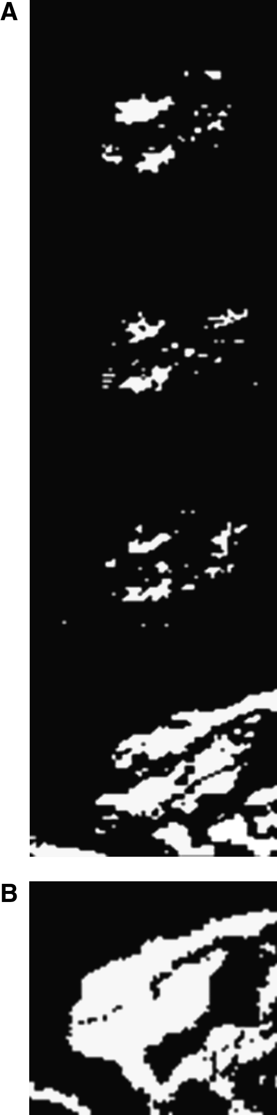
The aim of the algorithm was to compute a probability distribution of the pixel intensity frequency (change of color). The collection of frames (C) was preprocessed in order to eliminate noisy features. For each frame, the area corresponding to the heart region was cropped and converted into a grayscale image using the BT.709-5 algorithm by ITU-R (Fig. 2).
17
The new collection of preprocessed frames was used to perform a frame-by-frame image subtraction. The subtraction operation compares two consecutive frames and subtracts pixel by pixel the color components. A threshold value of 5 was used in order to discard insignificant color changes due to the noise of the environment.
12
If the difference in color intensity was more than the threshold value, the pixel coordinate was considered active and a pixel value of 255 (white) was assigned. For static pixels, a value of zero was assigned. The resulting image set (C*) is a set of black and white maps of dynamic pixels in two consecutive frames (Fig. 2A). A matrix (M) was then created. The number of times that a given pixel has been marked as active by the pixel-to-pixel subtraction was stored for every pixel coordinate Mx,y. When a value of 255 (white) was obtained, M was converted into a bitmap representation if Mx,y was more than a threshold value (threshold=10% of the total number of frames). The image generated was a black and white map of the dynamic pixels across the entire set C* (Fig. 2B). This map could be considered a representation of the most active heart regions captured by the video. For every active pixel in the matrix M (Mx,y>threshold), the average pixel intensity value was calculated from the initial image set C*:
Pixel average intensity frequency (PI) was obtained by counting the number of downward crossings of the line represented by the average pixel intensity values of each M*x,y and dividing this by the duration in seconds of the video.
A probability distribution was computed by counting the number of pixels at the same frequency, using buckets of 0.1 beats per second.
Malathion exposure and visual heartbeat counting
Embryos were treated acutely with malathion concentrations (33.3 and 50 μg/mL malathion) at 52, 76, and 96 hpf (n=27 embryos for each concentration at each time period). The concentrations of Malathion were based on previous toxicity studies conducted at Manhattanville College. Eggs were allowed to stabilize in aged tap water for 30 s in a single well of a six-well culture plate. Heartbeat was visually counted under a stereomicroscope before any chemical exposure. Eggs were then exposed to the corresponding concentration of malathion for 10 min. Heartbeat was counted for 15 s. An ulterior heartbeat count was performed to determine the steady rate. Malathion was subsequently removed, embryos were exposed for 10 min to aged tap water, and an additional measurement of heartbeat was taken.
A standard t-test was used to compare heart rate counting with FishBeat and also to compare control embryos heart rate with experimental embryo heart rate. Differences were significant at the p<0.05 level.
Results and Discussion
Automatic heart beat counting (FishBeat)
A comparison with manual stopwatch counting showed that FishBeat calculated heart rates of approximately one to two heart beats more than control embryos during the 15 s. period. There was no statistical difference between manual counting and FishBeat at 52 and 76 hpf (p>0.05). (Fig. 3). There was a slightly greater difference at 96 hpf (p=0.04). All embryos used to video record at 96 hpf were hatched. However, due to the fact that embryos at 96 hpf were free from their chorionic membrane, a significant increase in movement was observed due to the embryos being free to move. The software has a threshold value for minimal movement. According to Chan et al. 12 , a threshold value of 5 was used in order to discard insignificant color changes due to the noise of the environment. If the difference in color intensity was more than the threshold value, a pixel value of 255 (white and active) was assigned. For static pixels, a value of zero was assigned. 12

Comparison of manual counting to FishBeat shows no significant difference during each of the developmental time periods. FishBeat averages 1–2 more per count. As the embryos develop, the heart rate increases. There was a slight difference at 96 hpf (p=.059), but this may be due to a slight difference in embryo orientation.
However, some movements might have been significant enough to cross the threshold value and trigger the software to compute the pixel average intensity frequency, including those movements as well. Due to the nature of our test, we only wanted to observe the effects of a specific chemical (malathion) on the development of zebrafish embryos and, therefore, we strictly avoided any immobilizing agent or anesthetic in order to make sure that any change in heartbeat was indeed due to malathion and not to any other chemical applied to immobilize embryos. Researchers investigating other topics, however, might find the software extremely useful at 96 hpf (and beyond) in case they decide to anesthetize the embryos for heartbeat recording. Ultimately (and very importantly), this small statistical difference detected at 96 hpf was only slightly higher than our measurements at 52 and 76 hpf. The difference is still incredibly small. Even with a slight discrepancy at 96 hpf, these results show that pixel intensity frequency, at its maximum probability, is a good approximation of zebrafish heartbeat frequency, and FishBeat is as accurate, if not better, as manual stopwatch counting.
Exposure to 33.3 μg/mL malathion
Once collected, eggs were staged according to Kimmel et al. 1 at 52, 76, and 96 hpf. Out of the total number of eggs collected, there was very little mortality and a mean viability of 93.9%. The majority of the eggs hatched at 72 hpf in accordance with Kimmel et al. 1 and the previous studies conducted at Manhattanville College, Purchase, NY.
As an acetylcholinesterase inhibitor, Malathion was speculated to induce bradycardia. Zebrafish embryos exposed to acute 33.3 μg/mL malathion treatment showed a significant decrease in heartbeat at 52 and 76 hpf (p<0.01). Malathion-exposed embryos had mean heart rates of approximately two to three beats less than control embryos per 15 s (Fig. 4). Specifically, control embryos showed mean heart rates of 38, 42, and 44 at 52, 76, and 96 hpf; whereas malathion-exposed embryos showed mean heart rates of 36, 40, and 43 at 52, 76, and 96 hpf.

The lower dose of malathion had a significant effect on embryo heart rate during the 52 and 76 hpf time period (p<0.001).
Exposure to 50 μg/mL malathion
Zebrafish embryos exposed to acute 50 μg/mL malathion treatment showed a significant decrease in heartbeat at all hpf (p<0.01). Malathion-exposed embryos had mean heart rates of approximately 23–27 beats less than control embryos per 15 s (Fig. 5). Specifically, control embryos showed mean heart rates of 38, 42, and 44 at 52, 76, and 96 hpf; whereas malathion-exposed embryos had heart rates of 15, 16, and 17 at 52, 76, and 96 hpf.

The stronger dose of malathion had a significant effect on embryo heart rate at all time periods of development (p<0.001).
Other observations
Since the embryos were exposed to a brief, acute dose of malathion, no long-term developmental effects were observed. Once the embryos were removed from the malation, their heartbeat returned to normal and there were no lasting effects. However, in a current study at Manhattanville College, a number of deformities that are a direct result of chronic exposure have been observed. These include kinked spine, curved tail, thoracic edema, and absence of eye development or the development of only one eye. In addition, we also just recently observed erratic heartbeat in some of the exposed embryos, which supports the results reported here (Cheng and Todd, unpub. data).
Conclusion
Preliminary observations have successfully demonstrated that FishBeat is able to detect the heart rate of zebrafish embryos and is as accurate, if not more than manual counting. Furthermore, we have showed that malathion, acting as an acetylcholinesterase inhibitor, induces bradycardia in zebrafish embryos. Exposure to malathion decreased the heart rate at all hpf. Increased concentrations of malathion induced a more significant bradycardia. The difference between FishBeat and manual counting at 96 hpf may be due to the uncurled position of the embryo after hatching, and more observations should be performed during this time period to ensure consistency in heart beat quantification.
Additional studies that investigate the specific toxic effects of malathion are necessary, and Danio rerio is an excellent model organism for examining these effects and interpreting their impact on aquatic ecosystems and, ultimately, humans.
Footnotes
Acknowledgments
The authors would like to thank Beta Beta Beta Biological Honor Society for partially funding this research, and the Biology department at Manhattanville College for funding. Dr. Hiroshi Osaka provided valuable mentorship and advice during the early stages of this project.
Disclosure Statement
No competing financial interests exist.