
Abstract
Abstract
One attractive quality of zebrafish as a model organism for biological research is that transparency at early developmental stages allows the optical imaging of cellular and molecular events. However, this advantage cannot be applied to adult zebrafish. In this study, we explored the use of contrast-enhanced X-ray micro-computed tomography (microCT) on adult zebrafish in which the organism was stained with iodine, a simple and economical contrasting agent, after fixation. Tomographic reconstruction of the microCT data allowed the three-dimensional (3D) volumetric analyses of individual organs in adult zebrafish. Adipose tissues showed a higher affinity to iodine and were more strongly contrasted in microCT. As traditional histological techniques often involve dehydration steps that remove tissue lipids, iodine-contrasted microCT offers a convenient method for visualizing fat deposition in fish. Utilizing this advantage, we discovered a transient accumulation of lipids around the heart after ventricular amputation, suggesting a correlation between lipid distribution and heart regeneration. Taken together, microCT is a versatile technique that enables the 3D visualization of zebrafish organs, as well as other fish models, in their anatomical context. This simple method is a valuable new addition to the arsenal of techniques available to this model organism.
Introduction
Z
Traditionally, the anatomy of zebrafish is studied by the three-dimensional (3D) reconstruction from histological sections, 3 which can be tedious and inaccurate. More recently, multiphoton confocal and light sheet microscopy techniques provide 3D information with good spatial resolutions. However, these techniques are limited by the penetration power of light. “Transparent” zebrafish with mutations in pigmentation genes have been generated, 4 but the large body size of adult zebrafish still presents challenges for optical imaging. Individual organs often need to be dissected and clarified before imaging,5–7 preventing the in situ visualization of the tissues within their anatomical context.
Computed tomography (CT), and its high-resolution variation micro-computed tomography (microCT), is a tomographic technique that is widely used in medical imaging. 8 This technique has been increasingly used for nonclinical research in the recent years.9–11 Samples with mineralized tissues are clearly defined under CT scanning.12–14 However, due to the high penetration power of X-ray, nonmineralized organs are often very poorly contrasted. Therefore, staining agents are used to enhance the contrast of internal structures in scanning microCT.15,16 For example, osmium compounds were used to visualize the soft tissues in mouse embryo 15 and honeybee brain. 16 Phosphotungstic acid has also been used in small experimental animals. 11 However, the widespread use of these reagents is often limited by their high toxicity and relative low contrasting efficiency. Safer reagents, such as iodine-based compounds, have been explored as alternatives, 11 but their staining properties are still not fully evaluated.
This study aims to establish the use of contrast-enhanced microCT in adult zebrafish. After optimizing the concentration and duration of the iodine-based contrasting agent in zebrafish, we demonstrated the use of microCT imaging in revealing the detailed internal anatomy of intact zebrafish. Applying this method, we analyzed zebrafish hearts in situ at different stages after ventricular amputation. These data show that contrast-enhanced microCT is a versatile technique for assessing the 3D anatomical information of zebrafish and other small fish model systems.
Materials and Methods
The study was approved by the committee on the ethics of animals of the Department of Health of the Hong Kong SAR government (Reference number: 14–15 in DH/HA&P/8/2/5 Pt2). All the experiments and surgeries were done under complete anesthetization of the fish to minimize suffering.
Fish
One year old wild-type (AB line) fish were used for this study. We have used age-, weight-, and gender-matched (male) fish for this experiment to minimize individual variations. Animals were maintained under standard laboratory conditions at 28°C ± 0.5°C with a 14-h light/10-h dark cycle. 17 Fish were fed three times per day with fish food from TetraMin made by Tetra, as well as live brine shrimps newly hatched from Brine Shrimp Eggs produced by Brine Shrimp Direct once per day. Other species of aquarium fishes such as medaka (Oryzias latipes), guppy (Poecilia reticulata), bronze corydoras (Corydoras aeneus), celestial eye goldfish (Carassius auratus), and green spotted puffer (Tetraodon nigroviridis) were obtained from a local aquarium shop.
Heart injury
The amputation procedures followed the protocol from Poss et al. 18 Zebrafish were anesthetized by MS-222 (ethyl-3-aminobenzoate methanesulfonate salt, E10521; Sigma-Aldrich) 0.04 mg/mL for 3–5 min and placed ventral side up on a sponge. The scales were removed and ventral cavity opened by forceps. When the heart was exposed, the apex of the heart was clamped with forceps and amputated using a very fine surgical scissor. Fish were returned to a recovery tank and as the fish were still under anesthesia, the gills were ventilated with fresh water to stimulate recovery, and once swimming normally, fish were returned to the home tank and monitored daily. To further analyze, the fish were euthanized at day 1, 3, 5, 7, and 14 postinjury by an overdose of MS-222 (0.13 mg/mL).
Fixation and staining
Fixation of animals was performed with 4% paraformaldehyde (Sigma) in water at 4°C for 48 h followed by rinsing with Phosphate Buffer Saline (10 min, two times) and undergo staining with iodine (Riedel-de Haen) dissolved in aqueous iodide solution (Riedel-de Haen), ethanol and methanol at concentrations indicated in Results and Discussion. For the samples that went through the iodine alcoholic staining, they were incubated in absolute alcohol for 10 min two times before incubating in staining agents and those with iodine staining incubated in 1% iodine metal (I2) and 2% potassium iodide (KI) in water. 11 Staining was done by immersing the animals in staining agents in glass tubes at 4°C in rotator at our favorable periods of time (Fig. 1).
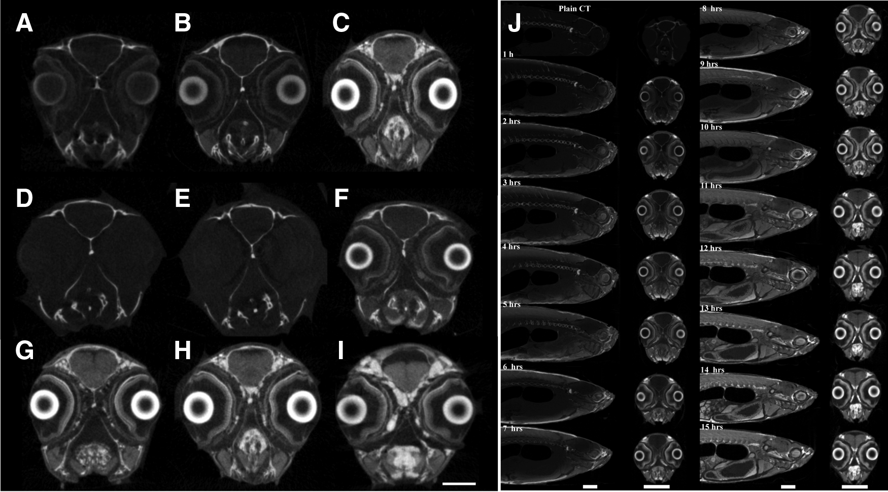
Optimization of iodine contrasting of zebrafish in microCT. Coronal sections of the head of zebrafish labeled with
Histological staining
To compare the microCT images of the fish with the traditional histological staining, whole mount histology was performed. The zebrafish and medaka were dissected to remove the otoliths and operculum, and gas from the swim bladder was released. They were fixed in a GPHS fixative (0.05% glutaraldehyde, 2% paraformaldehyde, 80% HistoChoice MB fixative, and 1% calcium chloride) overnight at 4°C, and processed and embedded in paraffin as previously reported. 19 Sections that were 5 μm in thickness were cut on a Leica RM 2125 RT microtome (Leica Instruments). The slides were deparaffinized in xylene (5 min, three times) and hydrated in 100%, 95%, and 70% ethanol (5 min each) and rinsed with distilled water until the ripples disappeared from the slides. The slides were stained in Mayer's hematoxylin (Dako Cytomation) for 1 min. They were then rinsed with distilled water and decolorized in 1% acid alcohol, with three to six quick dips followed by bluing with 0.2% ammonia water, and rinsed with distilled water. The slides were then stained with eosin (Electron Microscopy Science) for 1 min, dehydrated in 95% and 100% ethanol (two times in each type for 5 min), cleared with three changes of xylene for 5 min each, and mounted under a glass coverslip with Permount (Fisher Chemicals).
MicroCT scanning
All samples were scanned by microCT scanner model Skyscan1076 (Bruker). X-ray parameters for volume imaging were 59 kV voltage, 149 μA current, and 2400 ms exposure time. A 10.5 mm filter was applied. Samples were stabilized in the polystyrene tube to avoid any movement during scanning and scanned with a 0.7° step angle to 180° for 25 min. All data were saved as raw data.
Reconstruction
Cross sections of samples were reconstructed using the NRecon Reconstruction software. NRecon Server was used as the reconstruction engine, and 4 computers were run together to reconstruct the raw data made from the microCT scanner. Reconstructed images are 1216 × 1376 pixel with one pixel equal to 8.666 μm in BMP format.
Results and Discussion
Optimization of microCT contrast enhancement for zebrafish
In this study, we chose to focus our optimization efforts on iodine as a contrasting agent for microCT due to its general ease of use. First, we tested the effect of solvent in the contrasting performance of iodine. Paraformaldehyde-fixed zebrafish were incubated in 1% iodine dissolved in aqueous potassium iodide solution, in methanol or in ethanol, and the resulting contrast enhancement in microCT was examined. Figure 1A–C shows the coronal sections of the head of zebrafish stained by these reagents. Iodine dissolved in ethanol (Fig. 1A) or methanol (Fig. 1B) could also produce distinguishable tissues, but the contrast was not as good as the aqueous solution (Fig. 1C). The anatomy of zebrafish head could be clearly distinguished by the staining of iodine dissolved in potassium iodide. It is possible that the aqueous solvent favors the partitioning of iodine into the organic matters of the zebrafish, making the tissue uptake of iodine more efficient. Besides, obvious shrinkage artifact was produced by ethanol (Fig. 1A). These results suggest that iodine in aqueous solution is the best format for contrasting enhancement in zebrafish. Next, we optimized the concentration of iodine for contrast enhancement. Iodine at 0.1% has been used as a staining agent for a variety of animal embryos. 11 However, our initial observations showed that this concentration failed to generate high contrast images for adult zebrafish. Figure 1D–I shows images of the coronal sections of zebrafish head incubated with iodine at concentrations ranging from 0.1% to 2%. At the lowest iodine concentrations (Fig. 1D, E), no distinguishable features in the head could be identified. Tissue contrast became progressively enhanced with increasing iodine concentrations. At concentrations above 0.75% (Fig. 1F), clear images of brain anatomy were visible. The contrast was more or less similar in the section stained by 1% or 2% iodine (Fig. 1H, I). Therefore, iodine at a concentration of 0.75% or above is suitable to stain zebrafish for microCT scanning.
We then studied the effect of iodine staining duration on its contrasting efficiency. Zebrafish were incubated in 2% iodine and were scanned by microCT every hour for 15 h (Fig. 1J). As expected, only hard tissues in unstained fish could be seen. As the staining time increased, more anatomical details were visible. After 12 h of contrast enhancement, the in situ detail of tissues in the whole zebrafish could be visualized. Using the optimized protocol, we explored the use of microCT in visualizing the internal structure of adult zebrafish. As microCT is a tomographic technique, the volumetric reconstructions of the microCT data allow the virtual sectioning of the fish at any chosen angle and orientation (Fig. 2). This not only enables the 3D measurement of individual organs without dissection but also allows the analyses of the spatial and structural relationship of organs in their native anatomical context.
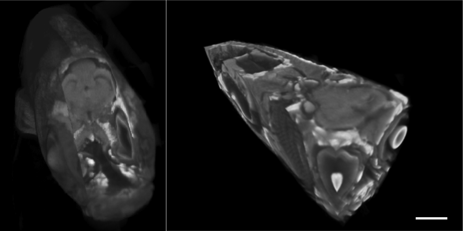
Tomographic reconstruction and rendering of the microCT data of whole mount adult zebrafish. Scale bar: 1 mm.
Iodine-contrasted microCT reveals adipose distribution in fish
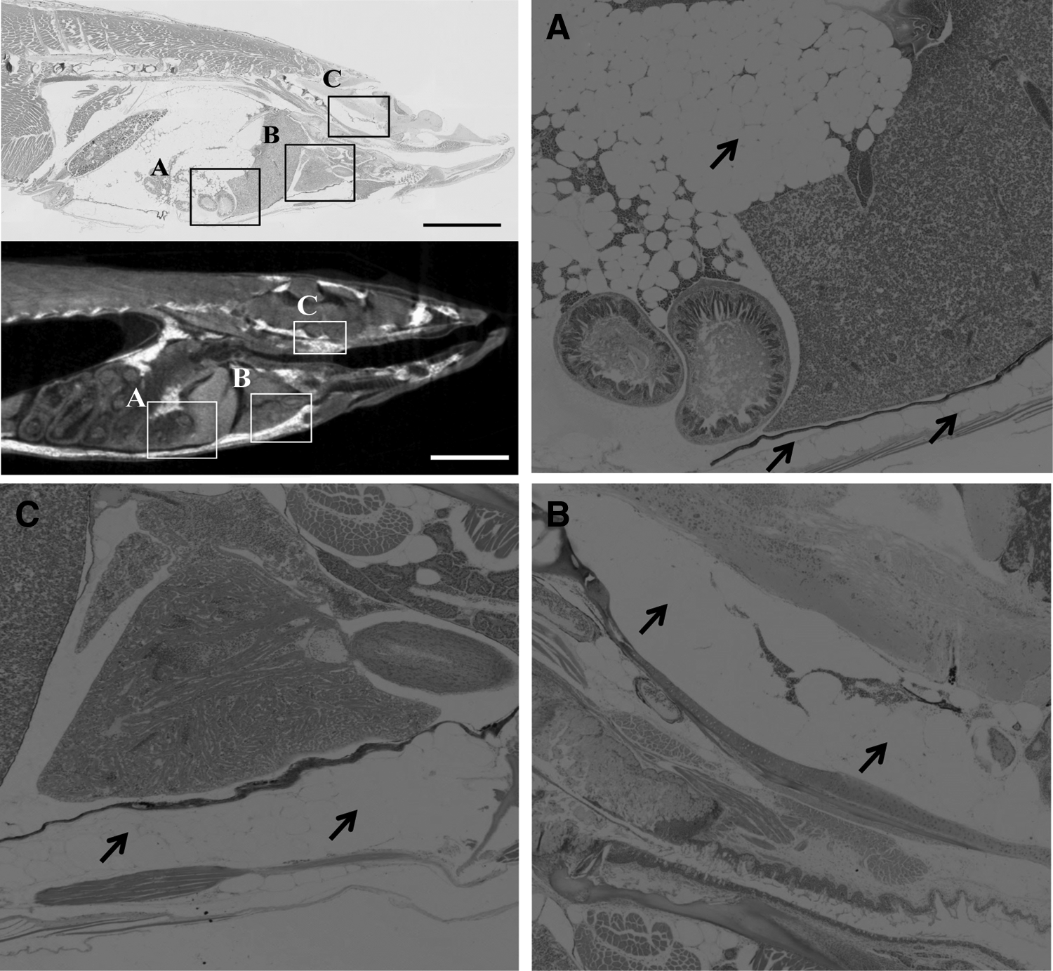
Although iodine provided a good contrast enhancement for the whole fish, some tissues consistently exhibited a higher contrast than others. It is possible that these tissues have a high affinity to iodine. To identify the strongly iodine-stained tissues, we compared the microCT images of the sagittal plane of an iodine-contrasted zebrafish with a histological section of an age- and gender-matched zebrafish (Fig. 3). The microCT image indicated the presence of strongly iodine-contrasted areas on the dorsal and abdominal walls and around the eye. The corresponding micrograph of a whole mount zebrafish section stained with hematoxylin and eosin (H&E, Fig. 3A–C) shows that these areas correspond to the adipose tissues (arrows). As iodine is more soluble in lipids, it is possible that fatty tissues have a higher affinity to iodine than other tissues. Hence, iodine-contrasted microCT provides a convenient method for the visualization of adipose tissues in zebrafish. For instance, a coronal section of the head of a female fish reveals the presence of adipose tissues around the brain (Fig. 3C, arrows). Fat tissues are usually poorly defined in paraffin sections, which involved dehydration by organic solvents during the preparation. Imaging of the adipose tissues requires the use of specific fluorescent dyes, 20 which is not applicable to nontransparent adults. Therefore, iodine-contrasted microCT offers a useful method for analyzing fat deposition in zebrafish. This potentially opens up a new avenue for quantitating the adipose content and distribution in zebrafish in different genders and ages and under various conditions or genetic manipulations.

Hematoxylin and eosin (H&E) staining and its correlated microCT images of male zebrafish. Boxed area
Monitoring zebrafish heart regeneration by iodine-contrasted microCT
Zebrafish is one of the most widely used organisms for the study of tissue regeneration. 21 In particular, the regeneration of zebrafish hearts is distinguished by its unparalleled speed and efficiency: the removal of as much as 20% of the ventricular tissue can be fully regenerated in 60 days. Although the cellular and molecular events during heart regeneration have been investigated in great detail,22,23 these studies are mostly based on histological characterization of dissected hearts. As microCT allows for the 3D analysis of anatomical features, we were able to image the location and size of heart lesions in situ and to observe the regeneration process in 3D within the anatomical context.
We monitored the heart regeneration process by iodine-contrasted microCT. Figure 4 shows sagittal and coronal sections of heart region of zebrafish on different days after ventricular amputation (0, 1, 3, 5, 7, and 14 days postamputation, dpa). Regenerating zebrafish hearts show a different absorption of iodine compared with that of control heart. Interestingly, a tissue strongly contrasted by iodine was observed around the regenerating heart starting from 3 to 5 dpa and then disappeared at 7 dpa. As iodine labels predominately the adipose tissues, these results suggest that adipose tissues and lipid metabolism play a role in heart regeneration in zebrafish.
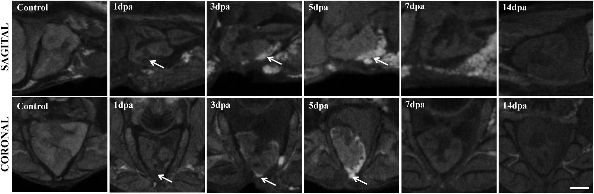
Anatomical changes of the regenerating heart of zebrafish by microCT scanning. Sagittal and coronal sections of zebrafish heart at different days postamputation. Bars: 1 mm. Arrows are showing the areas that are corresponding to the adipose tissues as mentioned in the text.
Iodine-contrasted microCT can be generally applied to other small fish models
The sample preparation and imaging protocol that we developed for zebrafish could readily be adopted for other small fish models. Figure 5 shows the coronal and sagittal sections of five different fish species as follows: O. latipes (Fig. 5A), C. auratus (Fig. 5B), P. reticulata (Fig. 5C), C. aeneus (Fig. 5D), and T. nigroviridis (Fig. 5E). The diversity of anatomical structures in fish is beautifully demonstrated by these images. Like in zebrafish, iodine differentially contrasted different tissues in these fishes. We compared the images of whole mount O. latipes (medaka) imaged by iodine-contrasted microCT and by H&E histology (Fig. 6). As in zebrafish, regions in medaka that were strongly contrasted by iodine also coincided with the adipose tissues. Hence, iodine-contrasted microCT can be used to visualize the adipose tissues in fish. Taken together, microCT, in combination with a simple iodine-based contrast enhancement, is generally applicable to small fish models.
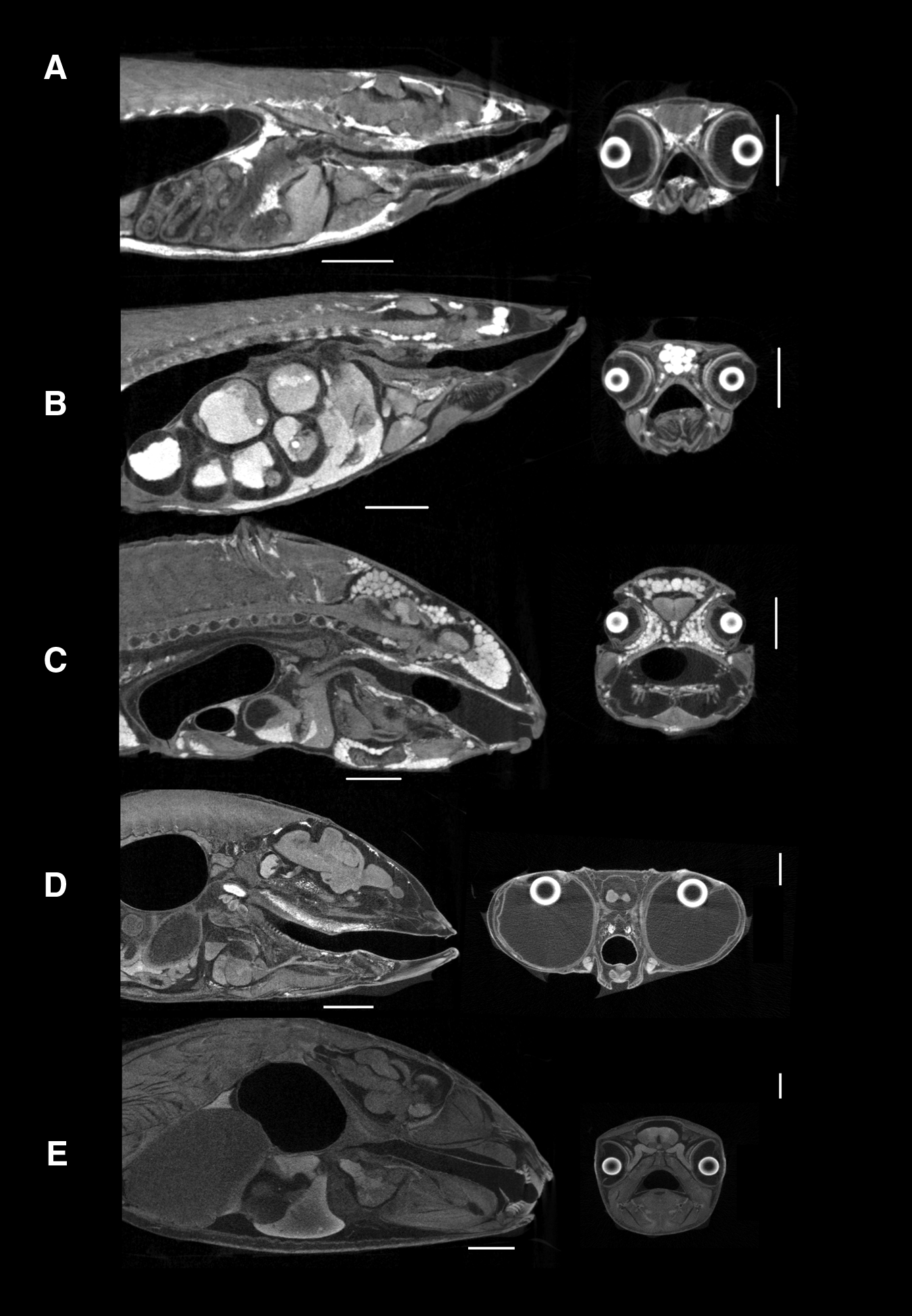
Anatomical differences among aquarium fishes were found by the scanning of microCT. The sagittal and coronal section of
Hematoxylin and Eosin staining and its correlated microCT images of male medaka (O. latipes). Boxed area
Conclusion
In this study, we reported an optimized protocol of contrast-enhanced microCT that can be applied to zebrafish and other small fish models. This simple technique allows the 3D visualization of the internal anatomy in adult zebrafish that was not accessible to light microscopy before. As the fat tissues have a higher affinity to iodine, this technique reveals the adipose distribution in fish.
Footnotes
Acknowledgments
The authors thank Miss Li Yuen Ha for the maintenance of the zebrafish and Dr. Kai Hung Fung (Radiology Department, Pamela Youde Nethersole Eastern Hospital, Hong Kong) for his advice on interpreting the microCT data. The authors also thank the members of the Lam laboratory for their generous ideas. FB was supported by a PhD studentship from the University Grant Council, Hong Kong. The work described in this article was substantially supported by a grant from the Research Grants Council of the Hong Kong Special Administrative Region, China (Project No. CityU 160110).
Disclosure Statement
No competing financial interests exist.