
Abstract
Abstract
Thrombosis is a leading cause of death and the development of effective and safe therapeutic agents for thrombotic diseases has been proven challenging. In this study, taking advantage of the transparency of larval zebrafish, we developed a larval zebrafish thrombosis model for drug screening and efficacy assessment. Zebrafish at 2 dpf (days post fertilization) were treated with phenylhydrazine (PHZ) and a testing drug for 24 h. Tested drugs were administered into the zebrafish either by direct soaking or circulation microinjection. Antithrombotic efficacy was quantitatively evaluated based on our previously patented technology characterized as an image analysis of the heart red blood cells stained with O-dianisidine staining. Zebrafish at 2 dpf treated with PHZ at a concentration of 1.5 μM for a time period of 24 h were determined as the optimum conditions for the zebrafish thrombosis model development. Induced thrombosis in zebrafish was visually confirmed under a dissecting stereomicroscope and quantified by the image assay. All 6 human antithrombotic drugs (aspirin, clopidogrel, diltiazem hydrochloride injection, xuanshuantong injection, salvianolate injection, and astragalus injection) showed significant preventive and therapeutic effects on zebrafish thrombosis (p < 0.05, p < 0.01, & p < 0.001) in this zebrafish thrombosis model. The larval zebrafish thrombosis model developed and validated in this study could be used for in vivo thrombosis studies and for rapid screening and efficacy assessment of antithrombotic drugs.
Introduction
T
New scientific models have been established in the past few years to identify novel factors of hemostasis and thrombosis and to analyze their functions in greater detail.4–7 One novel animal model is the zebrafish that could mimic mammalian hemostasis and thrombosis since it shares most of the central factors of platelet adhesion, activation, aggregation, and release reaction with humans, and possesses coagulation factors and thrombocyte receptors, and responds to anticoagulant and antiplatelet drugs commonly used in clinical treatment.4,5,7 In the earlier work, investigators have used the chemical ferric chloride (FeCl3) or a nitrogen pulsed dye laser to cause thrombus formation in the caudal vessels of larval zebrafish 5 and found that the zebrafish present a vertebrate model, in which both large scale screening is feasible and the hemostatic pathways are similar to those found in humans.4,8,9
Phenylhydrazine (PHZ) is known to cause externalization of phosphatidylserine (PS) on plasma membrane of erythrocytes and generate superoxide radicals, which cause oxidative damage to the lipid membrane of erythrocytes and increase the rate of thrombin generation, leading to thrombosis in rats.10,11 Sato et al. reported that PHZ induces acute thrombosis with regional stasis as a trigger, and subsequent hemostatic disruption is involved as an accelerator in the pathogenesis of rat thrombosis. They also found that PHZ caused dysfunction of endothelial cells, which resulted in a hypercoagulable state and it would act as one of the contributors of acute thrombosis. 11 In addition, the pro-inflammatory condition observed at the early stage of PHZ treatment was considered to play an important part in the development of thrombosis due to endothelial dysfunction. 12
Treatment of larval zebrafish with PHZ could cause zebrafish vascular occlusion in the caudal artery. 5 However, PHZ-induced vascular occlusion in zebrafish has not been characterized as a thrombosis model and not validated for drug screening yet.
Aspirin has been recognized to inhibit platelet activation through suppressing both the cyclooxygenase system and thromboxane A2 production, as well as the synthesis of prostacyclin. 13 Clopidogrel (Plavix) selectively and irreversibly inhibits adenosine diphosphate (ADP)-induced platelet aggregation. 14 Diltiazem is a calcium channel blocker that could potentially block thrombosis. 15 With a long history, the Traditional Chinese Medicines (TCMs) have been proved effective in preventing and treating thrombotic diseases. 16 Xueshuantong, an injectable drug extracted and prepared from an Chinese herb Panax notoginseng, has been used to treat thrombotic diseases in China for more than 30 years and has been proved highly effective. 17
Salvianolate prepared from a Chinese herb Salvia miltiorrhiza is a commonly used medicine for “invigorating” the blood and reducing blood clotting in eastern countries, particularly in China, and many studies have shown that salvianolate can inhibit platelet aggregation and adhesion and reduce thrombus formation. 18 Astragalus as a TCM has been used for thousands of years, and recently, mainly for treating thrombosis. 19 Numerous studies have shown that astragalus has obvious effects of inhibiting thrombus formation in vivo and resolving blood clot in vitro. 20
In the present study, we developed a zebrafish thrombosis model that utilized PHZ as a thrombosis inducer in combination with O-dianisidine staining to quantitatively assess antithrombotic effect of antithrombotic drugs. This model was further validated with three Federal Drug Administration (FDA)-approved antithrombotic drugs (aspirin, clopidogrel, and diltiazem hydrochloride injection) and three China FDA (CFDA)-approved antithrombotic medicines (xuanshuantong injection, salvianolate injection, and astragalus injection). Our results indicate that the zebrafish thrombosis model developed and validated in this study is a convenient and predictive animal model for rapid in vivo screening and efficacy assessment of antithrombotic drugs.
Materials and Methods
Zebrafish care and maintenance
Adult AB strain zebrafish were housed in a light and temperature controlled aquaculture facility with a standard 14 h light photoperiod/10 h dark photoperiod and fed with live brine shrimp twice daily and dry flake once a day. Four to five pairs of zebrafish were set up for natural mating every time. On an average, 200–300 embryos were generated. Embryos were maintained at 28°C in fish water (0.2% Instant Ocean Salt in deionized water, pH 6.9–7.2, conductivity 480–510 ms/cm, and hardness 53.7–71.6 mg/L CaCO3). The embryos were washed and staged at 6 and 24 hpf (hours post fertilization). 21 The zebrafish facility at Hunter Biotechnology, Inc. is accredited by the Association for Assessment and Accreditation of Laboratory Animal Care (AAALAC) International. 22
Chemicals and drugs
PHZ (lot #: G1324044) and O-dianisidine (lot #: 119-90-4) were purchased from Aladdin company of Shanghai, China. Aspirin and clopidogrel were bought from Sigma-Aldrich, diltiazem hydrochloride injection was from Mitsubishi Tanabe Pharma Corporation, xuanshuantong injection was from Wuzhou Pharmaceutical Corporation, salvianolate injection was from Shanghai Green Valley Pharmaceutical, and astragalus injection was from Chiatai Qingchunbao. Drug stock solutions were prepared in either 100% dimethyl sulfoxide (DMSO) or 0.9% sodium chloride, and serial dilutions were made before each experiment. Zebrafish treated with 0.1% DMSO or 0.9% sodium chloride served as a vehicle control. Untreated zebrafish were used to confirm that the vehicle solvents did not have an adverse effect on the zebrafish.
Zebrafish thrombosis model development
The zebrafish exhibit functional platelets and coagulation factors by 36 hpf,4,5,7 and consequently, we chose 2 dpf zebrafish as an appropriate stage to start PHZ treatment for the thrombosis model development. To optimize the PHZ treatment concentration and treatment time period, thirty 2 dpf zebrafish were distributed into six-well plates (Nest Biotech.) in 3 mL fresh fish water.23,24 Zebrafish were treated with 0.75, 1.5, and 3 μM PHZ for 6, 12, 24, and 48 h, respectively, to induce thrombosis. Zebrafish treated with 0.1% DMSO served as a vehicle control. Untreated zebrafish were used to confirm that vehicle solvent did not have adverse effect on zebrafish. After PHZ treatment, 10 zebrafish from each group were randomly selected for visually qualitative assessment of the thrombus formation in the caudal vein at the lateral view under a dissecting stereomicroscope. The resting zebrafish were stained with O-dianisidine for quantitative analysis of thrombosis. Based on the qualitative and quantitative results of thrombosis, the optimal PHZ treatment concentration and treatment time period were selected.
Zebrafish thrombosis model characterization
To characterize PHZ-induced zebrafish thrombosis model developed above, we used a well-known anticoagulant drug warfarin, 25 a free radical scavenger glutathione, 26 and a PS externalization inhibitor Z-Asp 27 to address the mechanisms of PHZ-induced thrombosis. Thirty 2 dpf AB strain zebrafish were distributed into six-well plates in 3 mL fresh fish water. Zebrafish were cotreated with 1.5 μM PHZ (the optimum PHZ concentration) and warfarin, glutathione, or Z-Asp for 24 h (the optimum PHZ treatment time period) at a serial concentration as indicated in Table 1. Zebrafish treated with 1.5 μM PHZ alone for 24 h were used as thrombosis model. Zebrafish treated with 0.1% DMSO served as vehicle controls. Untreated zebrafish were used to confirm that vehicle solvent did not have adverse effect on zebrafish. After treatment, zebrafish thrombus was quantified using a method as described below.
Compared with vehicle control: ap < 0.001.
Histopathological examination
To confirm PHZ-induced thrombosis, zebrafish histopathology was performed. After treatment, zebrafish were fixed in 4% paraformaldehyde in 0.1 M phosphate-buffered saline for 4 h at 4°C, dehydrated in graded series of ethanol solutions before paraffin embedding. Embedded zebrafish were longitudinally sectioned at 5 μm and stained with hematoxylin and eosin. Thirty zebrafish were used for each group. Histological images were obtained using a histological microscope (Leica) and pathological diagnosis was completed by a certified pathologist.
Quantitative analysis of zebrafish thrombosis
For quantitative analysis of thrombosis, zebrafish were stained with O-dianisidine using a method as reported before 28 to quantify the heart red blood cells (RBCs) (hemoglobin level) of zebrafish. Our previously patented zebrafish thrombosis assay technology suggested that the heart RBC amount is reversely correlated with the thrombus length/severity and, thus, measuring heart RBCs could quantify thrombosis in zebrafish (China patent number: 201110126427.2).
After O-dianisidine staining, 10 zebrafish from each group were randomly chosen for the heart RBC image acquisition. Zebrafish were immobilized in 3% methyl cellulose and images were acquired in the identical lighting intensity at a 56 × magnification under a dissecting stereomicroscope installed with a high-speed video camera (JVC). Quantitative image analysis of RBCs was performed by measuring heart RBC intensity (S) using the NIS-Elements D3.10 image analysis software (Nikon) and the data are expressed as mean ± SEM. The effect of a test drug was calculated based on the formula below:
A positive percentage means that a tested drug could prevent and/or treat thrombus and a negative percentage suggests that the tested drug had no effect on thrombosis in the zebrafish model.
Determination of no observed adverse effect level
To determine no observed adverse effect level (NOAEL) of a testing drug, 2 dpf zebrafish were treated with a testing drug for 24 h, and mortality and toxicity were recorded at the end of the treatment. In the initial tests, five concentrations (0.1, 1, 10, 100, and 500 mg/L for soaking drugs) or doses (0.1, 1, 10, 100, and 500 ng/zebrafish for microinjected drugs) were used for each drug. If a NOAEL could not be found from the initial tests, additional concentrations or doses within the range of 0.01–2000 mg/L or 0.01–2000 ng/zebrafish were tested. The NOAEL of a test drug was defined as a maximum concentration or maximum dose that did not induce any observable adverse effect on zebrafish and was determined under a dissecting stereomicroscope by a well-trained zebrafish toxicologist.
Drug delivery by circulation microinjection
Four injectable drugs (diltiazem hydrochloride injection, xuanshuantong injection, salvianolate injection, and astragalus injection) were delivered into zebrafish by circulation microinjection as we reported previously. 29 Drugs were dissolved in 0.9% sodium chloride and diluted to proper concentrations for zebrafish circulation microinjection. Before microinjection, zebrafish were anesthetized with 0.03% tricaine (Sigma) and loaded on a customized microplate designed specifically for zebrafish microinjection. A drug at a designated concentration was loaded into a pulled glass capillary (World Precision Instruments) that was drawn on an electrode puller (NARISHIGE) and then trimmed to form a needle with a resulting internal diameter of approximately 15 μm and the outer diameter of approximately 18 μm. The microneedle was attached to an air-driven CellTram (NARISHIGE). The tip of the needle was inserted into the circulation of zebrafish under a dissecting stereomicroscope and the pulse time was controlled to deliver 10 nL of the drug solution into the circulation through the glass capillary. Injected zebrafish were transferred to six-well plates, 30 zebrafish per well with 3 mL of fish water for a treatment period of 24 h. Zebrafish injected with 10 nL 0.9% sodium chloride served as a vehicle control and untreated zebrafish were used to confirm that the vehicle solvent did not have an adverse effect on the zebrafish. 29
Assessment of preventive effect of drugs on zebrafish thrombosis
To assess preventive effect of the tested drugs on thrombosis in zebrafish, six known human antithrombotic drugs (aspirin, clopidogrel, diltiazem hydrochloride injection, xuanshuantong injection, salvianolate injection, and astragalus injection) were selected for the validation of zebrafish thrombosis model. Thirty 2 dpf AB strain zebrafish were distributed into six-well plates in 3 mL fresh fish water. Zebrafish were cotreated with 1.5 μM PHZ and a test drug for 24 h at serial concentrations or dosages as indicated in Table 2. Zebrafish treated with 1.5 μM PHZ for 24 h were used as thrombosis model. Zebrafish treated with 0.1% DMSO or 0.9% sodium chloride were used as vehicle controls. Untreated zebrafish used to confirm vehicle solvent did not have adverse effect on zebrafish. After treatment, zebrafish thrombus was quantified using a method as described above.
Compared with vehicle control: ap < 0.05, bp < 0.01, cp < 0.001.
Assessment of therapeutic effect of drugs on zebrafish thrombosis
Therapeutic effect of each of the six tested drugs above on thrombosis was further assessed in the zebrafish model. Thirty 2 dpf AB strain zebrafish were distributed into six-well plates in 3 mL fresh fish water. Zebrafish were first treated with 1.5 μM PHZ for 24 h, followed by treatment with a testing drug for another 24 h at serial concentrations or dosages as indicated in Table 3. Zebrafish treated with 1.5 μM PHZ for 24 h were used as thrombosis model. Zebrafish treated with 0.1% DMSO or 0.9% sodium chloride were used as vehicle controls. Untreated zebrafish were used to confirm that vehicle solvent did not have adverse effect on zebrafish. After treatment, zebrafish thrombus was quantified.
Compared with vehicle control: ap < 0.001, bp < 0.01, cp < 0.05.
Statistical analysis
One-way ANOVA followed by the Dunnett's test was used to compare differences among groups. All statistical analyses were performed using the SPSS 16.0 software (SPSS), and p < 0.05 was considered statistically significant. For quantitative analysis, all data are presented as mean ± SEM, and results were statistically compared between drug-treated and vehicle-treated zebrafish groups. All experiments were repeated at least thrice.
Results
To develop a zebrafish thrombosis model for drug screening and efficacy assessment, the optimal concentration and treatment time period of the thrombosis inducer PHZ were first determined. Zebrafish at 2 dpf were treated with three various concentrations of PHZ (0.75, 1.5, and 3 μM) for a variety of time periods from 6 to 48 h, and the thrombosis was visually confirmed under a dissecting stereomicroscope and quantified after O-dianisidine staining, followed by image analysis of the heart RBCs as measured by hemoglobin levels.
We found that treatment with 0.75 μM PHZ for 6–48 h did not induce thrombosis in zebrafish at all. Various degrees of cardiovascular toxicity, including pericardial edema, arrhythmia (bradycardia and atrial fibrillation), or no blood circulation, were observed in all zebrafish treated with 3 μM PHZ for 6–48 h. Time-dependent thrombus formation was demonstrated in zebrafish treated with 1.5 μM PHZ, of which treatment for 24 h induced thrombosis in the caudal vein in 100% zebrafish without observable toxicity, and the thrombus formation was markedly reduced when the zebrafish were cotreated with a well-known antithrombotic drug clopidogrel (Fig. 1).Treatment with 1.5 μM PHZ for 48 h induced severe thrombosis with obvious cardiovascular toxicity.
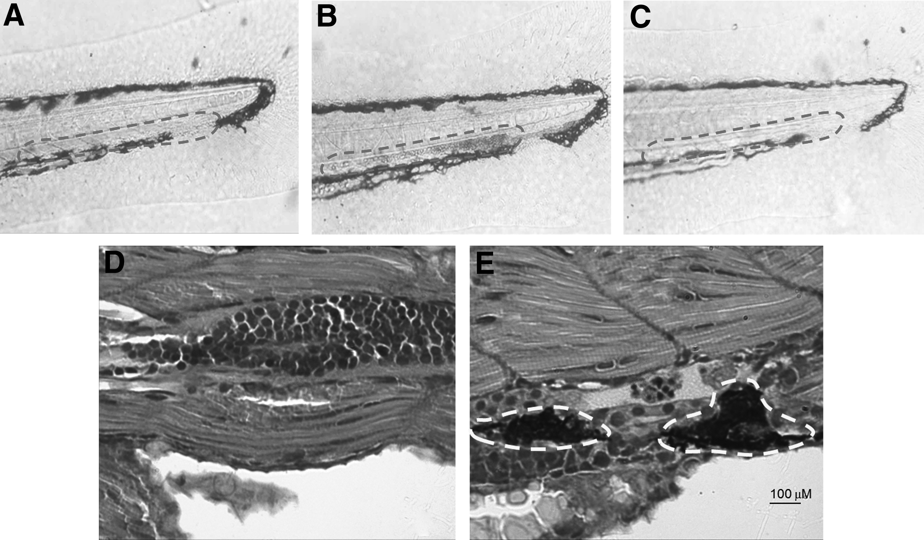
Determination of PHZ treatment concentrations and treatment time periods, as well as histopathological confirmation for PHZ-induced zebrafish thrombosis. Untreated zebrafish indicated no thrombus
Thrombus formation was confirmed by histopathological examination in zebrafish treated with 1.5 μM PHZ for 24 h (Fig. 1E). In quantitative analysis of thrombosis, time-dependent heart RBC intensity reduction (Fig. 2) was observed in zebrafish treated with 1.5 μM PHZ, further supporting our patented technology that indicates the heart RBCs intensity is reversely correlated with the thrombus severity and measuring heart RBCs could quantify thrombosis in zebrafish (China patent number: 201110126427.2). Based on these results, zebrafish treated with 1.5 μM PHZ for 24 h were selected as the optimum treatment concentration and treatment period for subsequent experiments.
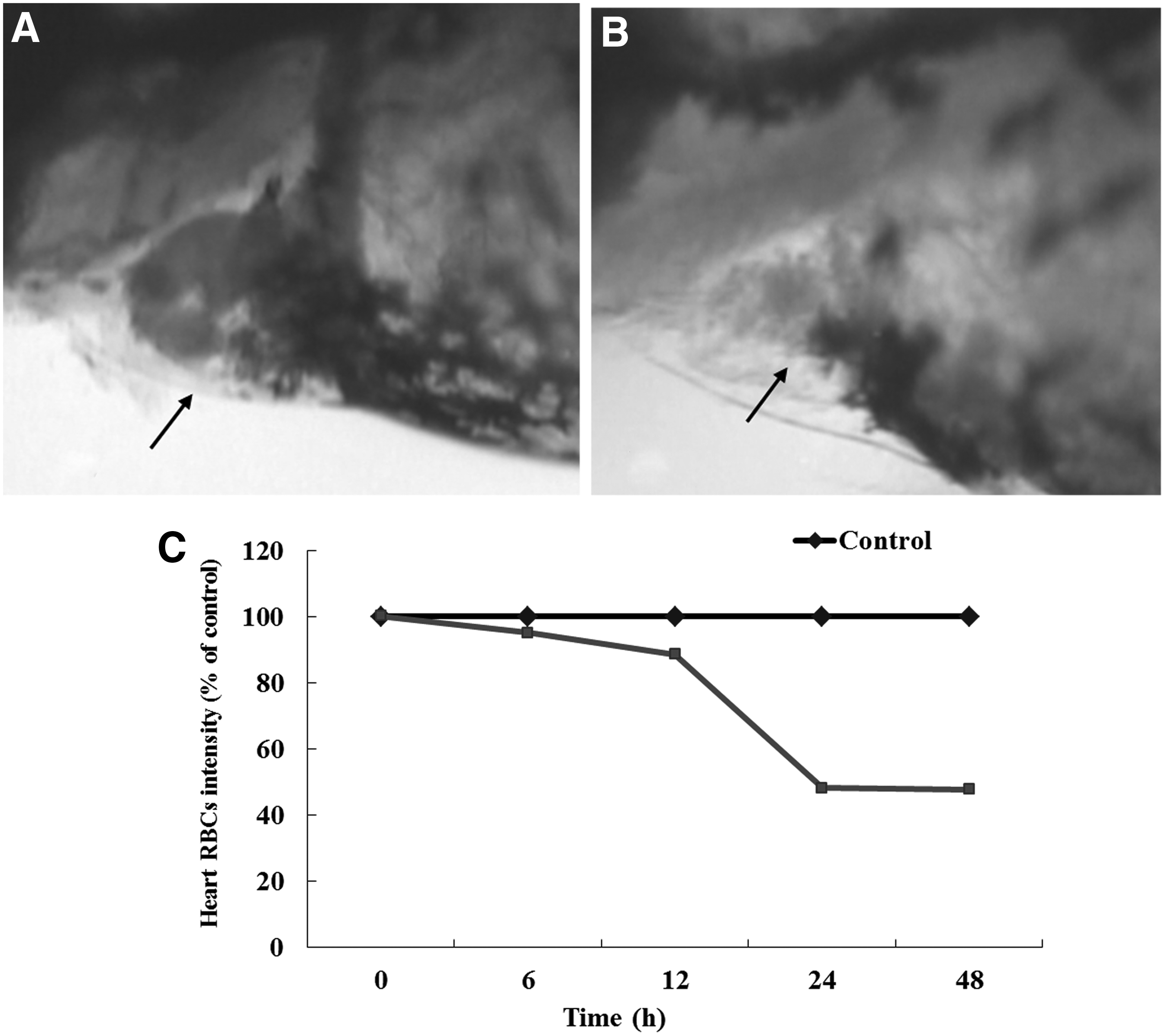
O-dianisidine staining of the heart RBCs of zebrafish for quantitative assay of thrombosis. The RBCs' intensity of zebrafish at 2 dpf treated with 1.5 μM PHZ for 24 h
Three target-specific drugs or chemicals warfarin, glutathione, and Z-Asp were used to characterize potential mechanisms responsible for PHZ-induced thrombosis in zebrafish. In a pilot study, we found that NOAEL was 10 μg/mL for warfarin, 500 μg/mL for glutathione, and 100 μg/mL for Z-Asp, respectively. Zebrafish were cotreated with 1.5 μM PHZ and warfarin, glutathione, or Z-Asp at a series of concentrations as indicated in Table 1. After cotreatment, we found that warfarin had no effect on the heart RBCs/hemoglobin staining intensity, whereas glutathione and Z-Asp significantly increased the heart RBCs/hemoglobin staining intensity (Fig. 3). The preventive efficacy of thrombosis was 9%–86% for glutathione and 4%–63% for Z-Asp. Statistically significant preventive effects on the zebrafish thrombosis were observed at 167 μg/mL (p < 0.001) and 500 μg/mL (p < 0.001) for glutathione and 33 μg/mL (p < 0.001) and 100 μg/mL (p < 0.001) for Z-Asp (Table 1 and Fig. 3).
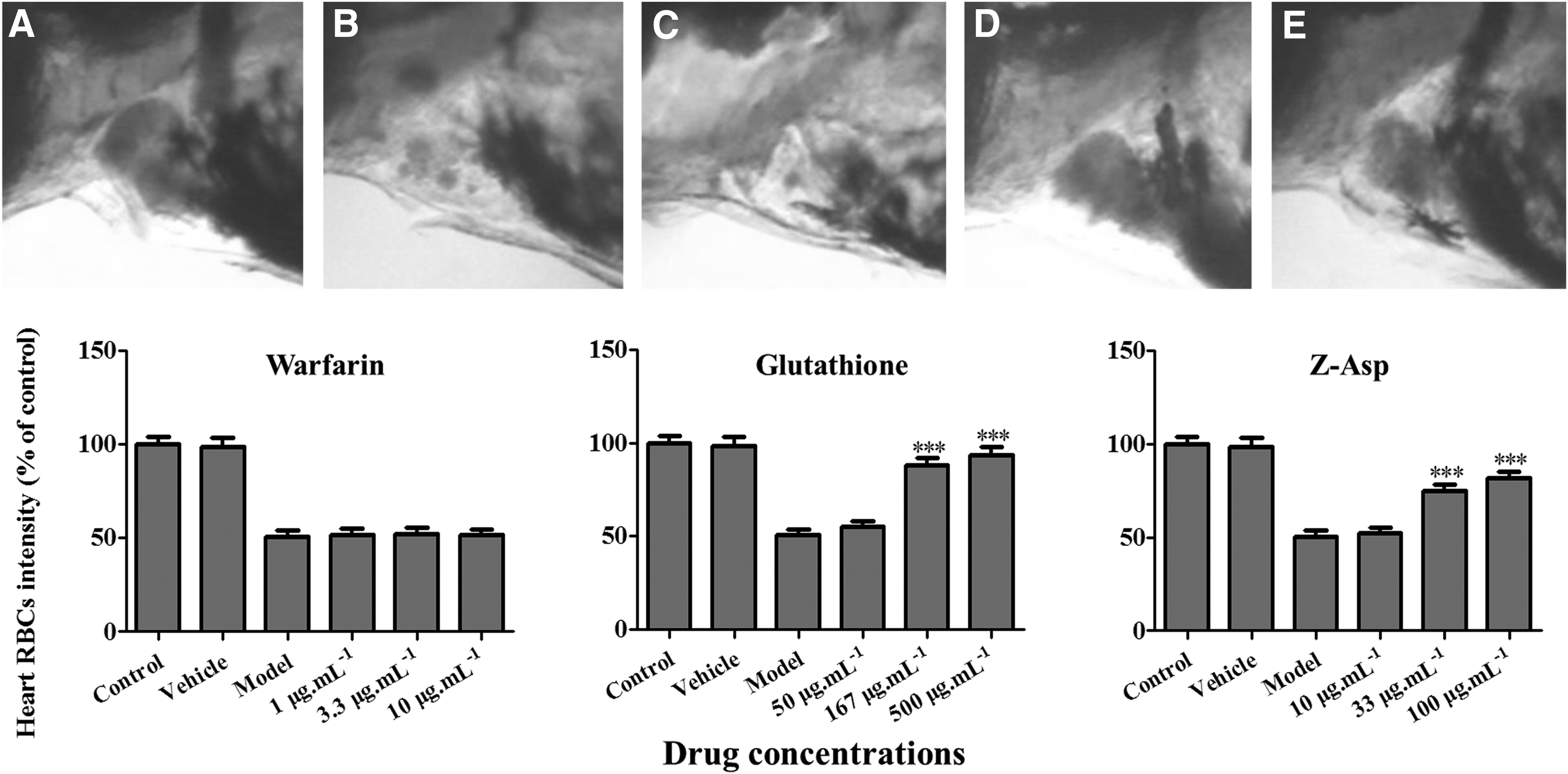
Heart RBCs and quantitative analysis of preventive efficacy in thrombotic zebrafish cotreated with a target-specific drug or chemical warfarin, glutathione, or Z-Asp for 24 h.
To determine whether the zebrafish response to antithrombotic drugs is similar to the response of mammalian model systems, we cotreated the zebrafish with 1.5 μM PHZ and each of the six known drugs (aspirin, clopidogrel, diltiazem hydrochloride injection, xuanshuantong injection, salvianolate injection, and astragalus injection) currently used in clinical practice. Furthermore, we used 1.5 μM PHZ to induce zebrafish thrombosis first and then treated the thrombosis zebrafish with each of the six drugs to evaluate whether or not these drugs could resolve the thrombosis. Three concentrations or dosages were assessed for each drug (Tables 2 and 3). NOAEL was 15 μg/mL for aspirin, 7.5 μg/mL for clopidogrel, 22.2 ng for diltiazem hydrochloride injection, 167 ng for xuanshuantong injection, 250 ng for salvianolate injection, and 3.3 ng for astragalus injection, respectively.
As expected, after a 24 h treatment, human antithrombotic drugs aspirin, clopidogrel, diltiazem hydrochloride injection, xuanshuantong injection, salvianolate injection, and astragalus injection significantly increased the heart RBCs/hemoglobin staining intensity (Fig. 4), implying the reduced thrombosis in zebrafish. The preventive efficacy of thrombosis was 19%–28% for aspirin, 23%–46% for clopidogrel, 37%–66% for diltiazem hydrochloride injection, 24%–47% for xuanshuantong injection, 5%–39% for salvianolate injection, and 19%–39% for astragalus injection, respectively. Statistically significant preventive effects on the zebrafish thrombosis were observed at 5 μg/mL (p < 0.05) for aspirin; 2.5 μg/mL (p < 0.01) and 7.5 μg/mL (p < 0.001) for clopidogrel; 2.5 ng (p < 0.001), 7.4 ng (p < 0.001), and 22.2 ng (p < 0.001) for diltiazem hydrochloride injection; 18.6 ng (p < 0.05), 55.7 ng (p < 0.001), and 167 ng (p < 0.01) for xuanshuantong injection; 250 ng (p < 0.001) for salvianolate injection; and 0.4 ng (p < 0.05), 1.1 ng (p < 0.001), and 3.3 ng (p < 0.01) for astragalus injection (Table 2 and Fig. 5).
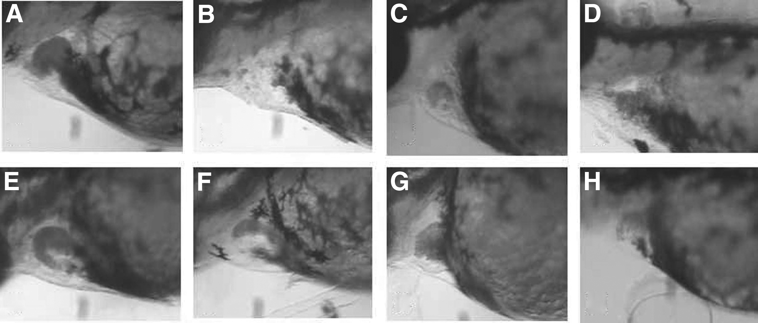
Increased heart RBCs in thrombotic zebrafish cotreated with antithrombotic drugs for 24 h.
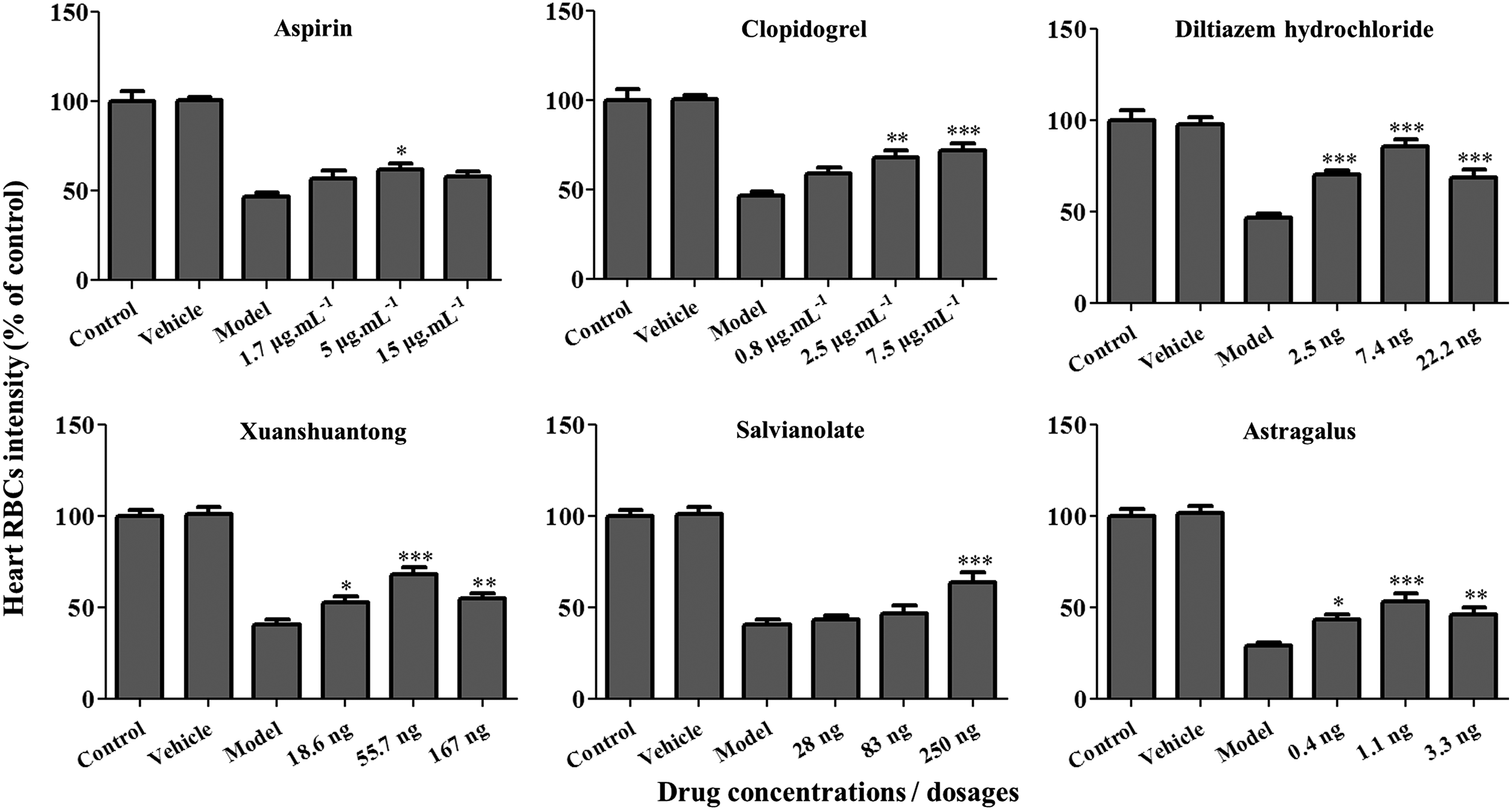
Quantitative analysis of preventive efficacy in zebrafish cotreated with PHZ and antithrombotic drugs for 24 h. Drug effect was measured based on quantitative image analysis of the heart RBC intensity in zebrafish. Compared with vehicle control: *p < 0.05, **p < 0.01, ***p < 0.001.
The therapeutic efficacy of thrombosis was 3%–41% for aspirin, 5%–41% for clopidogrel, 33%–49% for diltiazem hydrochloride injection, 16%–37% for xuanshuantong injection, 4%–37% for salvianolate injection, and 26%–37% for astragalus injection, respectively. Statistically significant therapeutic effects on the zebrafish thrombosis were observed at 5 μg/mL (p < 0.001) for aspirin; 2.5 μg/mL (p < 0.01) and 7.5 μg/mL (p < 0.001) for clopidogrel; 2.5 ng (p < 0.001), 7.4 ng (p < 0.001), and 22.2 ng (p < 0.01) for diltiazem hydrochloride injection; 55.7 ng (p < 0.01) and 167 ng (p < 0.05) for xuanshuantong injection; 83 ng (p < 0.001) and 250 ng (p < 0.001) for salvianolate injection; and 0.4 ng (p < 0.01), 1.1 ng (p < 0.001), and 3.3 ng (p < 0.01) for astragalus injection (Table 3 and Fig. 6). The reduced thrombus formations in the caudal vein of the zebrafish treated with antithrombotic drugs were confirmed by visual observations (data not shown).
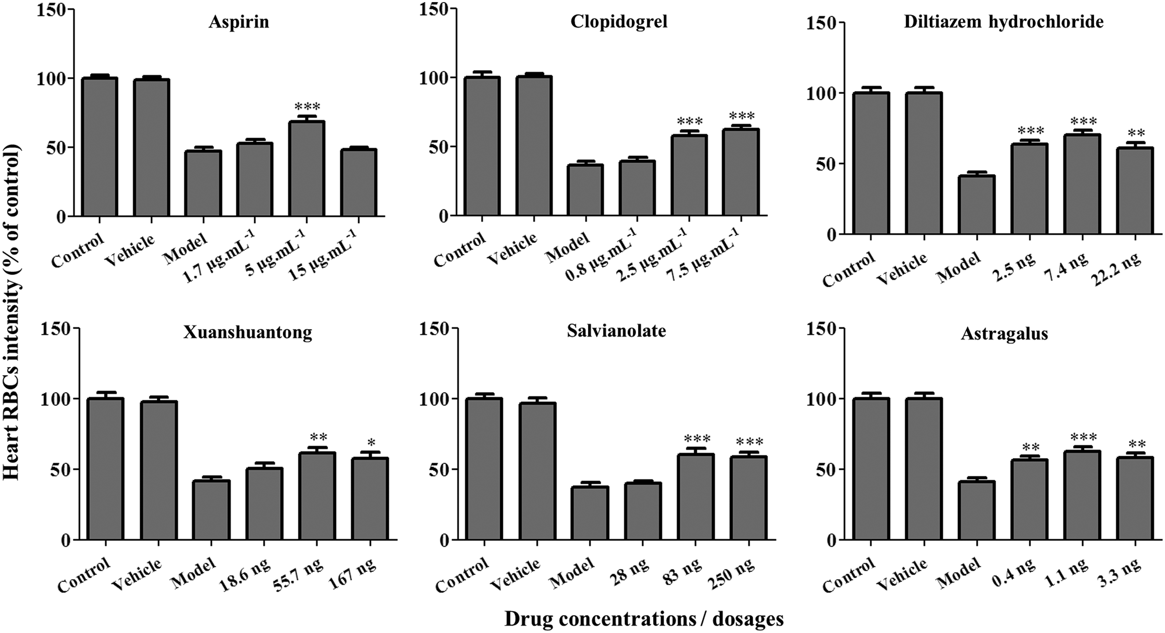
Quantitative analysis of therapeutic efficacy in thrombotic zebrafish after drug treatment for 24 h. Drug effect was measured based on quantitative image analysis of the heart RBC intensity in zebrafish. Compared with vehicle control: *p < 0.05, **p < 0.01, ***p < 0.001.
Discussion
This study was aimed to develop and validate a live zebrafish thrombosis model for rapid in vivo screening and efficacy assessment of antithrombotic drugs. Zebrafish is emerging as a predictive animal model for in vivo assessment of drug efficacy, toxicity, and safety.23,29–32 We use zebrafish because of their many advantages such as short generation times, amenability for large-scale screening, high fecundity, ease of in vitro fertilization, and transparency.4,33,34 Other important advantages of the zebrafish as an animal model are that the morphological and molecular basis of tissues and organs is either identical or similar to other vertebrates, including humans.23,35 Research regarding the zebrafish hematological system shows that human platelets and coagulation factors share many characteristics with their zebrafish counterparts. 6 Therefore, the zebrafish could be a new useful model for basic research on thrombocyte function, mechanisms of aggregation, release reactions of platelet granules, and thrombus formation, as well as antithrombotic drug screening.
The developing embryonic and larval zebrafish are small and transparent, enabling nonintrusive visualization of organs and biological processes in vivo with a high resolution. PHZ was reported to induce thrombus in rats. 10 A preliminary study found that treatment of zebrafish at 3–5 dpf with 20 mM PHZ could cause irreversible vascular occlusion in the caudal artery, and this study also found that adult zebrafish treatment with 5 mM PHZ for 5 min led to PS exposure on erythrocytes and that erythrocytes from PHZ-treated zebrafish showed an increased rate of thrombin generation. 5 O-dianisidine staining has been used for identification of the RBCs in developing zebrafish. 28 In this study, we have optimized PHZ treatment concentration and treatment time period to develop a live zebrafish thrombosis model with quantitative thrombosis analysis based on our previously patented technology (China patent number: 201110126427.2). Zebrafish at 2 dpf treated with PHZ at a concentration of 1.5 μM for a time period of 24 h were determined as the optimum conditions for the zebrafish thrombosis model development.
The thrombosis process in the live zebrafish was visually tracked and confirmed under a dissecting stereomicroscope. The thrombus size or severity in the caudal vein of zebrafish was consistent with the quantitative image assay of the heart RBCs. PHZ-induced zebrafish thrombosis was further confirmed by histopathological diagnosis. Sato et al. 11 observed histopathological time course of thrombosis in the lungs of PHZ-treated rats and they found focal deposition of eosinophilic material in the alveolar septa and positive immunohistochemical reactions with fibrin corresponding to eosinophilic material, indicating fibrin thrombi. This zebrafish thrombosis model is stable and highly reproducible with CV values of intraexperiments, interexperiments, and daily-to-daily variations ≤25% (data not shown).
Sato et al. suggested that endothelial dysfunction, pro-inflammatory condition, as well as regional stasis, and subsequent hemostatic disruption may contribute to PHZ-induced thrombosis in rats.11,12 In this study, we characterized possible mechanisms that is responsible for the pathogenesis of PHZ-induced thrombosis in zebrafish. A clinically extensively used anticoagulant drug warfarin was unable to block PHZ-induced zebrafish thrombus formation, whereas either a free radical scavenger glutathione or PS externalization inhibitor Z-Asp significantly prevented thrombosis in PHZ-induced zebrafish model, implying that PS externalization on RBCs and free radical generation may play major roles in PHZ-induced thrombosis of zebrafish. We postulate that PS externalization could result in cell clumping and free radical generation, leading to endothelial cell injury and thrombosis.
To validate zebrafish thrombosis model for pharmaceutical testing, six marked human antithrombotic drugs aspirin (thromboxane A2 inhibitor), 13 clopidogrel (ADP inhibitor), 14 diltiazem hydrochloride injection (calcium channel blocker), 15 xuanshuantong injection (inhibiting platelet aggregation, blood coagulation, fibrinolysis activation, and reducing plasma viscosity), 17 salvianolate injection (antiplatelet aggregation, anticoagulant, and antithrombosis), 36 and astragalus injection (restraining thrombosis and platelet aggregation) 37 were chosen and tested in this model for preventive and therapeutic effects on thrombosis. All six human antithrombotic drugs significantly reduced zebrafish thrombosis after 24 h cotreatment (thrombosis prevention) or treatment (thrombus therapy), indicating that the zebrafish thrombosis model developed in this report is suitable for in vivo screening and assessment of oral and injectable antithrombotic drugs no matter as a single compound medicine or a herb extract. Our results also further support that drugs derived from TCMs are valuable in the prevention and treatment of thrombotic diseases. The zebrafish as a model organism offer opportunities for rapid in vivo drug discovery and development. Larval zebrafish thrombosis model developed in this study was a live and physiology-associated whole animal assay. This model is a useful tool for whole animal thrombosis studies and for screening preventive and therapeutic agents of thrombus. The use of zebrafish as an alternative animal model for antithrombotic drug screening and assessment could save time, decrease costs, and reduce drug failure at later stages of drug development.
Extended studies are in progress to define endothelial dysfunction in PHZ-treated zebrafish using gene expression profiling and other biochemical and molecular biology techniques in this thrombosis model. We are also further validating this model using more human drugs and developing this model into a higher throughput screening or even an automatic assay system. We believe that the power of the zebrafish models and assays could be better utilized to advance the basic and translational research and drug discovery and development, including in the field of hemostasis and thrombosis.
Conclusion
This study developed and validated a zebrafish thrombosis model that could be used for in vivo screening and efficacy assessment of antithrombotic drugs. This conventional zebrafish thrombosis model is predictive, easily available, and less expensive with a short testing time and could speed up antithrombotic drug research and development.
Footnotes
Acknowledgments
Chun-Qi Li and Xiao-Yu Zhu designed the research; Xiao-Yu Zhu, Hong-Cui Liu, Sheng-Ya Guo, Bo Xia, and Ru-Shun Song performed the research; Xiao-Yu Zhu, Sheng-Ya Guo, Li-Jiang Zhang, Qiao-Cong Lao, and Yao-Xian Xuan analyzed the data; and Xiao-Yu Zhu and Chun-Qi Li wrote the article. This work was sponsored, in part, by the Zhejiang Provincial Key Science & Technology Project grant (2014C03009) and the National Key Science & Technology Project grant of the 12th 5-year program of China (2011ZX09301-003).
Disclosure Statement
No competing financial interests exist.