




Abstract
Abstract
Recently, the indiscriminate use of antibiotics in the aquaculture sector has raised public concern because of possible toxic effects, development of bacterial resistance, and accumulation of residues in individual tissues. Even if several countries have developed regulations about their use, it is clear that long-term growth of the aquaculture industry requires both ecologically sound practices and sustainable resource management. Alternative strategies for better management of antibiotic administration are of primary interest to improve absorption rates and, as a consequence, to reduce their release into the aquatic environment. The present study investigates, for the first time to our knowledge, a new methodology for oxytetracycline (OTC) administration through the use of iron oxide nanoparticles (NPs) (made of maghemite γ-Fe2O3) in zebrafish (Danio rerio). Fish were divided into 4 experimental groups: control; group A exposed to 4 mg/L OTC (through water); group B exposed to the 100 mg/L SAMNs@OTC complex (equivalent to 4 mg/L OTC), and group C exposed to bare NPs. No detoxification processes or anatomical alterations were observed in fish exposed to bare NPs. Exposure of fish to the SAMNs@OTC complex resulted in a 10 times higher OTC accumulation with respect to using water exposure. This new OTC administration method seems much more efficient with respect to the traditional way of exposure and has the potentiality to reduce antibiotic utilization and possible environmental impacts. However, the dynamics related to OTC release from the SAMNs@OTC complex are still not clear and need further investigations.
Introduction
T
The considerable release of antibiotics in the aquatic environment has been associated with the emergence of antibiotic resistance in bacterial fish pathogens, with the alteration of the microbiota of the aquatic ecosystems 5 and a constant presence in surface waters in the ng/L to mg/L range. 6 These relatively low concentrations can cause uncertain ecological effects and be a potential threat to aquatic organisms as a consequence of chronic exposure to these substances. 1
These concerns have led to the development of more strict regulations for the use of antibiotics in the aquaculture sector and, at date, some countries (Europe, North America and Japan) have licensed the use of a few antibiotics for fish treatment. 2
Among the approved antibiotics, oxytetracycline (OTC) is one of the most commonly used in aquaculture. 7 It belongs to the tetracycline class of broad-spectrum antibiotics with bacteriostatic effects and can be administered both orally or added to the water. 8 OTC is poorly absorbed by fish and Yonar 8 reported that 70%–80% of OTC was intact in exposed animals. OTC generally accumulates in skin, liver, and intestine.9–11 For example, Malvisi and collaborators, 9 in 1996, reported a 1.82 ± 1.26 μg/g OTC skin concentration in seabass exposed for 14 days to a medicated diet containing 75 mg OTC/kg body weight, while a 13.9 ± 9.5 μg/g OTC concentration was detected in the liver of seabass (Dicentrarchus labrax) 128 h after intravascular administration of 40 mg/kg of OTC. 11
It is clear that alternative strategies for better management of antibiotic administration are of primary interest to improve absorption rates and, as a consequence, to reduce the release in the aquatic environment.12–14 Metal nanoparticles (NPs), including superparamagnetic iron oxide NPs, are interesting materials that can be used in a wide range of biomedical applications, including drug delivery, magnetic resonance imaging, and hyperthermal treatment of tumors. 15
Among these materials, the two magnetic forms of iron oxide, magnetite (Fe3O4) and maghemite (c-Fe2O3), are considered suitable for in vivo applications15,16 since their surface can be easily functionalized to meet a number of requirements. Besides enabling selective binding of different bioactive molecules, magnetic NPs functionalized for biomedical applications must retain their magnetic properties, be nontoxic, hydrophilic, and biocompatible, and remain stable in aqueous colloidal suspensions.
Many drug nanocarriers were proposed to protect the drug from rapid degradation and/or clearance by enhancing drug concentration in target tissues. 17 As a consequence, a lower drug dose is usually required with a reduction of toxic collateral effects for patients or damage to the environment. 18
The present study investigates for the first time to our knowledge a new methodology for OTC administration through the use of iron oxide NPs (constituted of maghemite γ-Fe2O3) in zebrafish. zebrafish is a widely used model for biomedical, developmental biology, genetics, toxicology, and aquaculture studies due to its high reproduction rate and the abundant information that has recently become available from genome sequencing.19,20
OTC was administered in water solution or NPs bound at a final concentration of 4 mg/L for 28 days. OTC accumulation in different tissues was determined by high-pressure liquid chromatography (HPLC) analysis; liver, gills, and intestine integrity was examined by histological analysis and stress and growth markers were analyzed by real-time polymerase chain reaction (PCR).
Materials and Methods
Ethics
All procedures involving animals were conducted in line with the Italian legislation on experimental animals and were approved by the Health Ministry's Department of Veterinary Public Health and by the Ethics Committee of Università Politecnica delle Marche (Authorization No. 640/2015-PR). Optimal rearing conditions were applied throughout the study, and all efforts were made to minimize animal suffering by using an anesthetic (MS222; Sigma Aldrich).
Synthesis of surface active maghemite NPs
Bare NPs called surface active maghemite nanoparticles (SAMNs) constituted of stoichiometric maghemite (γ-Fe2O3) were synthetized as described in Refs.21,22 Briefly summarizing: FeCl3·6H2O (10.0 g, 37 mmol) was dissolved in MilliQ grade water (800 mL) under vigorous stirring at room temperature, to allow the reduction reaction occurrence NaBH4 solution (2 g, 53 mmol) in ammonia (3.5%, 100 mL, 4.86 mol/mol Fe) was quickly added to the solution. The final synthesis product resulted in a red brown nanopowder composed of maghemite NPs with a mean diameter of 11 ± 2 nm that showed a magnetic response when exposed to a magnetic field.
Formation of the complex magnetic NPs and OTC (SAMNs@OTC)
OTC hydrochloride (20 mg/L, O5875; Sigma Aldrich, Italy) was incubated with various NP concentrations (0.1, 0.2, 0.5, 1.0, 1.5, and 2.0 g/L) in H2O at room temperature. This solution was stored under overnight constant agitation to promote complex formation. Then, magnetic NPs were separated from the aqueous solution with the assistance of a magnet. Finally, OTC concentration in the supernatants was checked by spectrophotometry using the Varian Cary 60 instrument (Agilent Technologies, CA).
Fish
Adult 6-month-old female AB zebrafish were kept in a zebrafish system (Tecniplast, Varese, Italy) at 28°C, 14L/10D photoperiod, pH 7.0, NO2 and NH3 < 0.03 mg/L, and fed with a commercial pellet (Blue Line, Italy) twice daily (2% body weight) for 1 month. The experiment started after this first period of acclimation. Females were chosen because of the complexity of the reproductive cycle and because of the fact that oocytes are able to accumulate some molecules, including antibiotics.
Experimental design
Adult zebrafish (600 animals) were kept in 12 tanks (100 L) divided into 4 experimental groups, each of 150 fish (50 fish per tank), and exposed to the following conditions for 28 days: control (3 × 50 fish tanks) (28°C, 14L/10D photoperiod pH 7.0, NO2 and NH3 < 0.03 mg/L; total 150 fish); group A (3 × 50 fish tanks): fish exposed to 4 mg/L OTC through water (same chemical–physical conditions of control group); group B (3 × 50 fish tanks): fish exposed to 100 mg/L SAMNs@OTC equivalent to a concentration of 4 mg OTC (same chemical–physical conditions of control group); group C (3 × 50 fish tanks): fish exposed to 100 mg/L SAMNs (same chemical–physical conditions of control group).
Samplings were performed at 0, 14, and 28 days after the beginning of treatment. Fish were collected and anesthetized with a lethal dose of MS222 (1 g/L) (Sigma Aldrich) and the liver, brain, digestive tract, skin, ovary, and in toto fish were sampled and properly stored for further analysis.
In addition, water samples (30 mL in three replicates) were collected from all tanks at 0, 7, 14, 21, and 28 days from the beginning of the experiment to determine the presence of OTC in the water (see next paragraph).
HPLC analysis
Extraction procedure
About 100 mg of each tissue sample (in triplicate) was weighed, triturated, and homogenized in a blender, placed in a plastic tube, and dissolved in 1 mL of ultrapure water acidified with 1% phosphoric acid. The tube was vigorously mixed for 2 min, treated in ultrasonic bath for 5 min, and then centrifuged at 13,000 rpm for 10 min. The supernatant was collected, filtered through 0.2-μm membrane filters, and stirred before injection into the chromatograph. A 20 μL aliquot was injected.
The separation conditions for the OTC were a mobile phase flow of 0.8 mL/min containing acetonitrile acidified with 0.1% phosphoric acid and ultrapure water acidified with 1% phosphoric acid (15:85, v/v) filtered through a 0.45-μm nylon filter under vacuum and degassed by ultrasonication. Column oven temperature was 25°C, and ultraviolet detector was operated at wavelength of 360 nm. The quantitation was accomplished by using an analytical calibration curve built with six concentration levels in the range 20–250 μg/L (OTC). Extraction recoveries were determined by spiking untreated biological samples (100 mg) with a fortification solution at a limit of quantification (LOQ) and LOD levels: 500 and 100 μg/kg.
The HPLC system (Agilent 1100 series with Chemstation) used for analysis consists of a quaternary pump (Agilent 1100 series), a vacuum degasser, an injector, and a wavelength ultraviolet detector (Agilent 1100 series). The chromatographic column was an analytical reversed-phase ZORBAX Eclipse XDB C18, 4.6 × 150 mm, 5 μm.
Method validation
The method was in-house validated using the following performance criteria: linearity, sensitivity and selectivity, detection and quantification limits, and accuracy and precision. The solution for calibration and fortification was prepared in ultrapure water acidified with 1% phosphoric acid and stored at temperature ≤ −18°C.
Linearity, sensitivity and selectivity, and detection and quantification limits were established by the analytical curve (20, 40, 50, 80, 100, and 250 μg/L of OTC). The LOQ was obtained from the calibration curve and calculated using the following expression: y = ax + b, where y is the peak area of OTC and x is the amount of OTC in μg/mL; then, the LOQ was expressed in μg/kg by the following formula: OTC (μg/L) × Vex (mL)/W (g), where Vex is the extraction volume and W is the untreated sample weight. The limit of detection (LOD) was the lowest amount of an analyte in a sample that can be detected but not necessarily quantitated as an exact value and it was 100 μg/kg. The precision of the method, expressed as the relative standard deviation of peak area measurements (n = 5), was evaluated by the recovery data obtained (accuracy) at the LOQ concentration level (RSD% = 6.51). The accuracy of the method, expressed as percent recovery, was determined at 500 μg/kg, the LOQ concentration level (mean recovery 95.5%).
Histological analysis
Ten samples of the intestine, liver, and gills, randomly collected at each sampling time from control and groups A, B, C, were fixed by immersion in 4% paraformaldehyde and stored at 4°C overnight. Samples were then washed in PBS 1 × , dehydrated in an ethanol series, and embedded in paraffin. Slides of 4 μm sections (cut with a microtome LEICA) were stained with Mayer's hematoxylin and eosin stain and examined under a photomicroscope (Olympus Vanox photomicroscope, Japan).
RNA extraction and cDNA synthesis
Total RNA extraction from single zebrafish livers (five replicates randomly collected at each sampling time from control, A, B, and C groups) was optimized using the RNeasy Mini Kit (Qiagen, Italy) following the manufacturer's protocol. Total RNA extracted was eluted in 20 μL RNase-free water (Qiagen). Final RNA concentration was determined by the NanoPhotometer P-Class (Implen, Germany). RNA integrity was verified by ethidium bromide staining of 28S and 18S ribosomal RNAbands on 1% agarose gel. RNA was stored at −80°C until use. Total RNA was treated with DNAse (10 IU at 37°C for 10 min; MBI Fermentas). Then, 3 μg RNA was used for cDNA synthesis with the iScript cDNA Synthesis Kit (Bio-Rad) following the producer's instructions.
Real-time PCR
PCRs were performed with SYBR Green in an iQ5 iCycler thermal cycler (both from Bio-Rad) in triplicate. Reactions were set on a 96-well plate by mixing for each sample 1 μL cDNA diluted 1:20, 5 μL of 2 × concentrated iQ™ SYBR Green Supermix containing SYBR Green as the fluorescent intercalating agent, 0.3 μM forward primer, and 0.3 μM reverse primer. The thermal profile for all reactions was 3 min at 95°C, followed by 45 cycles of 20 s at 95°C, 20 s at 60°C, and 20 s at 72°C. Fluorescence was monitored at the end of each cycle. Dissociation curve analysis showed a single peak in all cases.
Relative quantification of the expression of genes involved in fish stress response (hnf4a, hsp70.1, sod1, sod2, and gsta.1) and growth (igf1, igf2a, and mstnb) was performed using rplp0 and actbl as the housekeeping genes to standardize the results (Table 1). Data were analyzed using the iQ5 optical system software version 2.0, including Genex Macro iQ5 Conversion and Genex Macro iQ5 files.
Amplification products were sequenced and homology was verified. No amplification product was detected in negative controls and no primer–dimer formation was found in control templates. Data were analyzed using the iQ5 optical system software version 2.0, including Genex Macro iQ5 Conversion and Genex Macro iQ5 files (all from Bio-Rad). Modification of gene expression is reported with respect to controls. Primer sequences were designed using Primer3 (210 v. 0.4.0) starting from zebrafish sequences available in ZFIN. Primers were used at a final concentration of 10 pmol/μL.
Statistical analysis
HPLC and real-time PCR data were analyzed by two-way analysis of variance and analysis of variance, both followed by Tukey's post-test. The statistical software package Prism5 (GraphPad Software) was used for analyses. Significance was set at p < 0.05.
Results
OTC content by HPLC analysis
At T0, OTC was not detectable in all analyzed tissues (control, group A, B, and C) (Fig. 1a–f). In addition, the same result was observed in all control and group C samples at T1 and T2 (Fig. 1a–f).
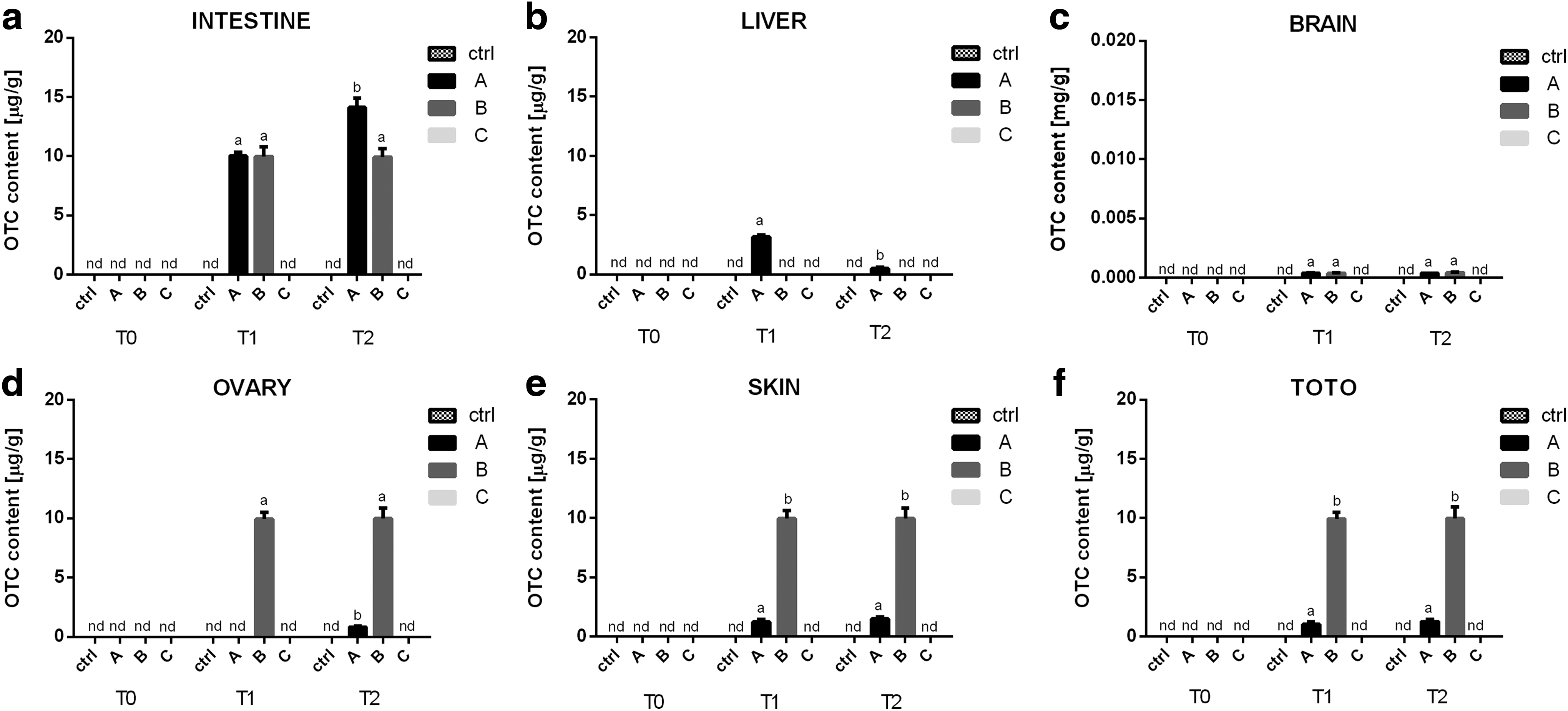
Mean OTC concentrations (expressed in μg/g of body weight) detected in ZF intestine
On the contrary, during the whole experimental time, in all group A and B samples, an OTC accumulation was evident (Fig. 1a, c–f), with the exception of group B livers exposed to SAMNs@OTC complex, in which OTC was not detectable (Fig. 1b).
The highest concentration of OTC was recorded in group A and B intestine samples (Fig. 1a). At T1 there were no significant differences (p > 0.05) between the two experimental groups and the mean concentration was 10.00 ± 0.30 and 9.97 ± 0.80 μg/g, for group A and B, respectively. At T2 the mean concentration was 14.10 ± 0.800 and 9.95 ± 0.67 μg/g, for group A and B, respectively. Group A concentration was significantly (p < 0.05) higher than group B.
In brain samples (Fig. 1c) of both group A and B, no significant differences (p > 0.05) in OTC concentration were detected at both sampling times (0.039 ± 0.02 and 0.37 ± 0.04 μg/g at T1, 0.37 ± 0.03 and 0.43 ± 0.06 μg/g at T2 in group A and B, respectively).
On the contrary, group B ovary, skin, and in toto samples (exposed to SAMNs@OTC complex) (Fig. 1d, e, f, group B) showed a significantly higher (∼10 times magnitude increase) OTC concentration (p < 0.05) with respect to those exposed to OTC through water (Fig. 1d, e, f, group A).
Histological analysis
The histological analysis of liver (Fig. 2A–D), gills (Fig. 2E–H), and intestine (Fig. 2I, L–N) did not show any significant difference in the morphological structure of the analyzed tissues. All the samples showed a normal structural and morphological integrity without evidence of steatosis, inflammation, vacuolation, edema, cellular atrophy, or necrosis.
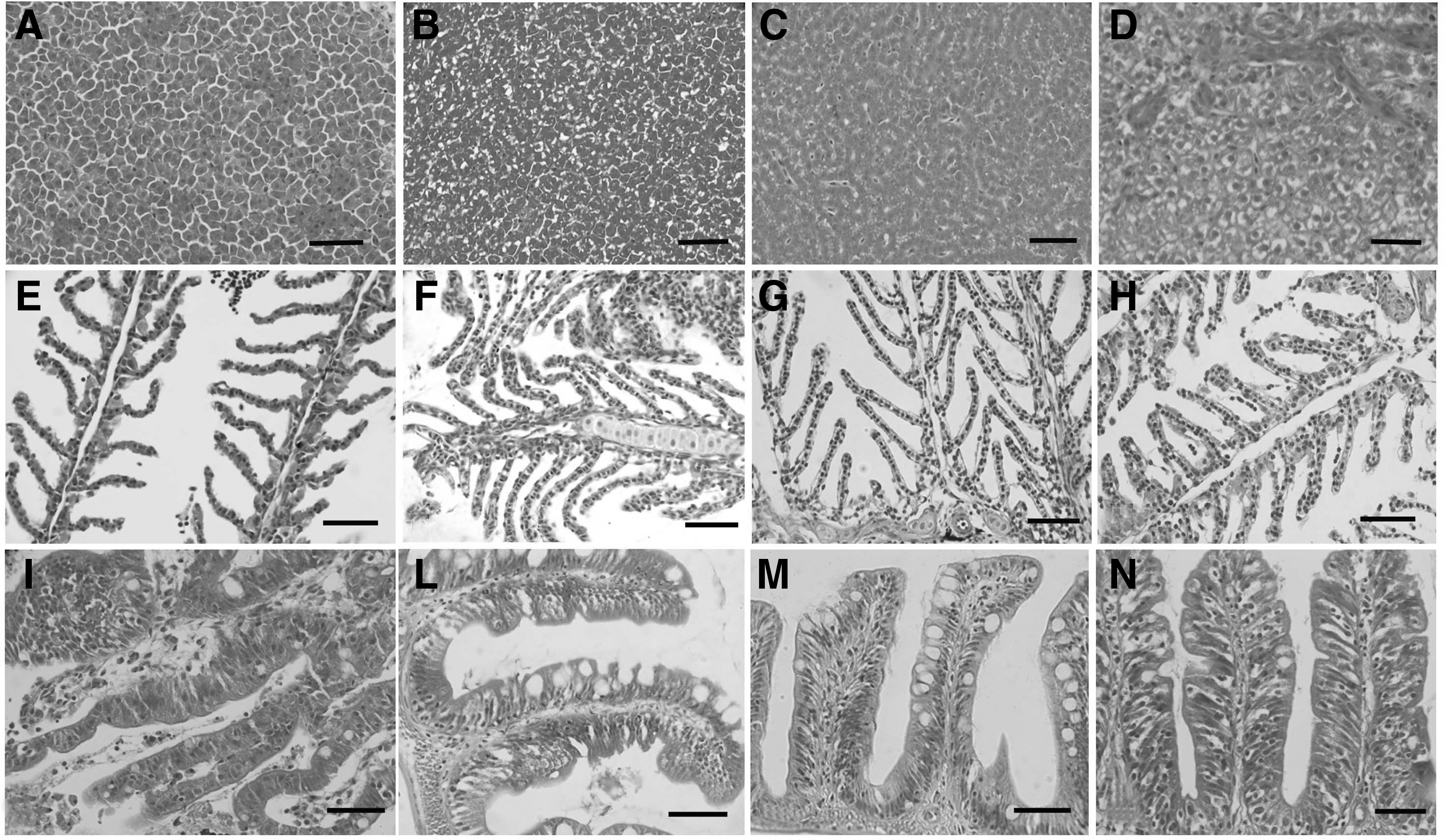
Example of histomorphology (day 28) of ZF: liver ctrl
Real-time PCR results
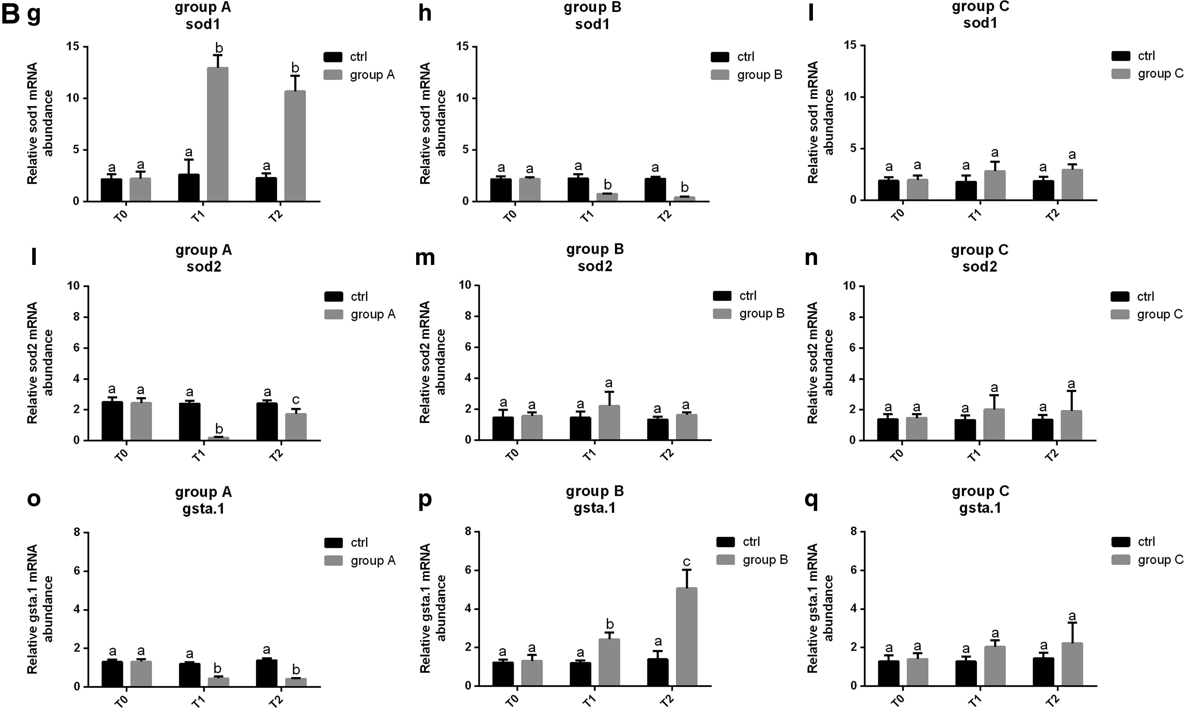
Real-time PCR analysis was performed on genes involved in fish growth (igf1, igf2a, and mstnb) and stress response (hnf4a, hsp70.1, sod1, sod2, and gsta.1). Results showed that naked NP administration (group C) did not cause significant variations in the expression of all the analyzed genes with respect to control during the whole experimental period (Fig. 3c, f, i), (Fig. 4c, f), (Fig. 4i, n, q).
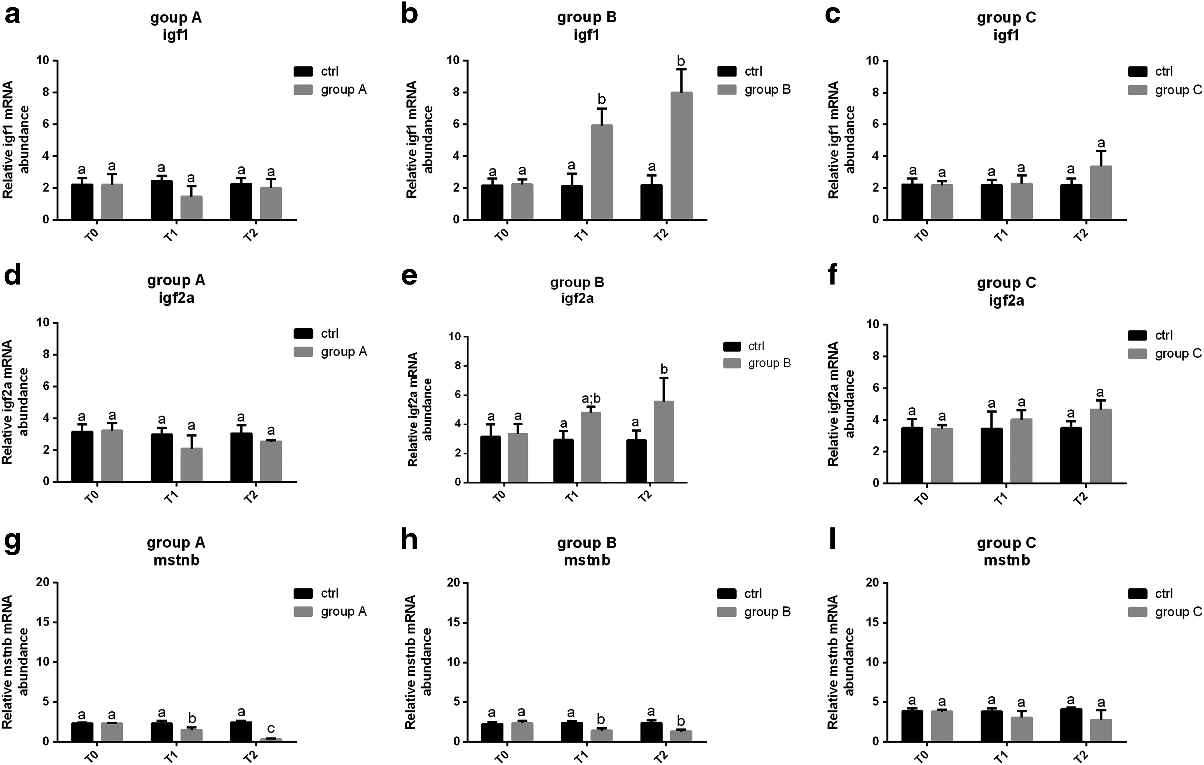
Relative mRNA levels of genes involved in fish growth (igf1, igf2, and mstnb) analyzed in the liver at 0 (T0), 14 (T1), and 28 (T2) days of exposure. Igf1, igf2a, and mstnb expression was analyzed in control (ctrl) and group A

On the contrary, through both water and SAMNs@OTC complex, OTC administered was able to act on the expression of some of the genes analyzed.
Growth factors
Regarding Igf1 gene expression, no significant (p > 0.05) differences with respect to control were detected in all group A samples (Fig. 3a). Conversely, in group B, exposed to the SAMNs@OTC complex, a time-dependent significant increase in igf1 gene expression was observed (p < 0.05) (Fig. 3b) during the experiment, with respect to control. A similar result was observed for igf2a gene expression for both group A and B (Fig. 3d, e, respectively). Finally, both through water (group A) and SAMNs@OTC (group B) exposure induced a significant (p < 0.05) time-related decrease of mstnb mRNA levels (p < 0.05) compared to control (Fig. 3g, h).
Stress and detoxification stress markers
In group A samples, hnf4a gene expression showed a significantly lower gene expression at both T1 and T2 with respect to control (p < 0.05) (Fig. 4a), while in group B, no significant differences were observed with respect to control at T1 and T2 (p > 0.05) (Fig 4b).
Regarding hsp70.1, a significantly lower (p < 0.05) gene expression was observed in both group A (Fig. 4d) and B (Fig. 4e) with respect to control at both T1 and T2. In particular, group B showed a significantly time-related decrease of this gene expression (p < 0.05).
As concerns sod1 gene expression, an opposite change in gene expression was detected in group A and B. A significant increase (p < 0.05) of sod1 gene expression was observed in group A with respect to control (Fig. 4g), while for group B samples, a significant reduction (p < 0.05) in sod1 gene expression was detected both at T1 and T2 (Fig. 4h).
With regard to sod2 gene expression, group A samples were characterized by a significant downregulation (p < 0.05) compared to control, at both T1 and T2 (Fig. 4l). Group B gene expression did not show any significant difference (p > 0.05) with respect to control during the whole time of exposure (Fig. 4m).
Finally, an opposite trend in gsta.1 expression was observed between group A and B; a significant decrease was observed in group A (p < 0.05) (Fig. 4o), while a significant time-related increase of gsta.1 expression was evident in group B (p < 0.05) (Fig. 4p).
Discussion
In the present study, the possible application of synthetic iron oxide NPs (SAMNs) for OTC administration in zebrafish was evaluated. At date, there is a strong need for the development of new and alternative administration methods for this antibiotic in the aquaculture sector because of its wide use, 23 very low absorption rate, 8 and its high release into the environment through the feces and waste waters. 24
NPs are materials characterized by different chemical compositions, which have been reported to be potentially toxic for aquatic organisms.25,26 However, toxicity relies on their composition, size, and physical and chemical properties. 27 In particular, iron oxide NPs have been reported to have no toxicity in in vivo and in vitro systems.15,16,28 The same results were confirmed in the present study, evidencing, by a multidisciplinary approach, that iron oxide NP (naked or as a complex with OTC) exposure did not cause any significant stress response in treated fish.
All the different experimental conditions tested in this study had no effects on the structural and morphological integrity of gills, liver, and intestine of treated zebrafish. These tissues were selected because of their importance in ecotoxicological studies.29,30
In addition, as expected, 31 molecular analysis revealed that especially SAMNs@OTC complex exposure had a greater growth promoter action with respect to using water treatment. This result can be easily related to the OTC concentrations detected by HPLC in the different organs, which were higher in fish exposed to the SAMNs@OTC complex with respect to using water exposure and are discussed later on in this same section.
Previous studies showed that OTC can be responsible for the induction of toxicity in fish liver by altering the activity of enzymes involved in stress response.32,33 As already demonstrated by Nakano et al., 34 OTC exposure is able to cause a significant decrease in hsp70 gene expression; in the present study, the same result was observed in the liver of both group A and B fish.
The liver is in fact an important storage organ, the primary function of which, in fish, is detoxification. 35 Regarding detoxification genes, it is clear that in the present study, naked NPs had no effect on their gene expression. However, group A and B results often show opposite trends. This could be related to the activation of different defense/detoxification mechanisms related to the administration method and/or the different OTC concentrations detected in the samples analyzed.
As expected, OTC administration, both through water and through NPs, caused an accumulation in target tissues of zebrafish. For both treatments, HPLC analysis showed that the intestine was the main accumulation organ. On the contrary, the lowest OTC concentrations were observed in the brain and, surprisingly, in the liver of the treated fish. The low OTC concentration detected in the brain can easily be explained by the hydrophilic characteristics of OTC, 36 its high molecular weight (496.89 g/mol, CAS#O5875-50G; Sigma-aldrich), and the presence of a blood–brain barrier characterized by tight junctions and selective transporters, which restrict paracellular diffusion of molecules with high molecular weight. 37
Results obtained from liver samples were not obvious. No OTC was detected in all liver samples of SAMNs@OTC (group B) exposed fish. In addition, in fish exposed to 4 mg/L OTC (using water), the liver antibiotic concentration was very low during the whole experimental time, especially if compared to previous experiments reporting that the liver is one of the main accumulation organs of OTC.38–40 These results, as already demonstrated in grass carp, 41 could be explained by the occurrence of a very active and fast detoxification mechanism in the liver, and its high vascularization that guarantees a fast absorption rate as well as fast disposal mechanisms. 41 However, the total absence of OTC in the liver of group B fish exposed to SAMNs@OTC complex lays the foundation for new hypotheses. First, it has to be underlined that the binding of OTC on SAMNs is very stable. Attempts to obtain the release of OTC from SAMN surface were unsuccessfully carried out using absolute ethanol, and 2 M NH4OH that led only to a partial release of OTC, as already tested for the release of bound substances on iron oxide NPs.42,43 As a consequence, a first hypothesis is that OTC was not released by the NPs because the liver did not provide the proper chemical–physical environment for its release. For this reason, the absence of OTC in the liver could be explained by the inability of HPLC to detect the complex SAMNs@OTC. The second hypothesis, which is now under study, is that SAMNs@OTC complex, within the organism, was able to bind to other macromolecules as already observed in complex media.17,44,45 Some of these macromolecules are released into the blood stream and could serve as a carrier for SAMNs@OTC promoting their accumulation in other organs. In the present study, while using water treatment did not cause a significant OTC accumulation in the ovary and skin of treated fish, SAMNs@OTC treatment caused a 10 times higher OTC concentration in the same target organs. It is known that zebrafish presents an asynchronous ovary and spawns every day. 46 Oocyte maturation is supported by vitellogenesis that occurs in the liver through the production of vitellogenin that is secondly released into the blood stream before reaching the ovary. 1 Vitellogenin is a high molecular mass glycolipophosphoprotein, which is usually encoded by multiple vg genes in several species, including fish. 1 It could be speculated that SAMNs@OTC could bind to vitellogenin, and secondly be incorporated into the oocytes, after reaching the ovary through the blood stream.
This new administration method is thus very promising for the treatment of those diseases characterized by vertical transmission. An example is the treatment of Piscirickettsia salmonis for which OTC is usually used. 47 In addition, nanotechnology and in particular the NPs developed in this study, which have a high surface/volume ratio, a colloidal stability, a proper surface reactivity, and superparamagnetism, can easily be collected through the use of a magnet and may represent an efficient treatment technology (bioremediation) to remove OTC from waste waters.
Conclusions
The present study represents an important first step for the development of a new OTC administration method able to guarantee a higher absorption of the antibiotic in zebrafish. In fact, group B fish revealed a 10 times higher OTC accumulation with respect to group A (exposed through water). As a consequence, this new administration method seems much more efficient with respect to the traditional way of exposure, reduces antibiotic utilization and possible environmental impacts. However, the dynamics related to OTC release from the SAMNs@OTC complex are still not clear and need further investigations.
Footnotes
Acknowledgments
The authors are grateful to Dr. Giorgia Gioacchini and Dr. Francesca Maradonna for their help during sampling. The project was supported by Fondi di Ateneo 2015 to IO and by Biotecnologie B.T. Srl, Pantalla di Todi, Perugia, Italy.
Disclosure Statement
No competing financial interests exist.