
Abstract
Abstract
Microinjection of zebrafish larvae is an essential technique for delivery of treatments, dyes, microbes, and xenotransplantation into various tissues. Although a number of casts are available to orient embryos at the single-cell stage, no device has been specifically designed to position hatching-stage larvae for microinjection of different tissues. In this study, we present a reusable silicone device consisting of arrayed microstructures, designed to immobilize 2 days postfertilization larvae in lateral, ventral, and dorsal orientations, while providing maximal access to target sites for microinjection. Injection of rhodamine dextran was used to demonstrate the utility of this device for precise microinjection of multiple anatomical targets.
Introduction
Z
More recently, microfabrication and microfluidic approaches have provided useful devices to study small animal models, including Drosophila, Caenorhabditis, and zebrafish larvae.3,4 Early approaches achieved positioning of zebrafish embryos to visualize development, with the ability to add chemical stimuli at will.5,6 More recently, this concept has been scaled up to enable high-throughput chemical screening. 7 High-resolution three-dimensional (3D) printing has also been used to create detailed casts designed to orient zebrafish embryos and larvae for confocal imaging, without requiring them to be mounted in agarose. 8 In combination with in vivo microscopy, microfluidic systems have been used to monitor neutrophil chemotaxis migration in anesthetized zebrafish larvae. 9 Microfluidic approaches can achieve immobilization of larvae in the absence of anesthetic, which has been useful for studying physical reactions of larvae to stimuli such as hypoxia 10 and to measure brain activity following drug treatment.11–13 However, these devices have not effectively addressed the issue of microinjection. Although some microfluidic device designs for immobilizing larvae included a window to allow putative access for microinjection, 9 this approach does not provide the flexibility to target delivery to different tissues.
Microinjection of chemicals and dyes into zebrafish larvae requires small diameter needles, which can easily penetrate zebrafish tissues. For these experiments, simple techniques such as immobilization on an agarose bed by water tension are relatively efficient. 14 However, for delivery of larger particles, such as fungal conidia, cells, or microbeads, a larger diameter needle is required, which often makes penetration of the desired tissue more difficult. To address this issue, larvae can be mounted in agarose or methylcellulose or immobilized with a holding pipette, 15 but these approaches are time-consuming and technically challenging. Our attempts to utilize 3D printed molds 8 highlighted the resolution limitations of standard 3D printing for this application.
In this study, we utilized photolithographic methods to build microstructured surface arrays (MSAs) for optimal immobilization and positioning of dozens of zebrafish for microinjection. These structures allow alignment of 2 days postfertilization (dpf) larvae in one of the three standard orientations: dorsally for microinjection into the brain, ventrally for delivery to the circulation through the duct of Cuvier, and laterally for targeting of the otic vesicle and somites.
Materials and Methods
Device design and fabrication of the silicone mold
The master wafer was fabricated through standard photolithographic techniques using Mylar masks that were designed using AutoCAD and printed by Fineline Imaging (Colorado Springs, CO). Three masks were used to achieve three height layers with SU-8 100 during the photolithography process, which were aligned using a Quintel Q-2001CT mask aligner. Polydimethylsiloxane (PDMS) (Sylgard 184; Ellsworth Adhesives, Wilmington, MA) microfluidic devices were made by replica molding from the master wafer. Briefly, PDMS and curing agent were combined at a 10:1 ratio, mixed thoroughly, and poured over the master wafer. PDMS was then degassed for at least 30 min, then baked at 65°C for at least 3 h. PDMS arrays were then cut from the wafer and treated with oxygen plasma before wetting. To wet, the PDMS was submerged in E3 and bubbles removed by flowing E3 over the molds with a transfer pipette. Negative PDMS molds were made by treating PDMS casts with oxygen plasma and then exposing to silane vapor. 16 Fresh PDMS was then poured over the original cast, degassed, and baked. The negative PDMS mold was then peeled from the original cast and used as a negative for casting of agarose molds.
Fabrication of agarose molds
For casting the agarose molds, 2% low gelling temperature agarose was prepared by dissolving agarose powder in E3 and boiling. Hot agarose was then poured over the PDMS negative and bubbles removed by flowing hot agarose around the features with a transfer pipette. Once set, the agarose block was removed from the PDMS negative, placed in a Petri dish, and wetted with E3 (containing tricaine) before use.
Zebrafish husbandry
Zebrafish stocks were maintained and bred according to standard protocols. 2 The Casper (roy−/−nacre−/−) 17 Tg(mpx:GFPuwm1) 18 strain used was a kind gift from Elliott Hagedorn and Leonard Zon. Embryos were manually dechorionated at 30 hpf to allow straightening of larvae overnight before mounting. Microinjection of larvae was approved by the Massachusetts General Hospital Subcommittee on Research Animal Care under Protocol 2011N000127.
Microinjection of zebrafish larvae
For microinjection, larvae were anesthetized in E3 containing 168 mg/L Tricaine (MS222, pH 7.5) before mounting. Microinjection needles were prepared from glass capillaries (World Precision Instruments) using a Sutter P-1000 Micropipette puller. Microinjections were performed using an MPPI-1 pressure injector (Applied Scientific Instruments) and an MM33 micromanipulator (Sutter Instruments). Injection was traced using high-molecular-weight (70 kDa) rhodamine dextran (Sigma) at 2 mg/mL.
Results
Application of photolithographic approaches to fabricate silicone MSAs
We designed our MSA devices to immobilize 2 dpf zebrafish in orientations convenient for microinjection. To design the structures, we calculated dimensions based on the online zebrafish bioatlas database (http://bio-atlas.psu.edu/zf/view.php?s=529&atlas=49). We used these dimensions to model the basic shape of the larvae (Fig. 1A) and employed three different height layers (100, 200, and 400 μm) to model geometrical details that are specific to each desired orientation (Fig. 1C, D). Structures were designed to leave the target anatomy fully exposed, while confining the larvae as securely as possible to provide stabilization during the microinjection procedure.

Designing microstructures to orient zebrafish larvae.
Photolithography was utilized to generate a master wafer (Fig. 2), which was then used to cast the MSAs in PDMS (Fig. 3), which provides optical clarity, longevity, and is biologically compatible.
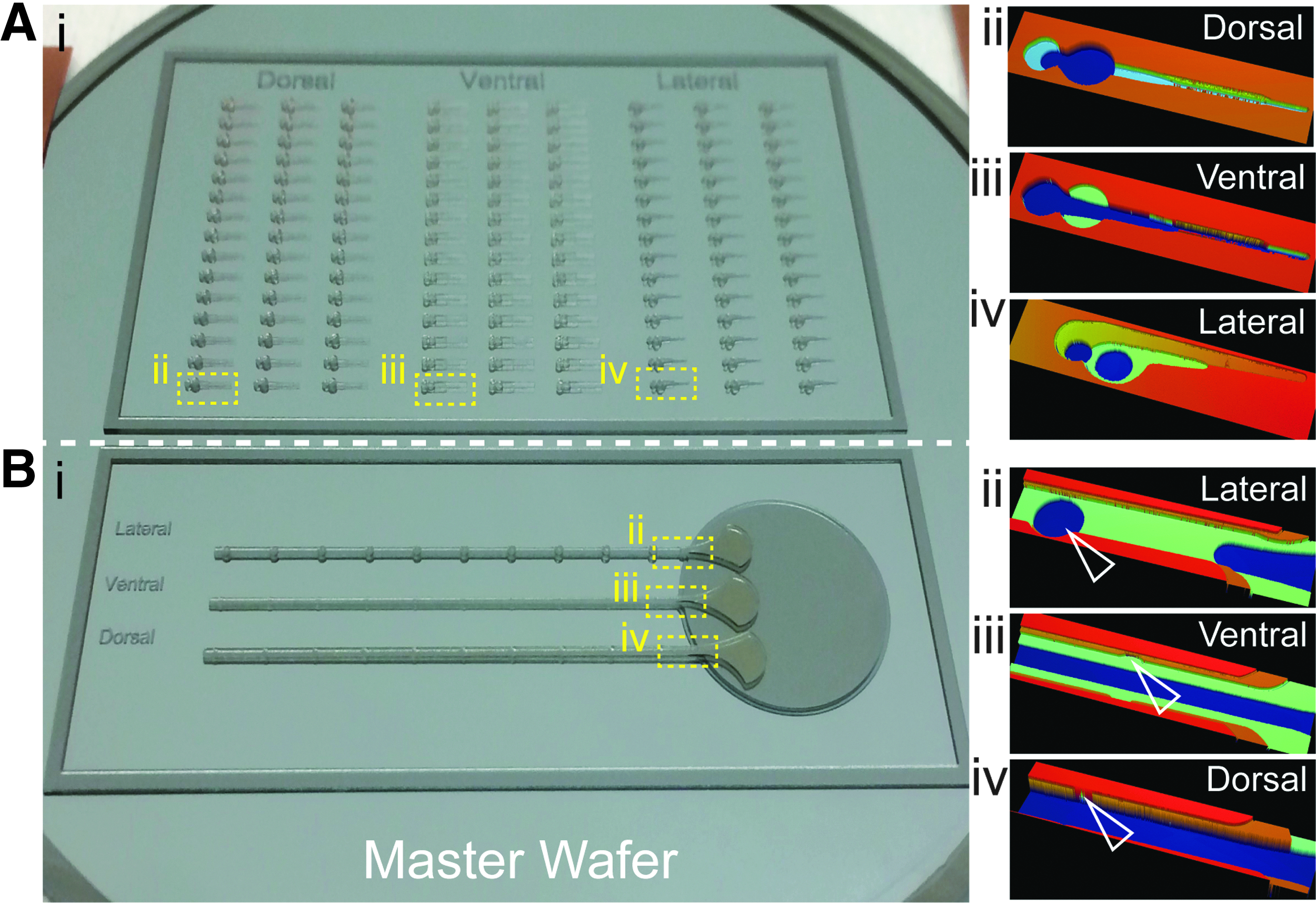
Fabricating the design using photolithography.
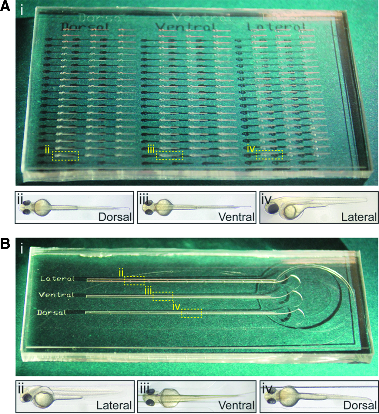
Using microstructured silicone surfaces to orient larvae.
MSAs for orienting individual larvae
To provide effective immobilization of 48–56 hpf zebrafish, MSAs were designed as an array of 180 individual casts, each of which provided a secure fit for an individual larva. To mount the larvae, they were first anesthetized, and then delivered onto the surface of the array by transfer pipette, covered by a shallow layer of E3 media. Larvae were then manually arranged into the casts using a standard zebrafish hair loop manipulator.
For lateral orientation, the deepest structures are situated at the yolk and eye, while the medium and shallow structures were designed to position the yolk extension and trunk, respectively (Figs. 1Ci and 2Aiv). For dorsal orientation, the deepest structures match the yolk and heart, while the medium depth matches the head, yolk extension, and fin, and the shallowest provides guidance for the trunk (Figs. 1Ciii and 2Aii). For the ventral orientation, the deepest structures run the length of the larva and orient by matching the width of the head, yolk extension, and trunk. The medium depth matches the yolk, and the shallowest again provides extra guidance for the trunk (Figs. 1Cii and 2Aiii).
Following orientation of the larvae, excess E3 was removed from the surface, providing additional immobilization, which was particularly useful for laterally mounted larvae. Lateral mounting was suitable for microinjection of the somite (Fig. 4A) and otic vesicle (Fig. 4B), while ventral mounting exposed the circulation valley beneath the heart for vascular delivery (Fig. 4C). Dorsal mounting provided excellent immobilization for microinjection into the hindbrain ventricle (Fig. 4D).

Targeted delivery to different zebrafish tissues by microinjection of oriented larvae.
Microstructured channels for linear alignment of multiple larvae
Individual pockets provided the greatest stability as they prevented the larva from sliding horizontally during microinjection. As an alternative, we designed microstructured channels, which we hypothesized might be easier to load. For this approach, sliding of the larvae during injection should be minimal, provided that the microinjection needle is applied at an angle perpendicular to the channel. Channels were designed using the same depths as the microstructure arrays, such that 10 larvae could be manually maneuvered from a shallow circular reservoir (Fig. 3B) and aligned tip to tail in parallel channels in the desired orientation. Deep funnel-shaped guides were included at the opening of the channels, allowing embryos to be correctly oriented before sliding them into the channel.
For lateral orientation, the channels are at medium depth to match the trunk, while deeper features are arrayed along the channel to align the larvae through their yolk (Figs. 1Di and 2Bii, open arrowhead). For dorsal orientation, the channel is deep to allow movement of the larvae yolk-side down, while features in the wall provide guides to array the larvae before microinjection (Figs. 1Diii and 2Biv, open arrowhead). For ventral orientation, a deep thin channel provides orientation of the larvae through their trunk, while features along the wall allow them to be arrayed according to positioning of their yolk (Figs. 1Dii and 2Biii, open arrowhead).
AutoCAD files containing the designs presented here will be permanently available to download at https://dx-doi-org-s.web.bisu.edu.cn/10.6084/m9.figshare.4282853 and on the ZFIN community protocols wiki page: https://wiki.zfin.org/display/prot/ZFIN+Protocol+Wiki. Updated files may also be added if improvements are made upon the existing design.
Practical application of silicone versus agarose MSAs
We investigated whether a microstructured surface in agarose could be used, providing an environment closer to that used in traditional microinjection methods. Rather than casting agarose directly from our master silicone wafer, which would risk contamination and deformation of the structures with agarose residues, we instead created a negative PDMS mold from an existing PDMS device. We prevented the bonding between the two PDMS layers by treating the existing device with oxygen plasma, followed by silane vapor, resulting in an inert surface. 16 Fresh PDMS was mixed with primer, poured on top, then degassed, and baked. This PDMS-PDMS casting approach resulted in a negative PDMS mold that could then be used to cast agarose.
Two percent (w/v) low-melting-point agarose was chosen for this process to allow more time for degassing while the agarose set. This approach provided excellent fidelity of the microstructure features on a surface that was hydrophilic, which facilitated easier wetting of the device before use.
Discussion
In this study, we present a range of silicone microstructures designed to immobilize 2 dpf zebrafish at different orientations for microinjection. Photolithographic techniques were utilized to provide structures with the spatial resolution required to effectively constrain larvae during the procedure. These MSAs provide superior larva orientation and stabilization compared with other methods. The flexibility to select different orientations provides increased accuracy for microinjection. Stabilization of the larvae in these orientations is particularly important when using larger bore needles to deliver particles such as fungal conidia or cells for xenotransplantation. Using other techniques, the needle often pushes the larvae across the surface rather than penetrating the tissue.
Most zebrafish microinjection channels are cast in agarose using a plastic mold. Agarose is affordable, easy to work with, and available within any laboratory that performs molecular biology. It is, however, easily damaged and susceptible to microbial contamination, rendering it less amenable to reuse.
PDMS is also relatively cheap and easy to prepare and provides robust structures that can be used multiple times. One drawback of using PDMS in the context described here is its hydrophobicity, which can make wetting of deeper features challenging. If not wetted properly, residual air pockets within the deeper features render them unusable. In the current study, this challenge was overcome by wetting the device immediately following oxygen plasma treatment, which makes the PDMS surface transiently hydrophilic. The microstructured surface was then stored submerged for the lifetime of the device.
The robust orientation of larvae using MSAs also provides opportunity for imaging, particularly with upright microscopes. Dorsal and ventral mounting of larvae, for brain or heart imaging, respectively, is notoriously time-consuming. The MSAs presented here would allow rapid orientation of dozens of embryos, which could be further stabilized for imaging with agarose. Use of inverted microscopes would require the PDMS to be cast in a very thin layer to create the shortest possible working distance between the lens and the larva.
The microstructured surfaces and channels presented here will increase the throughput of larval microinjection. Minor alterations of the flexibility to array dozens of larvae in each orientation for the individual casts and 10 per orientation per channel. By multiplexing of the basic design and homogenizing orientations, these devices could provide increased throughput for specific applications. Additionally, adjustment of the microstructure dimensions used here could also be used to achieve immobilization of larvae at other developmental ages, such as 3 or 4 dpf.
Zebrafish and microfabrication methodologies are well matched in scale and application. Used in parallel, they provide opportunities for comparative and translational studies of cell biology in vivo and in vitro. Used in conjunction, microfluidic approaches provide tools for manipulating zebrafish embryos and larvae, with the potential to customize devices for specific applications. The microstructured surfaces that we present here provide stable orientation of zebrafish larvae for enhanced microinjection accuracy and throughput, and are particularly useful for delivery of larger particles using wide-bore needles.
Footnotes
Acknowledgments
The authors would like to thank David Langenau for generously providing aquarium space; Eric Stone, John C. Moore, and Qin Tang for help with zebrafish maintenance and reagents; and Anne Robertson and Elliott Hagedorn from Leonard Zon's laboratory for procuring the zebrafish strain used here. They would also like to thank Octavio Hurtado for advice on photolithographic techniques. F.E. was funded by Fellowships from Shriners Hospital for Children and the American Australian Association. This work was funded by NIH grant GM92804.
Author Disclosure Statement
No competing financial interests exist.