




Abstract
In vivo imaging of adult zebrafish is challenging, particularly for dynamic or long acquisitions when using, for example, positron emission tomography, single photon emission computed tomography, computed tomography (CT), or magnetic resonance imaging (MRI). An aqueous environment is indispensable to ensure animal welfare, but commercial small-animal imaging chambers do not provide such conditions, as they are designed for rodents. In this study we present a dedicated flow-through chamber that includes fish immobilization and allows for the continuous supply of fresh water and anesthetics, as well as the removal of excretions. Both flow simulations and experiments, as well as first scans with MRI and CT, support the suitability of the chamber.
Introduction
In this study we present a novel imaging chamber and measurement procedure specifically designed for dynamic noninvasive in vivo imaging of adult zebrafish. The focus lies on positron emission tomography (PET), 1 single photon emission computed tomography (SPECT), computed tomography (CT),2,3 and magnetic resonance imaging (MRI),4,5 as well as their combination, for example, PET/CT and PET/MRI.
The goal is to allow the fish to remain anesthetized and immobilized in an aqueous environment 6 without stress or danger of injuries over the scan duration. To that aim, oxygen supply and excretion removal should also be guaranteed. The latter is particularly important to prevent image degradation when contrast media (CT and MRI) or radioactive molecules (PET and SPECT) are used.
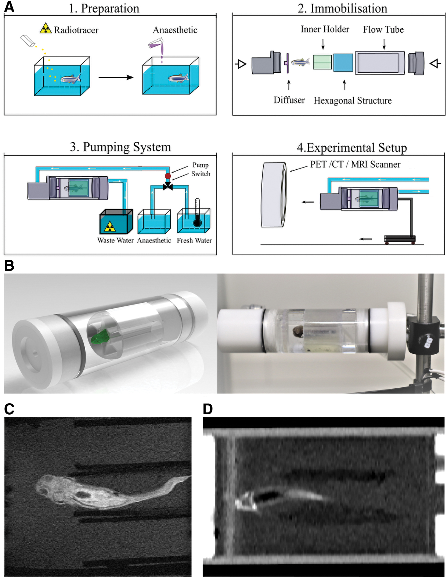
Our imaging procedure is divided into four main parts (Fig. 1A): (1) fish preparation, (2) immobilization, (3) pumping system, and (4) imaging. This study focuses on (2), (3), and (4). Experiments were performed using fish dummies and dead zebrafish; the success of these preliminary functional and proof-of-efficacy tests is a prerequisite for the authorization to validate our imaging chamber in living fish.
Methods
The imaging chamber consists of two main parts, an internal holder, made of flexible three-dimensional-printed material, for fish immobilization, and a flow-through chamber (Fig. 1B). The chamber is made of polymethylmethacrylate and is 14.4 cm long with a diameter of 38 mm. To reduce attenuation and scatter for CT, PET, and SPECT and for compatibility with MRI, the prototype contains no metal parts. The same internal holder can accommodate different fish sizes and prevents fish motion without harming the skin. A diffusor placed in front of the fish reduces the speed and pressure of the inflowing water and improves the distribution of anesthesia.
We developed a dedicated pumping system aimed to provide continuous flow of tempered fresh water and anesthetics, as well as to enable the removal of excreted radiotracer or contrast agents. It consists of three peristaltic pumps coupled to reach the required flow rates. They are connected to the chamber and water tanks using hoses with an inner diameter of 4 mm. Three water tanks are to be used: one for tempered oxygen-rich water, a second for water mixed with anesthetics, and the third for wastewater. The latter needs to be shielded for the use with PET or SPECT.
In this study, only one tank was employed as no living specimen was used. We have investigated water flow and pressure in the chamber and various diffusor designs using flow simulations, optical techniques, fish dummies, and ex vivo measurements. The chamber tightness was examined using a radiotracer method, able to detect very small leakages. Preliminary imaging tests with a clinical CT and a 1-Tesla small-animal MRI scanner were performed to check compatibility with those systems.
Results and Discussion
The chamber proved to be watertight. A flow rate of 8.4 mL/s provided the best results for inflow behavior and removal. This rate corresponds to the natural swimming speed of zebrafish, that is, the subsequent pressure is similar to its natural environment. The selected diffusor led to a homogeneous water flow, so that anesthetics could be administered in a controlled way. For that specific flow rate, the water in the chamber was exchanged within 15 s; this translates into efficient removal of potential residues.
Thanks to the immobilization holder, no displacements of either the dummy or the dead fish caused by the water flow were observed. The flexible material of the internal holder easily adapts to various fish sizes and shapes; further holders can also be printed very quickly and at low costs in slightly varying dimensions.
Images acquired with human CT and small-animal MRI showed no obvious artifacts (Fig. 1C, D), proving that the chamber is compatible with the two modalities. The hose arrangement should allow for placing the chamber within other small-animal scanners as well.
For step (1) of the imaging procedure, fish preparation, we plan the following: first, the specimen is anesthetized, for example, through the surrounding water. 7 For PET or SPECT, a radiotracer is administered before anesthesia, either though the surrounding water, oral gavage, or through labeled microalgae. 8 Specific CT and MRI studies might require the administration of contrast medium at this point. The type of anesthesia and the necessary dose for PET scan times up to 30-min scans are currently under investigation.
Future study will focus on further ex vivo and first in vivo experiments with continuous water flow and various scanners and modalities. As a next step, the development of a vital sign control system for guaranteed animal welfare is planned.
Footnotes
Disclosure Statement
No competing financial interests exist.
Funding Information
The project is funded by German Research Foundation (DFG) under grant agreement no. RA2830/5-1 and supported by the Cluster of Excellence Precision Medicine in Chronic Inflammation (PMI) under grant EXC216.