




Abstract
Zebrafish larva is a simple model system to study blood vessel development because of its excellent optical properties. Current methods to study blood vessels in zebrafish involve utilizing either blood vessel-specific transgenic lines or specialized techniques such as microangiography and video capillaroscopy. In this study, we have developed a simple protocol using ImageJ to observe the blood vessels of live zebrafish larva at different developmental stages, namely 1- to 4-day postfertilization (dpf). To validate this protocol, we treated 1 dpf zebrafish embryos with 100 μM quercetin, and analyzed the impaired development of subintestinal vein in the quercetin-treated 3 dpf larvae.
Introduction
The zebrafish is a versatile vertebrate model to study the development of cardiovascular system. 1 The available techniques to observe and study the blood vascular system in live zebrafish larvae are the following: (1) blood vessel-specific transgenic lines, for example, Tg(fli1a:EGFP)y 1 line, which has been extensively used for studies focusing on vasotoxicity, 2 tumor angiogenesis, 3 and pro- and antiangiogenic drug screening.4,5
Although this is a robust strategy, availability and maintenance of specific transgenic lines through multiple generations could be a challenging exercise for small laboratories; (2) microangiographic method, wherein fluoresceinated carboxylated latex beads are injected into the sinus venosus, and the imaging of blood vessels is done using confocal microscope. 6 This method has the limitation of blood vessels needing to be imaged within 15 min after the injection of the beads; (3) digital motion analysis and (4) video capillaroscopic methods, wherein the zebrafish larval blood vessels are observed by imaging the blood flow area as a series of image stacks and processed using a proprietary algorithm.7,8
The video capillaroscopic method has been successfully used to observe zebrafish larval blood vessels up to 2 dpf stage larvae only. 7 Hence, in this study, we have developed a simple method for blood vessel observation based on the blood flow-based image subtraction strategy using a mobile phone camera, simple light microscope, and ImageJ, a freely available image processing program.
A Brief Workflow
Zebrafish wild-type embryos/larvae were raised in optimal conditions at 28.5°C ± 0.5°C as per the standard protocol. 9 The zebrafish larvae were anesthetized using 0.168 mg/mL of tricaine 7 and their blood flow were recorded at specific developmental stages using a simple light microscope and a mobile phone camera. 10 Subsequently, the video files were converted into a series of individual images by DVDVideoSoft Free Studio v.6.6.28. Then the image sequences were uploaded to ImageJ, processed, and the blood vessels were observed by performing image subtraction.11,12 The detailed steps of the protocol are given in the Supplementary File S1.
Results and Discussion
Zebrafish is a useful model system to perform in vivo angiogenic assays because of its optical transparency during early developmental stages. The principle behind this protocol is the image subtraction analysis, wherein a field of moving objects (blood cells) in an image sequence can be obtained by subtracting the difference between two image frame fields. The moving object field, known as shifting vectors, is piled up from successive image sequences that will give the path (area of blood cell flow) of movement of blood cells. The deduced path of blood cell movement will be finally represented as blood vessels (Supplementary Video S1). 8 So, we adopted this principle and observed blood vessels in zebrafish larvae by performing the analysis of image sequences using ImageJ.
The overall work flow is represented in Figure 1A. The blood vessels of selected wild-type zebrafish larvae (1–4 dpf) were observed using this protocol as shown in Figure 1B. The arrow marks in Figure 1B represent the following: development of main vasculature in 1 dpf larva, intersegmental vessel formation at 2 dpf, and subintestinal vein (SIV) development at 3 dpf. We could also observe angiogenic vessel extension in the tail region of the 3 and 4 dpf larvae (Fig. 1B). Subsequently, we demonstrated the inhibition of SIV angiogenesis by treating zebrafish larvae with 100 μM quercetin, a flavonoid with antiangiogenic potential that has been reported to inhibit the SIV angiogenesis in zebrafish larvae. 5
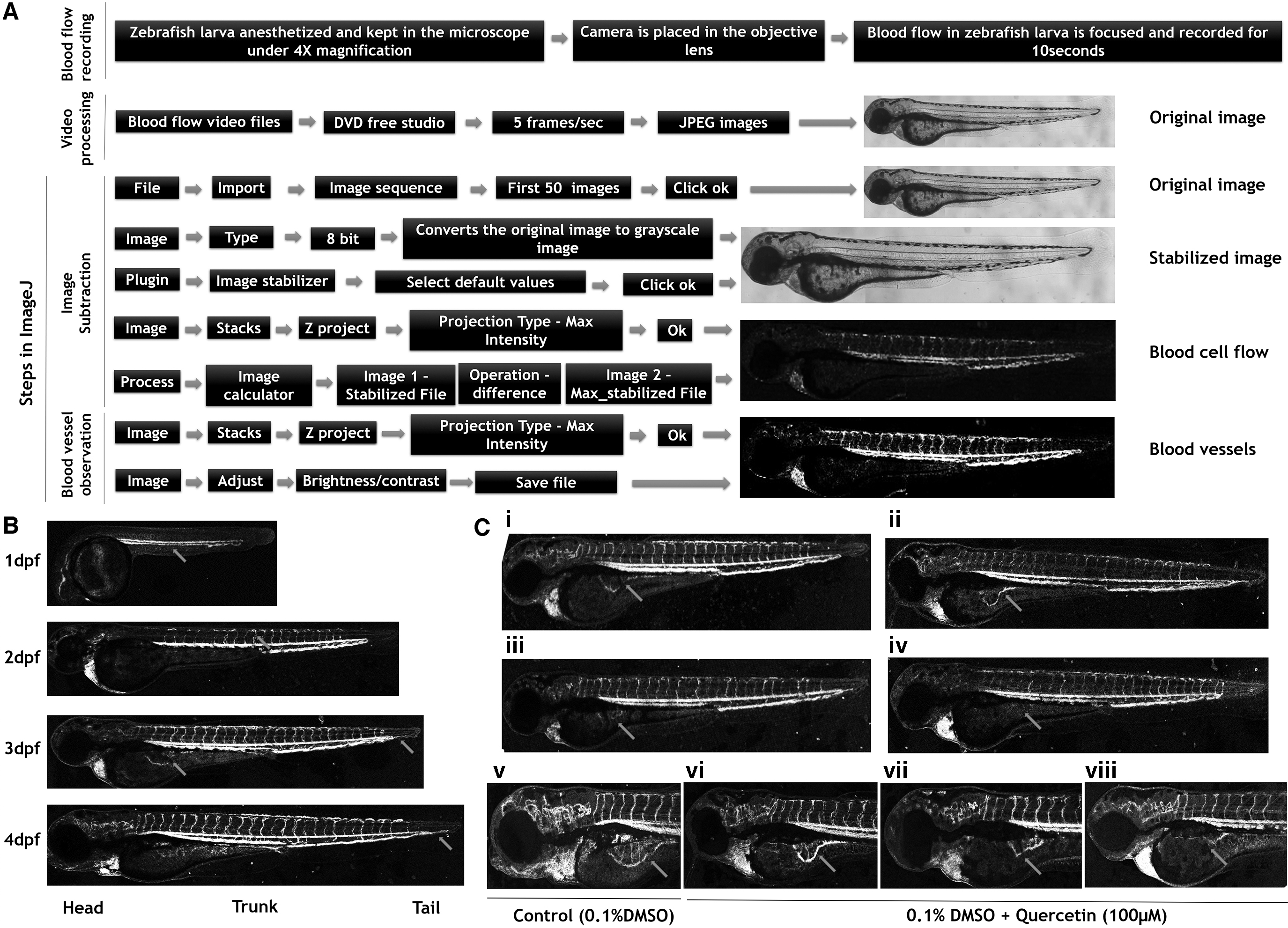
Our results showed impairment of SIV angiogenesis (11 out of 15 zebrafish larvae) (arrow marked region in Fig. 1C [i–viii] in the quercetin-treated 3 dpf zebrafish larvae). Thus, we have developed a very simple protocol, using ImageJ program, to observe zebrafish larval blood vessels that is affordable and that can easily be performed in any small laboratory setting.
Footnotes
Acknowledgment
The facilities provided by SRM Institute of Science and Technology is gratefully acknowledged.
Disclosure Statement
No competing financial interests exist.
Funding Information
The authors thankfully acknowledge the financial assistance received from the Department of Biotechnology (DBT), New Delhi (BT/PR26189/GET/119/226/2017) and DST-SERB, New Delhi (EMR/2017/000465).

References
Supplementary Material
Please find the following supplemental material available below.
For Open Access articles published under a Creative Commons License, all supplemental material carries the same license as the article it is associated with.
For non-Open Access articles published, all supplemental material carries a non-exclusive license, and permission requests for re-use of supplemental material or any part of supplemental material shall be sent directly to the copyright owner as specified in the copyright notice associated with the article.
