




Abstract
Zebrafish are widely used as experimental animal models. They are small and move fast in the water. Real-time imaging of fast-moving zebrafish is a challenge, and it requires that the imaging technique has higher spatiotemporal resolution and penetration ability. The purpose of this study was to evaluate the feasibility of dynamic phase retrieval (PR)-based phase-contrast imaging (PCI) for real-time displaying of the breathing and swimming process in unanesthetized free-moving zebrafish, and to evaluate the feasibility of PR-based phase-contrast CT (PCCT) for visualizing the soft tissues in anesthetized living zebrafish. PR was performed using the phase-attenuation duality (PAD) method with the δ/β values (PAD property) of 100 and 1000 for dynamic PR-based PCI and PR-based PCCT, respectively. The contrast-to-noise ratio (CNR) was used for quantitatively assessing the visibility of the adipose tissue and muscle tissue. The skeleton and swim bladder chambers in fast-moving zebrafish were clearly shown. The dynamic processes of breathing and swimming were visibly recorded. The respiratory intensity and frequency and the movement flexibility of the zebrafish could be dynamically evaluated. By producing more obvious image contrast, PR-based PCCT clearly showed the adipose tissue and muscle tissue. The CNRs from PR-based PCCT were significantly higher than those from PR-free PCCT for both adipose tissue (9.256 ± 2.037 vs. 0.429 ± 0.426, p < 0.0001) and muscle tissue (7.095 ± 1.443 vs. 0.324 ± 0.267, p < 0.0001). Dynamic PR-based PCI holds the potential for investigating both morphological abnormalities and motor disorders. PR-based PCCT offers clear visualization and the potential for quantification of soft tissues in living zebrafish.
Introduction
Zebrafish has emerged as a versatile experimental model organism for research into many diseases, such as abnormal behavior,1–3 respiratory injury,4–6 and bone disorders.7–11 Imaging provides helpful information for these researches. Video recording using camera has been commonly used for tracking the zebrafish movement. Some studies4,12,13 used analog signals that represented opercular displacements to explore respiratory responses to different external conditions. However, these observation modes cannot investigate the internal bone structures of organs that coordinate the zebrafish movement. Zebrafish have been visually evaluated using conventional X-ray planar imaging14,15 and micro-CT techniques.7,16,17 The imaging techniques can evidently display the static-state organs to provide morphological information.
However, it is more valuable to obtain a dynamic perspective of the internal organs in some studies, especially in behavioral physiology and kinematics of skeleton participation. Real-time imaging of the small and fast-moving zebrafish is still challenging. In addition, living zebrafish should stay in water, so more penetrating X-rays are needed to penetrate the water besides the zebrafish. Conventional absorption-contrast imaging (ACI) relies on the attenuation of X-rays to show different materials. There is a small difference in X-ray absorption between soft tissues, such as adipose tissue and muscle tissue. It is always difficult to obtain satisfactory image contrast to clearly distinguish the soft tissues using ACI.
Previous studies have demonstrated the brilliant experimental capabilities of synchrotron radiation (SR) for imaging objects with its high spatiotemporal resolution.18–21 Phase-contrast imaging (PCI) utilizes the phase shift of SR beams to increase the contrast between soft tissues.22–24 Phase retrieval (PR) is an image processing method to purify the phase information in PCI. PR can noticeably enhance the contrast between different tissues.25–27 PR-based phase-contrast CT (PCCT) can further reconstruct the extracted phase information to obtain detailed observation of the inner structures of objects. Based on CT images, the morphological parameters of inner structures can be measured and appropriately analyzed.
The purpose of this study was to evaluate the feasibility of dynamic PR-based PCI for real-time displaying of the breathing and swimming process in unanesthetized free-moving zebrafish, and to evaluate the feasibility of PR-based PCCT for visualizing the soft tissues in anesthetized living zebrafish.
Materials and Methods
SR parameters
In-line PCI was conducted at the X-ray Imaging and Biomedical Application Beamline (BL13HB) in Shanghai Synchrotron Radiation Facility (SSRF) in China. A diagram of the setup is given in Figure 1. X-rays were created from a 3.5 GeV electron storage ring and were monochromatized using a double-crystal monochromator with Si(111) and Si(311) crystals. The beamline covered an energy range of 7.7 to 40.4 keV. The main parameters are presented in Table 1.
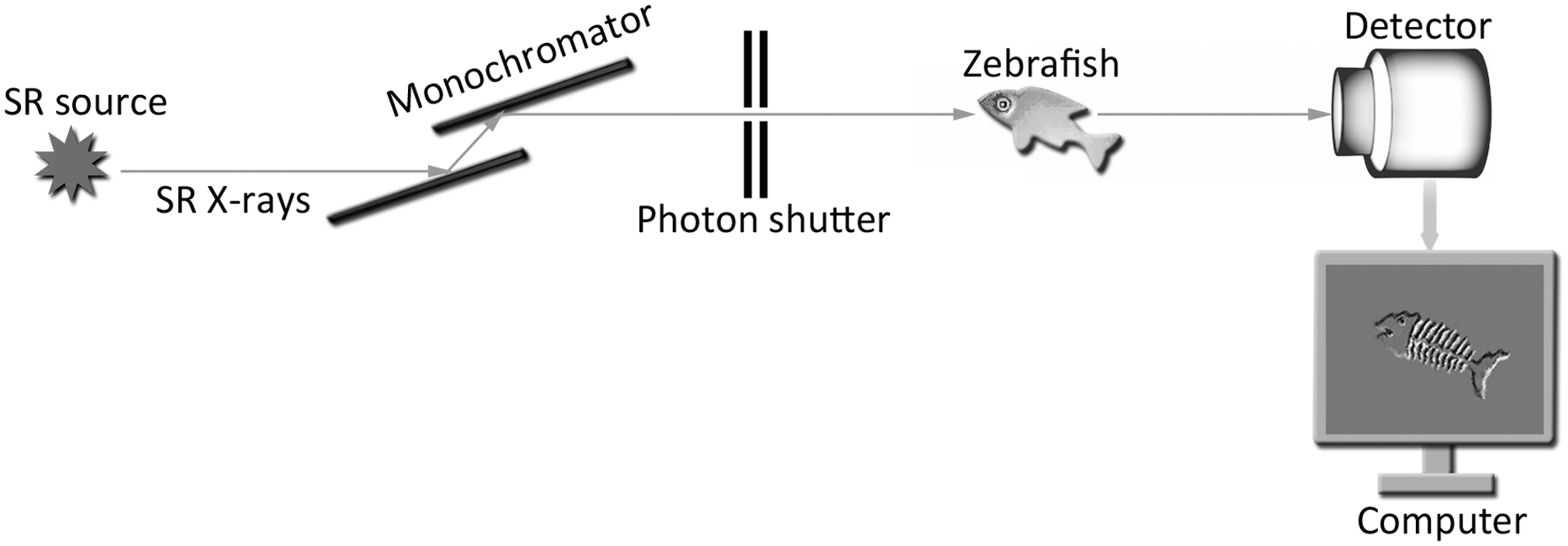
Schematic of the experimental setup. Prefiltered SR X-rays were monochromatized by a double-crystal monochromator. A detector captured the light and converted it into a signal that formed an image on the computer.
Main Parameters of the BL13HB at the Shanghai Synchrotron Radiation Facility
Dynamic PCI of the swimming zebrafish
All animal experiments were conducted in accordance with the guidelines established by the Institutional Animal Care and Use Committee of Shanghai Jiao Tong University, China. Zebrafish were raised under standard laboratory conditions of 27°C–28°C on a 12-h light/12-h dark cycle. Without anesthesia, zebrafish were placed in water in a polyethylene tube perpendicular to the X-ray beam (Fig. 2). Dynamic PCI was performed at an energy of 20 keV using a 9-μm-pixel detector (Photonic Science, East Sussex, United Kingdom). The sample-to-detector distance was 60 cm. The exposure times for dark field, flat field, and sample field images were 0, 450 μs and 100 μs, respectively. Five dark field images and 10 flat field images were attained before real-time imaging of the zebrafish.
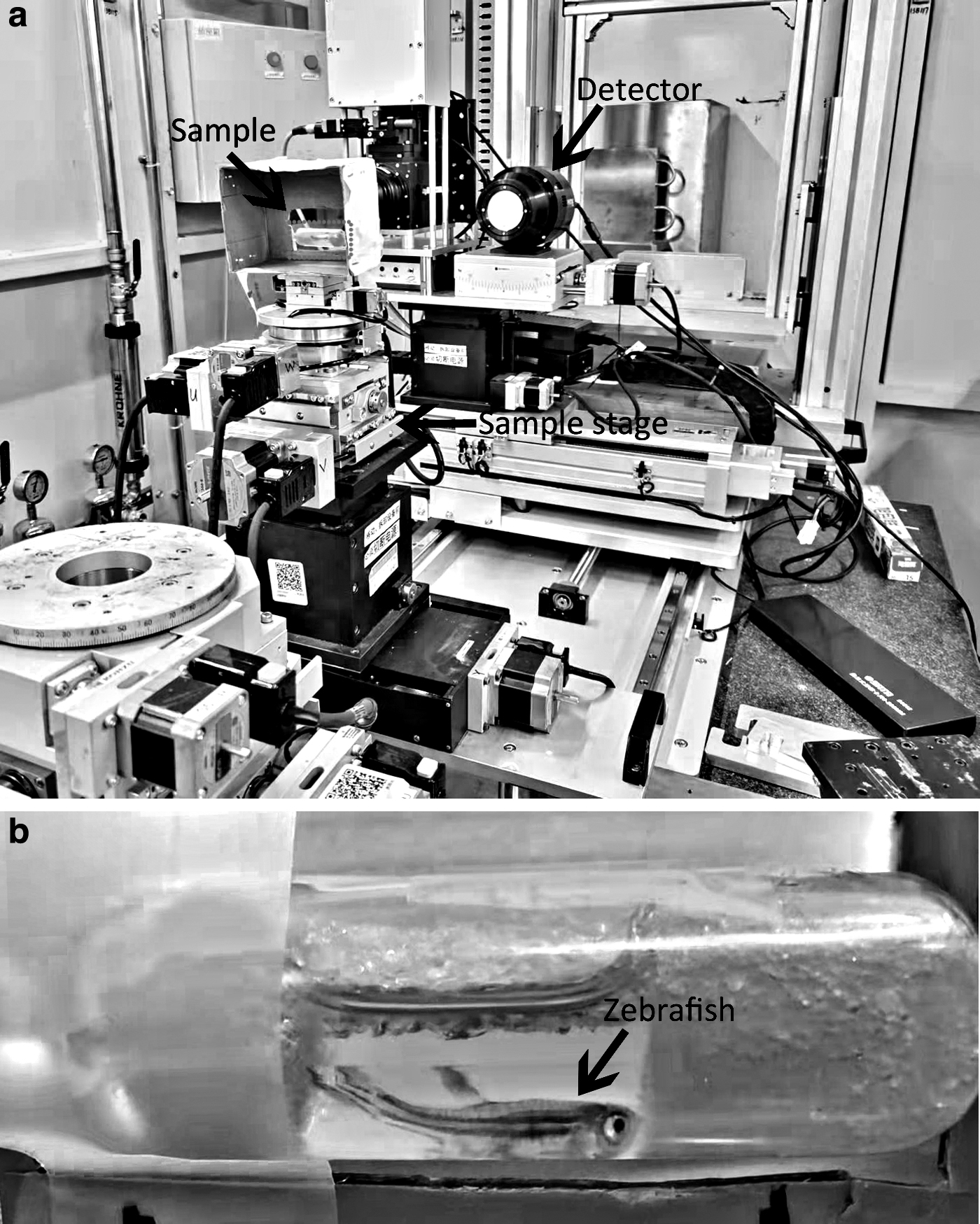
Position of the swimming zebrafish.
PCCT of the living zebrafish
Zebrafish were anesthetized with tricaine (MS-222; Sigma). PCCT was performed for the anesthetized zebrafish. The imaging parameters are presented in Table 2. Because the body length of the zebrafish exceeded the viewing field of PCCT, several separate CT scans were needed for completing the overall scan. To keep the fish motionless, a small amount of tricaine was added between CT scans.
Main Parameters for Phase-Contrast CT
Radiation dose measurement
Radiation dose was measured according to an ionization chamber (IC) method.
28
In our previous study,
25
we deduced the following equation for the measurement:
Here, I0 is the current measured by the IC, t is the total exposure time, the constant 33.7 is the mean excitation potential (W-value) of air, sslit × n is the total slit area, ρair is the air mass density at an atmospheric pressure of 1.29 × 10⁻³ g/cm3, and L is the length of the IC. After all the data were in SI units, the radiation dose of each snap for the swimming zebrafish was ∼5.42 mGy, and 0.16 Gy per CT scan.
Image postprocessing
PR processing was performed using Phase-sensitive X-ray Image processing and Tomography REconstruction (PITRE) software, which could be downloaded freely from the website provided in the Ref. 26 Dynamic phase-contrast images of swimming zebrafish were processed using the phase-attenuation duality (PAD) method with a δ/β value (PAD property) of 100. Projections of PCCT were processed using the PAD method with a δ/β value of 1000. Then, the projections were reconstructed into CT slices by applying the Gridrec algorithm. 29 8-bit CT slices, which had a gray value scaling from 0 to 255, were acquired by the PITRE software. The workflow is shown in Figure 3. The gray value scaling was adjusted to enhance the exhibited contrast of the images. Videos were made using ImageJ 1.51j8 (Wayne Rasband, NIH). Line profile analysis and intensity measurements were performed using Image Pro Plus 6.0 software (Media Cybernetics, Bethesda).
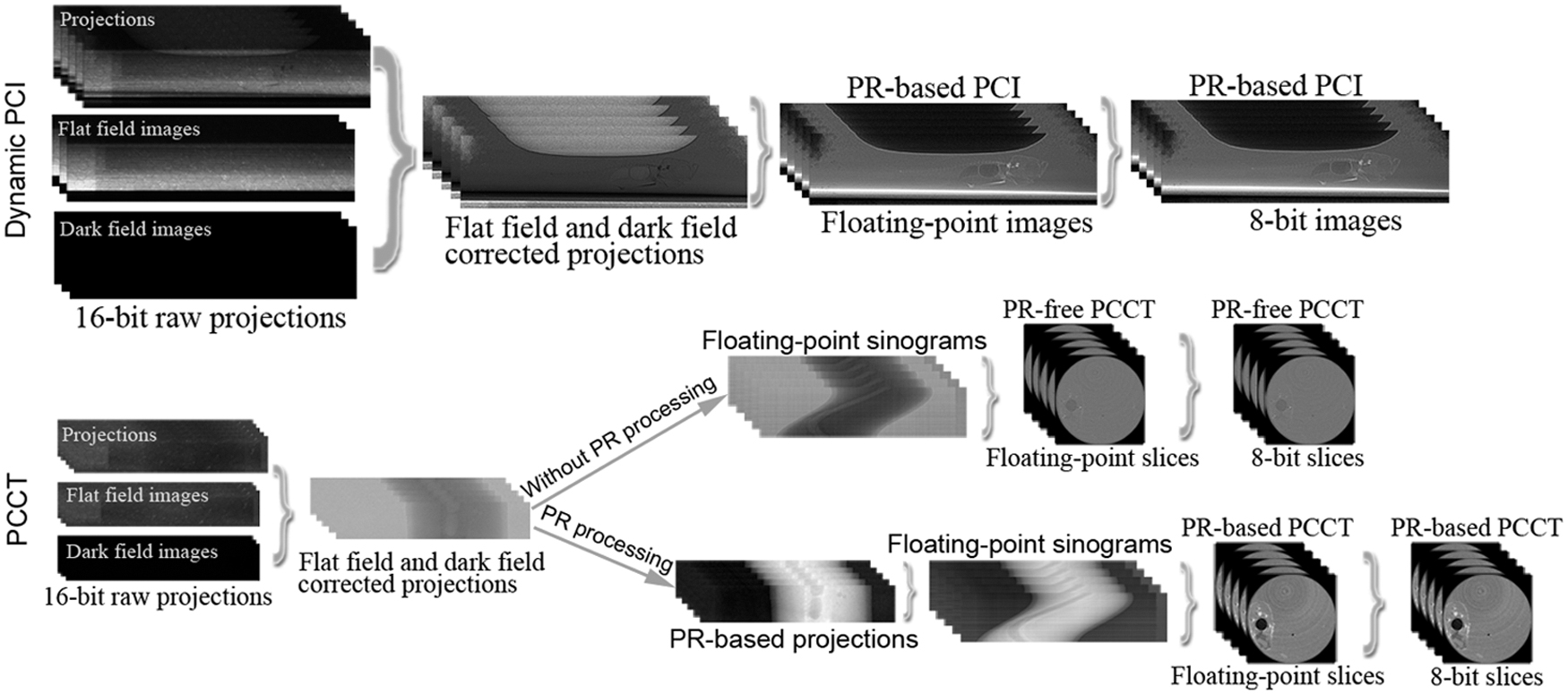
PR processing using PITRE software. The 16-bit raw projections were corrected to remove their background noise. For PR-based PCI, PR processing was performed to get the PR-based floating-point images. For PR-based PCCT, PR processing was performed to get PR-based projections before the gaining of the sinograms. The floating-point sinograms were then reconstructed into floating-point slices. Finally, the floating-point images or slices were converted to 8-bit. PCCT, phase-contrast CT; PCI, phase-contrast imaging; PITRE, Phase-sensitive X-ray Image processing and Tomography REconstruction; PR, phase retrieval.
Quantification of the tissue visibility
Ten CT slices were randomly selected for the comparison of the visibility of soft tissues between PR-free PCCT and PR-based PCCT. The relative contrast-to-noise ratio (CNR) was used to quantify the visibility. The CNR for the tested tissue was calculated using the following equation
30
:
where Itest and Iref represent the mean intensity in a round region of interest (ROI) located in the tested tissue and the reference tissue, respectively, and σref indicates the standard deviation (SD) of the corresponding signal in the ROI of the reference tissue, as a measure of image noise. Adipose tissue and muscle tissue were used as a reference for each other.
Statistical analysis
Statistical analyses were performed using SPSS Statistics software (Version 22; IBM, Armonk, NY). The CNRs for the adipose tissue and the muscle tissue on PR-free PCCT and PR-based PCCT were compared using an independent t test. Data are presented as the mean value ± SD, and p < 0.05 was considered statistically significant.
Results
Dynamic PR-based PCI
In Figure 4, the skeleton, such as cranial bones, fins, vertebrae, and ribs were clearly shown. The air-filled posterior and anterior chambers were also clearly shown (Fig. 4a). Including the image storage time, ∼9.88 images could be acquired per second using the detector. This image acquisition speed allowed observation of the coordinated movement of facial structures during breathing (Fig. 5 and Supplementary Video S1). For truly displaying the original movement, videos were made with a frame rate of 9.88 f/s, the same as the image acquisition speed of the detector. The included angle between the maxilla and dentary bone and the space between the operculum and dentary bone were found to increase and decrease periodically.

PR-based PCI of the swimming zebrafish.
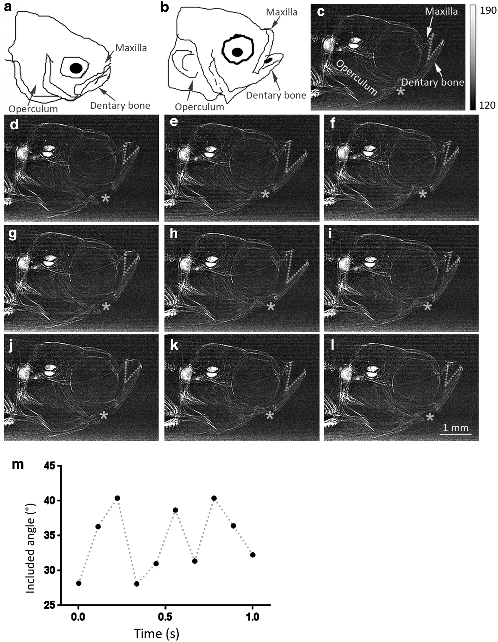
Dynamic PR-based PCI of zebrafish facial breathing movement in 1 s.
The real-time PR-based PCI of the swimming zebrafish is shown in Figure 6 and Supplementary Video S2. The dynamic motion of the skeleton was visible. Zebrafish were found to twist their bodies and swing their fins to generate turning propulsion during swimming. When the zebrafish were about to turn, their body bent sideways in the opposite direction.
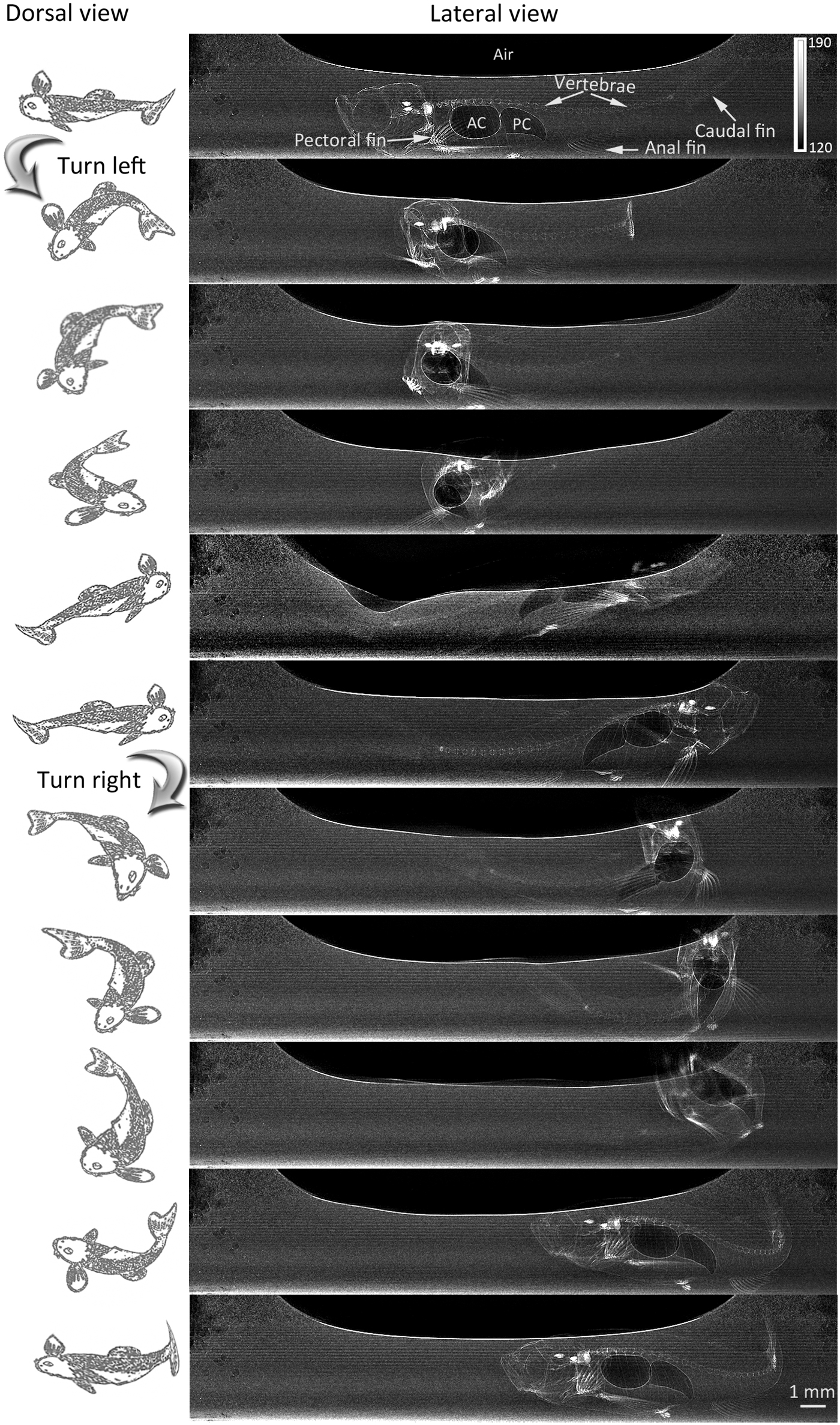
Dynamic PR-based PCI of the swimming zebrafish. Column 1 shows the dorsal view diagrams. Column 2 shows the X-ray lateral view of the swimming zebrafish. The dynamic process of twisting the body and swinging the fins to make turns during swimming was clearly shown. The adjusted gray value scaling is given on the right of the first image. The 8-bit images had an original gray value scaling from 0 to 255.
PCCT of living zebrafish
To confirm the superiority of PR processing for visualizing soft tissues, PR-based PCCT was compared with PR-free PCCT (Fig. 7). Air-filled posterior chamber was definitely shown on both PR-free PCCT image (Fig. 7c) and PR-based PCCT image (Fig. 7d). The soft tissues produced a more pronounced change in the gray value on the PR-based PCCT image (Fig. 7d) than on the PR-free PCCT image (Fig. 7c). When the adipose and muscle tissues were imaged by PR-free PCCT (Fig. 7f), they were hardly detected.
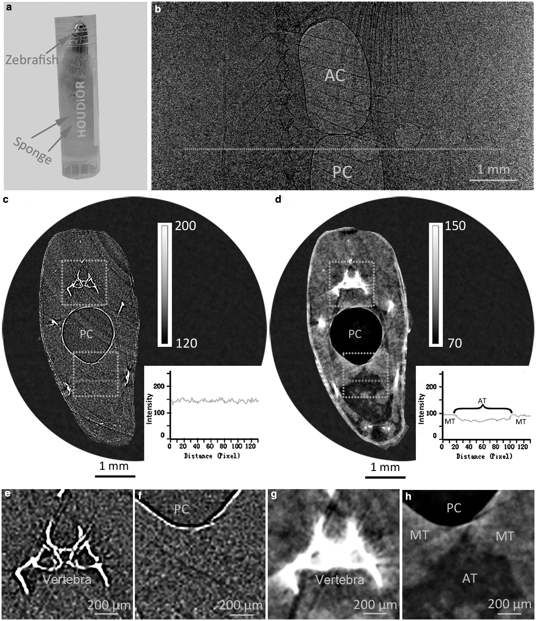
Comparison of the soft tissue visibility between PR-free PCCT and PR-based PCCT for living zebrafish.
PR-based PCCT, conversely, presented a clear discrimination of them after the gray value scaling was adjusted for better contrast (Fig. 7h). The vertebra was noticeably shown on PR-free PCCT (Fig. 7e), but the apparent thickness of the vertebra increased on PR-based PCCT images with soft-tissue gray value scaling (Fig. 7g). To solve this issue, gray value scaling was adjusted for better visualizing different tissues on PR-based PCCT images (Fig. 8). The vertebra (Fig. 8d), pectoral fin (Fig. 8e, f), adipose tissue, and muscle tissue (Fig. 8b) were markedly delineated at the appropriate gray value scaling. In Figure 9, the CNRs from PR-based PCCT were significantly higher than those from PR-free PCCT for both adipose tissue (9.256 ± 2.037 vs. 0.429 ± 0.426, p < 0.0001) and muscle tissue (7.095 ± 1.443 vs. 0.324 ± 0.267, p < 0.0001).

PR-based PCCT with different gray value scalings.
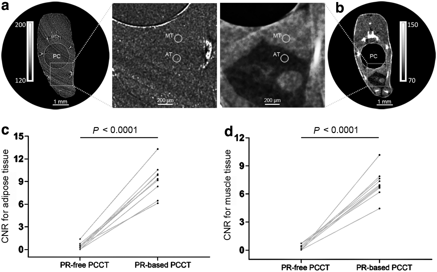
Comparison of the soft tissue CNRs between PR-free PCCT and PR-based PCCT.
Discussion
Zebrafish has developed into a useful animal model for understanding many human diseases and evaluating potential therapeutic efficacy.31–34 Different from the commonly used animal models of rats and mice, living zebrafish should be placed in water. Higher-penetration X-rays are necessary for acquiring sufficient image contrast. The penetrability of the SR X-rays is related to photon energy and luminous flux. As there were some fluctuations in the luminous flux at different experimental times in SSRF, we adjusted the photon energy according to the luminous flux at that time. Therefore, we chose two photon energies for better image contrast. The use of appropriate photon energy and luminous flux ensured sufficient penetration of SR X-rays.
Our results indicate that PR-based PCI is uniquely suited to be applied in some studies on zebrafish disease models. Respiratory intensity and frequency always need to be estimated in respiratory-related diseases. Because the temporal resolution of dynamic PCI reached the submillisecond level, the real-time process of zebrafish breathing was plainly recorded. This real-time recording could be used to measure respiratory intensity and frequency. Anesthesia can affect zebrafish breathing, and imaging without anesthesia can better reflect their real state. In addition, the coordinated movement of facial structures involved in breathing was clearly shown, which could supply more information for assessing the respiratory function.
Studies on behavior and skeleton kinematics also require live observation. We found that the head bones, vertebrae, ribs, and fins were remarkably presented, even when the zebrafish were fast moving. The body activities of zebrafish, for example, the bending curvature when turning, the turning speed, and the turning pattern, were visibly visualized. These indicators could reflect the zebrafish activity flexibility, which might act as a potential evaluation index to judge the new drug's effectiveness of the behavioral abnormality or spinal disorder models.
In addition to the high-speed image acquisition, the exploitation of the differences in the refractive index is another advantage of PCI. Because gas has a different refractive index than many tissues, the interfaces between the gas-filled anterior chamber and posterior chamber and their adjacent tissues generate distinct phase shifts. PCI can utilize these phase shifts and intensely highlight the interfaces. Therefore, the anterior chamber and posterior chamber were definitely shown on PR-free PCCT. Because the difference in density between soft tissues is relatively small, poor tissue discrimination is generally obtained on ACI. The image contrast between fish soft tissues was enhanced after applying grating-based differential phase-contrast (DPC) setup to conventional ACI. 35 However, the exposure time of DPC was up to 40 s, which might restrict its application for unanesthetized free-moving zebrafish. The difference in refractive index is often larger than the difference in density for soft tissues.
However, as the interfaces between different tissues are extremely accentuated by PCI, a substantial part of the phase contrast in the chosen area may be missing if the phase shifts vary slowly.27,36 Consequently, the soft tissues were barely visualized on PR-free PCCT. PR can extract the phase information from the mixed contrast displayed in a projection image.26,27,37,38 The lost phase contrast could be fully restored on the PR-based PCCT, which enabled the clear revelation of the adipose tissue and the muscle tissue. This result indicates the application of PR-based PCCT for imaging and potential quantification of the muscle tissue or adipose tissue in some zebrafish disease models, such as muscular dystrophy, fatty liver disease, obesity, or other metabolic diseases. In addition, the swim bladder chambers were more clearly displayed on the PR-based PCCT than on the PR-free PCCT.
Imaging the chambers has been used for disclosing the effects of some intervention factors, such as hypoxia 15 and triadimefon, 39 on the chambers. Zebrafish samples are often documented by using conventional micro CT or SR micro CT. 40 After stained with iodine contrast agent, paraformaldehyde fixed organism was clearly visualized by conventional micro CT. 17 However, this staining method cannot be available for living fish. Paraformaldehyde will cause fish death. In future studies, some safe strong-absorption contrast agents may be directly injected into zebrafish blood circulation using microinjection device. The image contrast will be undoubtedly improved by these contrast agents. Due to the movement, imaging the living zebrafish is much more challenging than ex vivo samples.
Anesthetized zebrafish should be placed in water for breathing; however, the zebrafish will wake up in a short time in the water. It requires the CT scan to be finished quickly. Here, we completed a CT scan within 60 s. The anesthetized zebrafish could remain motionless during each scanning. Different from soft tissues and chambers, PR-based PCCT had no remarkable superiority in showing bones. Bones are high-absorption material, and they can also be clearly shown in both ACI and PR-free PCCT. In PR-based PCCT, the gray value scaling could be adjusted separately for better visualization of bones and soft tissues, similar to the application of window width and window level in clinical CT.
Several limitations should be noted in this study. First, the vertical field of view was limited for PCCT. Multiple CT scans were needed to present the whole zebrafish, which increases the irradiation dose. Second, we only obtained a real-time lateral view of swimming zebrafish. It seems impossible to directly obtain the 3D visualization of swimming zebrafish by the CT system. When the water container rotates during the CT scanning, the position of the swimming zebrafish is not fixed. Therefore, it is difficult to reconstruct the zebrafish clearly on CT slices. Some image algorithms, such as 3D registration method, 41 may be applied to reconstruct the dynamic motion in 3D. Third, the image acquisition speed and image contrast are always in contradiction. The image acquisition speed could be further improved by reducing the exposure time, however, this would lead to an attenuated X-ray penetration, and consequently the reduction of the image contrast.
Conclusion
In conclusion, dynamic PR-based PCI has high spatiotemporal resolution for obtaining clear real-time observation of the skeleton and chambers in fast-moving zebrafish. Such an imaging advantage allows for evaluating zebrafish motor function. PR-based PCCT overcomes the limitations of ACI techniques and generates satisfactory image contrast for soft tissues, which holds high potential for studying many zebrafish disease models in the future.
Footnotes
Acknowledgments
The authors thank Yanan Fu, Yanling Xue, Guohao Du, and Han Guo from BL13HB for their technical support and assistance with image acquisition.
Disclosure Statement
No competing financial interests exist.
Funding Information
This study was supported by the Innovative research team of highlevel local universities in Shanghai.

References
Supplementary Material
Please find the following supplemental material available below.
For Open Access articles published under a Creative Commons License, all supplemental material carries the same license as the article it is associated with.
For non-Open Access articles published, all supplemental material carries a non-exclusive license, and permission requests for re-use of supplemental material or any part of supplemental material shall be sent directly to the copyright owner as specified in the copyright notice associated with the article.
