
Abstract
A facial differential pulse voltammetric procedure using a glassy carbon electrode modified with zeolite imidazolate framework-67/graphitic carbon nitride (ZIF-67/g-C3N4) for the diclofenac (DCF) determination is demonstrated. ZIF-67/g-C3N4 with different mass ratios of the components was synthesized in a self-assembly process. The obtained materials were characterized by using X-ray diffraction, scanning electron microscopy (SEM), EDX-mapping, and nitrogen adsorption/desorption isotherms. The peak current varies linearly with the DCF concentration in the range of 0.2–2.2 μmol·L−1 and has a detection limit of 0.071 μmol·L−1. The modified electrode exhibits acceptable repeatability, reproducibility, and selectivity towards DCF. The proposed electrode allows determining DCF in human urine without pretreatment, and the results are comparable with those determined with HPLC.
1. Introduction
Diclofenac, 2-(2
Zeolite imidazolate framework-67 (ZIF-67) is a subclass of metal-organic frameworks (MOFs). It is an organic-inorganic hybrid solid with infinite and uniform crystalline coordination networks consisting of cobalt ions (II) and imidazolate ligands [10]. ZIF-67 exhibits various unique properties, such as thermal and chemical stability, high surface area, large pores, accessible coordinative unsaturated sites, and excellent chemical and solvent stability [10]. At present, ZIF-67 attracts increasing attention from researchers in various fields, such as adsorption and separation [11], catalysis [12], and gas separation [13]. However, its poor electroconductivity leads to limited applications in electrochemistry [14]. Graphitic carbon nitride (g-C3N4) with graphitic-like structure possesses a unique ability to be simply prepared by thermally condensation of cheap nitrogen-rich compounds such as urea, melamine, and cyanimide [15, 16], and this is unlike graphene, reduced graphene oxide, and graphitic oxide that require the more complex synthesis processes. It has gradually attracted attention in multidisciplinary areas due to its characteristic physicochemical properties, such as ability to resist attacks from strong acid/alkaline solution [17], moderate bandgap (~2.7 eV), and superior electronic properties [18]. Therefore, the combination of highly electroconductive g-C3N4 with large surface area ZIF-67 might lead to versatile materials with properties of both components.
To the best of our knowledge, only a few papers have been reported on the voltammetric or amperometric detection of DCF on a ZIF-67/g-C3N4 modified electrode. Therefore, to fabricate new electrodes, we expand our research on modifying glassy carbon electrodes (GCE). In the present work, we describe the determination of DCF by using a GCE electrode modified with ZIF-67/g-C3N4. This modified electrode exhibits sound electrocatalytic and accumulative effects on DCF and enables us to determine the DCF content in human urine with satisfactory results.
2. Experimental
2.1. Materials
Melamine (C3N3(NH2)3), absolute ethanol (C2H5OH), methanol (CH3OH), cobalt nitrate hexahydrate (Co(NO3)2·6H2O), and 2-methylimidazole (CH3C3H2N2H) were purchased from Merck company. Diclofenac sodium salt (C14H10Cl2NNaNO2) was procured from Sigma-Aldrich. Acetic acid (CH3COOH), phosphoric acid (H3PO4), boric acid (H3BO3), and potassium hydroxide (KOH) were purchased from Daejung (Korea).
The phosphate buffer solution (PBS) with pH 7 was prepared from 0.5 M Na2HPO4, 0.5 M KH2PO4, 0.5 M NaCl, and 0.5 M KCl solutions. The Britton–Robinson buffer solution (B–RBS) with pH 3–9 was prepared from 1 M H3BO3, 1 M H3PO4, and 1 M CH3COOH solutions and adjusted with a 1 M KOH solution. The stock solution was prepared by dissolving 29.6 mg of diclofenac in a 10 mL volumetric flask containing a pH 6 buffer solution. The flask was subjected to ultrasonication in a cold water bath and stored in a refrigerator at 5°C. The stock sample was prepared 30 min before analysis.
2.2. Material Synthesis
ZIF-67 was synthesized according to Qian et al. [10]. Briefly, 1.16 g of Co(NO3)2·6H2O and 1.31 g of 2-methylimidazole (Hmim) were dissolved in 100 mL of methanol separately. These two solutions were mixed so that the resulting mixture has the following molar composition:
The g-C3N4 was synthesized according to Yan et al. [19]. In brief, 10 g of melamine powder was put into an alumina crucible with a cover and heated at 500°C in a muffle furnace for 2 h.
The ZIF-67/g-C3N4 was synthesized as follows: 0.5 g of the g-C3N4 and ZIF-67 mixture was distributed in 100 mL of ethanol under ultrasonication for 6 h. The ZIF-67/g-C3N4 mass ratio is as follows: 0 : 10, 3 : 7, 4 : 6, 5 : 5, 6 : 4, and 10 : 0. The samples are denoted as (0/10)ZIF-67/g-C3N4, (3/7)ZIF-67/g-C3N4, (4/6)ZIF-67/g-C3N4, (5/5)ZIF-67/g-C3N4, (6/4)ZIF-67/g-C3N4, and (10/0)ZIF-67/g-C3N4.
2.3. Instruments
The material was characterized by using X-ray diffraction (XRD) with a D8-Advance device (Bruker, USA; Cu Kα radiation,
2.4. Preparation of Electrode and Actual Sample
2.4.1. Electrode Preparation
Prior to modification, the GCE was first polished with alumina slurry (0.05 μm) on a polishing pad, followed by successive ultrasonication in ethanol and distilled water. Then, the electrode was washed with double distilled water and dried at ambient temperature. The suspension of ZIF-67/g-C3N4 was prepared by mixing 10 mg of ZIF-67/g-C3N4 with 10 mL of ethanol under ultrasonication for 24 h. Five microliters of suspension was cast dropwise on the GCE surface and dried in an oven for a few minutes. The as-prepared electrode was denoted as ZIF-67/g-C3N4-GCE.
2.4.2. Real Samples
Human urine samples were received from five healthy persons aged 25–30 and stored in a refrigerator at 5°C. About 5 mL of the sample was filtered through a 0.2 μm membrane. One milliliter of the supernatant was collected and diluted with 2 mL of pH 6.5 BRS to avoid complex interference. The resulting solution was transferred to a 100 mL electrochemical cell for analysis.
3. Results and Discussion
3.1. Characterization of Materials
The XRD patterns of g-C3N4, ZIF-67, and ZIF-67/g-C3N4 are shown in Figure 1. Two peaks at around 13.7 and 27.1° are found in g-C3N4 ((0/10)ZIF-67/g-C3N4)). It is well known that g-C3N4 consists of tri-s-triazine building blocks [20]. The higher peak at 27.1° is assigned to the characteristic interlayer of aromatic systems, indexed for graphitic materials as (002), while the peak at 13.7°, indexed as (100), is attributed to the periodic arrangement of the condensed tri-s-triazine units in the sheets [21] (the inset of Figure 1). For the XRD pattern of (10/0)ZIF-67/g-C3N4, the characteristic peaks for ZIF-67 at 18.20, 30.22, 35.68, 43.28, 53.68, 57.18, and 62.76°, indexed as (111), (220), (311), (400), (422), (511), and (440) according to CCDC 671073, are clearly observed indicating that the (10/0)ZIF-67/g-C3N4 is ZIF-67. The intensity of these peaks in ZIF-67/g-C3N4 increases with an increase in the ZIF-67/g-C3N4 fraction. This increase manifests the coexistence of g-C3N4 and ZIF-67 phases in the composite. Clear characteristic peaks of ZIF-67 witness that the hybrid of ZIF-67 with g-C3N4 has no significant effects on the crystal structure of ZIF-67.

XRD patterns of ZIF-67, g-C3N4, and ZIF-67/g-C3N (the inset presents the XRD partten of (0/10)ZIF-67/g-C3N4).
Nitrogen adsorption/desorption isotherms are employed to evaluate the samples’ specific surface area and porosity (Figure 2). The isotherm curves of ZIF-67 have a type I isotherm, indicating the presence of a microporous material. The g-C3N4 has a type IV isotherm with an H3 hysteresis loop at high relative pressure between 0.46 and 1.0, confirming the existence of a mesoporous structure. The BET surface area (S BET) and total pore volume (V pore) are listed in Table 1.

Nitrogen adsorption/desorption isotherms of ZIF-67, g-C3N4, and ZIF-67/g-C3N4.
Textural properties of ZIF-67/g-C3N4.
Compared with pure ZIF-67, the ZIF-67/g-C3N4 has the
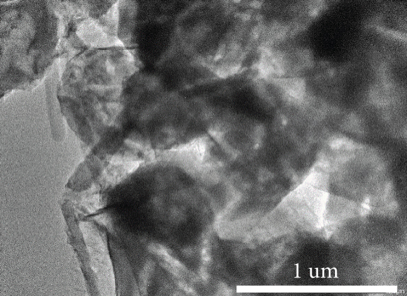
As shown in Figure 3(a), smooth rhombic dodecahedral-shaped crystals with 1.5 μm in width are observed for ZIF-67. The surface of g-C3N4 exhibits a layered and platelet-like morphology (Figure 3(b)). Although several inclusions of g-C3N4 appear on the ZIF-67 surface, the rhombic dodecahedral structure of ZIF-67 remains (Figure 3(c)). The TEM image illustrates a sheet-like structure of g-C3N4 embroiled with dark-colored ZIF-67 crystals (Figure 3(d)). These results indicate the coexistence of ZIF-67 and g-C3N4 via self-assembly.

(a) SEM image of ZIF-67, (b) TEM image of g-C3N4, (c) SEM image of ZIF-67/g-C3N4, and (d) TEM image of (5/5)ZIF-67/g-C3N4.


The ZIF-67/g-C3N4 structure is characterized by using Raman spectra (Figure 4). Typical characteristic peaks of g-C3N4 at 470, 543, 702, 746, 978, and 1147 cm–1 are observed, and they are consistent with those in a previously published report [22]. The peaks at 702 and 978 cm–1 indicate the existence of the heptazine ring structure [23]. The peak at 702 cm–1 is ascribed to the inplane bending vibrations of the heptazine linkages, whereas the 978 cm–1 peak is assigned to the symmetric N-breathing mode of the heptazine units [24]. For ZIF-67, characteristic peaks at 686, 831, 951, 1044, 1147, 1178, 1307, 1385, 1459, and 1506 cm–1 are attributed to the cobalt ion [25], imidazolium ring puckering, δ H (out of plane), bending C–H (out of plane) (C4–C5), bending C–H (out of plane) (C2–H), stretching C–N, N–H wag, bending of CH3, stretching of C2–N1, and stretching of C–H (methyl), respectively [26]. The characteristic vibrations of ZIF-67 and g-C3N4 in the Raman spectrum of ZIF-67/g-C3N4 confirm the coexistence of ZIF-67 and g-C3N4 again.

Raman spectra of (a) g-C3N4, (b) ZIF-67, and (c) ZIF-67/g-C3N4.


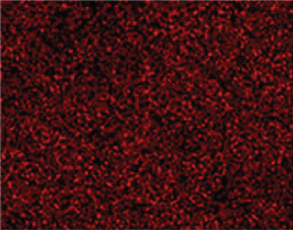
The elemental composition of ZIF-67/g-C3N4, derived from the EDX spectrum, is presented in Figure 5. As expected, the elements in (5/5)ZIF-67/g-C3N4 are only carbon (at 0.3 keV), nitrogen (0.4 keV), oxygen (0.6 keV), and cobalt (0.8; 7.0; 7.7 keV), indicating that the obtained material has high purity (Figures 5(a) and 5(b)). The EDX mapping of carbon (Figure 5(c)), cobalt (Figure 5(d)), nitrogen (Figure 5(e)), and oxygen (Figure 5(f)) on the ZIF-67/g-C3N4 surface shows that all these elements are not confined to a single site. Instead, they are distributed in the matrix.

EDX-mapping of electron image for ZIF-67/g-C3N4 (a, b), carbon element (c), cobalt element (d), nitrogen element (e), and oxygen element (f).


Although Figure 6 shows that ZIF-67 could attach to the surface of g-C3N4, such self-assembly can be further confirmed by studying the surface charges of ZIF-67, g-C3N4, and their composite (Figure 6). These values are determined with the drift pH method [27]. As seen from the figure, the material’s surface is positively charged when

Point of zero charge (pHPZC) of (5/5)ZIF-67/g-C3N4, ZIF-67, and g-C3N4.
The stability of (5/5)ZIF-67/g-C3N4 in the solutions with different pHs is essential for the electrochemical application. Figure 7 presents the XRD patterns of (5/5)ZIF-67/g-C3N4 immersed in water with different pHs for 5 hours. At pH less than 4, the characteristic diffractions of ZIF-67 are absent, revealing that the material is destroyed at these pHs. The peaks of ZIF-67 remain in the solutions with pH 5–10, indicating that ZIF-67 is stable in this pH range.

XRD patterns of (5/5)ZIF-67/g-C3N4 at pH 2–10.
3.2. Electrochemical Determination of Diclofenac by Using (5/5)ZIF-67/g-C3N4
3.2.1. The Cyclic Voltammetry of DCF on (5/5)ZIF-67/g-C3N4 Modified Electrode (ZC-GCE)
The cyclic voltammograms (CVs) (Figure 8) are obtained in the presence of 5 mM DCF on GCE, ZIF-67-GCE, g-C3N4-GCE, and ZIF-67/g-C3N4-GCE in 0.2 M BRS pH 6. The bare GCE does not exhibit any electrochemical signals, indicating that this electrode cannot detect DCF. The modified electrodes (with ZIF-67, g-C3N4, or ZIF-67/g-C3N4) exhibit an oxidation peak of DCF at around 0.68 V. In particular, the ZIF-67/g-C3N4 modified electrode significantly enhances the electrochemical response, and the intensity of peak current depends on the ZIF-67 and g-C3N4 mass ratio. The (5/5)ZIF-67/g-C3N4-GCE (henceforth denoted as ZC-GCE) provides the highest intensity with a peak potential of 0.671 V. The peak current is 2.1-fold and 2.12-fold higher than that of g-C3N4/GCE and ZIF-67-GCE, respectively. The substantial enhancement in the anodic peak current clearly shows the catalytic effect of ZIF-67/g-C3N4. No reduction peaks are observed on the reverse scan, indicating that the electron transfer on the ZC modified electrode is irreversible. In addition, the ZC-GCE does not exhibit an oxidation/reduction peak on the voltammogram in the solution without DCF, indicating the ZC-GCE is inactive in the studied potential range.

Cyclic voltammograms of 5 mM DCF in 0.2 M BRB pH 6 on GCE, ZIF-67-GCE, g-C3N4-GCE, and ZIF-67/g-C3N4-GCE.
(1) Effect of pH. The influence of pH on the oxidation peak current of the

CVs of 5 mM DCF with (a) pH 3–5, (b) pH 6–9 in 0.2 M BRB, (c)


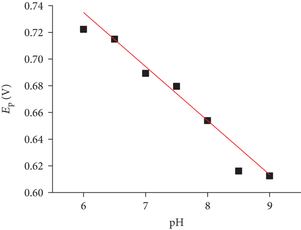
When pH is greater than 5, the peak potential decreases, suggesting the involvement of protons in the oxidation reaction (Figure 9(d)). The peak potential in the DCF oxidation on the ZC-GCE decreases linearly with the pH of the buffer solution according to the following equation:
(2) Effect of Scan Rate. The scan rate of CVs is chosen from 0.1 to 0.5 mV·s–1. In this range, the anodic peak current increases (Figure 10(a)). The highly linear relationship between

CVs of 5 mM DCF at various scan rates in 0.2 M BRS pH 6.5 on ZIF-67/g-C3N4 (a), the plot of


The relationship between the peak potential and the natural logarithm of the scan rate can provide the number of electrons transferred (
The value of
These results are consistent with those reported by Madsen et al. [33] and Goyal et al. [34], in which DCF is oxidized to 5-hydrodiclofenac via losing two electrons and two protons, as shown in Scheme 1.
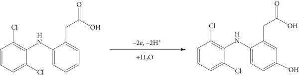
Oxidation mechanism of DCF on ZC-GCE.
The enhancement of electrochemical signals could result from the synergic effect of ZIF-67 and g-C3N4. The open Co(II) sites, as a Lewis acid [35], could attract the carboxyl group in DCF, and as a Lewis base via acid-base interaction, the graphitic rings in g-C3N4 could attract the benzene rings in DCF via π–π interaction. These arguments are schematically illustrated in Scheme 2.

Schematic illustration of the formation of ZIF-67/g-C3N4 modified GCE and its oxidation of DCF.
3.2.2. Linear Range, Detection Limit, Repeatability, and Interference
(1) Linear Range and Detection Limit. The relationship between the peak current and DCF concentration is studied by using differential pulse voltammetry (DPV). In this case, the peak current increases linearly with the concentration of DCF, from 0.2 to 2.2 μM (Figure 11). The regression equation is

DPV curves of DCF with different concentrations (0.2–2.2 μM) in 0.2 M BRS pH 6.5 at the modified electrode (a). The plot of

Comparison of the analytical performance of the different modified electrodes for the determination of DCF.
Square wave voltammetry: SWV.
(2) Reproducibility and Repeatability. The reproducibility is studied from four replicates measurements of DCF determination. To investigate the repeatability of this modified electrode, we conduct the measurements ten times with the sample electrode. Figure 12(a) shows very similar DPV curves of four scans on the same working electrode. The small RSD of 1.32% indicates good reproducibility of the proposed method. Figure 12(b) presents the values of peak currents measured on 10 distinct working electrodes under the same modified electrode procedure. The RSD varies from 0.151% to 5.315% in run 1 to run 10, manifesting excellent repeatability.

The DPV of four replicates measurement (a) and the values of peak current of DCF on ZC-GCE modified using (5 μL ZC (1 mg/mL) for ten times on the same electrode (b).

The biological sample is a very complex mixture consisting of various ions and molecules. Possible interfering electroactive species on the biological samples include inorganic salts (KHCO3, Na2SO4, CaCl2, and NH4NO3) and organic compounds (uric acid, caffeine, paracetamol, and ascorbic acid). Table 3 reveals that inorganic salts do not significantly interfere with the measurement even at extremely high content (100–320 fold). Ascorbic acid does not seem to affect the peak current at 480-fold concentration. Paracetamol, uric acid, and caffeine exert a medium effect on the peak current at a concentration of 40 to 80-fold higher than that of DCF. These results confirm the relevant selectivity of the proposed method for DCF detection.
Effect of some foreign substances on the determination of 1 μM DFC in 0.2 M BRBS pH 6.5.
3.2.3. Real Sample Analysis
This DPV method is used to analyze five actual human urine samples. To determine the method’s accuracy, we spike each sample with 50 μM of DCF, and the relative recovery (Rev.) is calculated. All the values fall in the expectable range of 95.4–105% (Table 4). The content of DCF in the samples is also determined with the HPLC method for comparison. When
The results of DCF analysis in urine by the proposed DPV method and HPLC.
(–): not found.
4. Conclusion
In this research, we successfully synthesized ZIF-67/g-C3N4 with the self-assembly method and used the material to modify a glassy carbon electrode for the electrocatalytic determination of diclofenac. The g-C3N4 is highly dispersed in ZIF-67 through an ultrasonic assisted stir to form a composite of g-C3N4 and ZIF-67. The enhancement of electrochemical signals is due to the synergic effect of ZIF-67 and g-C3N4. The oxidation of diclofenac on the modified electrode takes place with a two-electron-proton mechanism. The electrode process is controlled by adsorption. The proposed DPV method exhibits high sensitivity with a low detection limit and can be used to determine diclofenac at trace levels.
Footnotes
Data Availability
The data used to support the findings of this study are available from the corresponding author upon request.
Conflicts of Interest
The authors declare that they have no conflicts of interest.
Acknowledgments
This work was supported by the Hue University under the Core Research Program No. NCM.DHH.2019.08.