



Abstract
Objectives:
To assess the safety and efficacy of intrathecal fluorescein administration during transmastoid repair of temporal lobe encephaloceles and identify predictors for intraoperative fluorescein identification.
Methods:
This was a retrospective case-control study of consecutive adult patients with temporal lobe encephaloceles repaired via transmastoid approach from October 2012 to October 2024 at a single tertiary academic medical center. Medical records of qualifying patients were searched for demographics, diagnostic workup, disease characteristics, and intraoperative and postoperative outcomes. Analysis incorporated the Wilcoxon rank-sum, Pearson’s X2, and Fisher’s exact tests, along with unadjusted Kaplan-Meier estimates. Outcomes included encephalocele recurrence and symptomatic resolution rates, fluorescein complications, and rates and predictors of intraoperative fluorescein identification.
Results:
38 patients met inclusion criteria (age 59.8 + 11.9 years, BMI 35.8 + 8.7 kg/m2, 23 [60.5%] female). Recurrence (0% vs 11.5%, P = .54) and symptom resolution (83.3% vs 88.5%, P = .64) rates did not differ between patients who received and did not receive intrathecal fluorescein. No fluorescein-related complications were reported. Fluorescein was intraoperatively detected in six-twelfths cases, yielding a sensitivity of 50%. No demographic or clinical variables, including higher BMI, history of prior encephalocele, and size of largest defect, were associated with intraoperative fluorescein identification.
Conclusions:
Intrathecal fluorescein administration preliminarily appears to be a safe adjunctive tool to identify the leak source in the transmastoid repair of temporal lobe encephaloceles, but its risk-benefit ratio remains up for debate. Higher-powered studies are necessary to explore its utility in specific circumstances and to determine which patients may benefit most.
Introduction
Encephaloceles are a clinically significant condition involving the herniation of brain tissue, meninges, and cerebrospinal fluid (CSF) through a skull base defect.1-3 Although classically associated with errors in embryological neural tube development, encephaloceles have been increasingly observed in the adult population secondary to trauma, prior skull base surgery, and chronic changes related to idiopathic intracranial hypertension (IIH), chronic sinus disease, and connective tissue disorders.4-8 A significant portion of encephaloceles are temporal lobe encephaloceles herniating through defects in the tegmen tympani or tegmen mastoideum, and they can result in conductive hearing loss (CHL), otorrhea, aural fullness, and tinnitus.9-11 Given their potentially significant morbidity, treatment is recommended for many patients, with surgical repair being the gold standard.11,12
The transmastoid approach for encephalocele repair is generally successful in carefully selected patients, providing direct access to the tegmen mastoideum and, on occasion, tegmen tympani, while avoiding a craniotomy.13,14 However, encephalocele recurrence is not uncommon, and may result from difficulties with intraoperative localization of herniation sites and confirmation of adequate repair, particularly in cases of small defects and intermittent CSF leaks.13,15-17 As such, supplemental techniques, such as intrathecal fluorescein dye administration to allow for the real-time visualization of CSF leaks, have been proposed to address these challenges and improve surgical outcomes.18,19
While there are some potential adverse effects associated with fluorescein such as nausea and vomiting, most reports note a general lack of major adverse reactions, with low levels of toxicity.20,21 Reported complications of lower extremity weakness and numbness, seizures, anaphylaxis, and cranial nerve palsies are rare, especially at low doses used for this indication (<50 mg).19,22 Most literature on the use of fluorescein to detect CSF leaks is limited to endoscopic anterior skull base repairs.23-25 However, data is limited on the application of fluorescein for use in the transmastoid repair of temporal lobe encephaloceles. 26 Additionally, patient-specific variables that may affect diagnostic utility have not been fully explored.
Hence, this study aims to assess the safety and efficacy of intrathecal fluorescein administration during transmastoid repair of temporal lobe encephaloceles and identify predictors for intraoperative fluorescein identification. By doing so, it seeks to recognize patients who may benefit from the use of intraoperative fluorescein and thereby improve rates of temporal lobe encephalocele resolution.
Materials and Methods
Study Design, Setting, and Sample
This was a retrospective case-control study involving consecutive adult patients with temporal lobe encephaloceles repaired via transmastoid approach by 1 neurotologist at a tertiary academic medical center before and after implementing routine intrathecal fluorescein administration from October 2012 to October 2024. Preoperative diagnosis was established in all patients via radiologic confirmation on MRI with internal auditory canal protocol and/or high-resolution CT temporal bone, with independent review by both the operating neurotologist and a radiologist. All but 2 patients were also tested for beta-2 transferrin to confirm active CSF leak preoperatively. Per protocol, all patients after September 2021 who underwent transmastoid repair of temporal lobe encephaloceles had a lumbar drain placed and received intrathecal fluorescein, unless external circumstances unrelated to patient characteristics (eg, neurosurgery and/or anesthesia unavailable, contraindications to lumbar puncture, patient refusal) precluded lumbar drain placement. Lumbar drains were removed by postoperative day two. Selected patients received 10 mg fluorescein, prepared by diluting 0.1 mL of 10% fluorescein in 10 mL of the patient’s own CSF and slowly reinfusing over 10 minutes. Inclusion criteria were age > 18 years with a documented preoperative diagnosis of temporal lobe encephalocele. Patients were excluded if an encephalocele was incidentally found intraoperatively without appropriate preoperative workup or if no encephalocele was found intraoperatively. Clinical notes and operative documents from the electronic medical records of included patients were reviewed. This study was approved by the University of Pennsylvania Institutional Review Board (project #856621) and adheres to the “Strengthening the Reporting of Observational studies in Epidemiology” framework. 27
Study Outcomes and Variables
Primary outcomes of interest were rates of encephalocele recurrence and symptomatic resolution, intraoperative and postoperative fluorescein complications, and intraoperative fluorescein identification. Secondary outcomes of interest included demographic and clinical characteristics associated with fluorescein identification. Study variables included patients’ demographics, symptomology, medical comorbidities, encephalocele characteristics, and postoperative follow-up. Defect size was measured using the largest single linear dimension on standard orthogonal planes (axial, coronal, and sagittal).
Statistical Methods
Wilcoxon rank-sum test, Pearson’s X2 test, and Fisher’s exact test were used to conduct univariate analyses. Unadjusted Kaplan-Meier estimates were used for univariate comparison of 2-year recurrence rates. Significance was set at P < .05, P-values were two-sided, and 95% confidence intervals (CIs) were constructed where appropriate. Multivariable analyses, including linear and logistic regression models, were not performed to calculate predictive associations or compare 2-year recurrence rates given the sample size and concerns for overfitting. Given the descriptive nature of baseline comparisons and the exploratory intent of univariate predictor analyses, Bonferroni correction for multiple comparisons was not applied, as the primary concern in this cohort was minimizing Type II error rather than controlling family-wise Type I error rate. Statistical analyses were performed using R version 4.3.0 (R Project for Statistical Computing) via RStudio version 2023.06.0+421 (Posit Software, PBC).
Results
Of the 38 patients included in the final study population, intrathecal fluorescein was administered in 12 (31.8%) cases. The 2 cohorts had similar demographics, disease characteristics, and reason for surgery (including revision) aside from mean defect size, which was larger in the cohort that did not receive fluorescein (2.72 + 1.05 vs 4.64 + 2.58 mm; P = .023; Table 1).
Demographics and Disease Characteristics of Patients Who Did and Did Not Receive Fluorescein.

Note. Statistically significant differences are indicated in
Mean (SD); n (%).
Wilcoxon rank-sum, Pearson’s X2, or Fisher’s exact tests.
Surgical Outcomes
There was no significant difference in overall encephalocele recurrence rates between patients who did (0%) and did not (3 [11.5%]) receive intrathecal fluorescein (P = .54). Both cohorts also had similar rates of overall (10 [83.3%] vs 23 [88.5%], P = .64) and specific symptom resolution. No intraoperative or postoperative complications attributable to fluorescein were reported in the cohort that received fluorescein. These analyses are summarized in Table 2. Two-year rates of encephalocele recurrence (P = .40) and resolution of at least 1 preoperative symptom (P = .18) also did not differ between the 2 cohorts based on Kaplan-Meier analysis (Figure 1).
Surgical Outcomes and Surveillance of Patients Who Did and Did Not Receive Fluorescein.
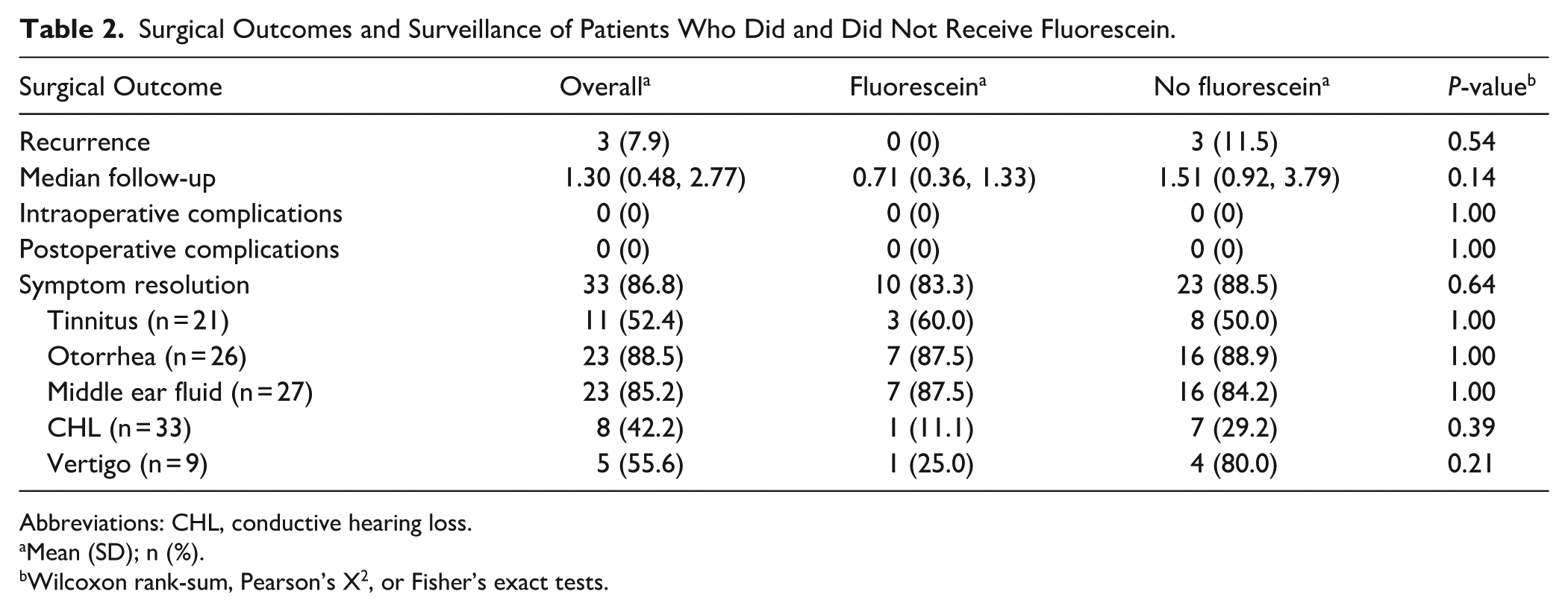
Abbreviations: CHL, conductive hearing loss.
Mean (SD); n (%).
Wilcoxon rank-sum, Pearson’s X2, or Fisher’s exact tests.

Kaplan-Meier curves depicting 2-year: (a) encephalocele recurrence rates and (b) symptom resolution rates of patients who did and did not receive fluorescein.
Intraoperative Fluorescein Identification
In accordance with our inclusion criteria, encephaloceles and tegmen defects were visually identified in all patients, but fluorescein was only detected intraoperatively in 6 cases, yielding a sensitivity of 50%. Mean BMI was higher in patients in whom fluorescein was detected (36.5 + 6.0 vs 33.1 + 4.0) but did not reach significance (P = .13). Similarly, history of prior encephalocele (3 vs 0, P = .18), involvement of tegmen tympani only (3 vs 0, P = .18), spontaneous etiology of encephalocele (2 vs 6, P = .061), and size of largest defect (2.23 + 0.97 vs 3.20 + 0.90, P = .15) were not significant. History of IIH was not significantly associated with fluorescein visualization. Proxies of active CSF leak, including otorrhea, middle ear effusion, and type B tympanogram, were not significantly associated with fluorescein visualization. No other demographic, clinical, or disease characteristics were associated with the identification of fluorescein. These analyses are summarized in Table 3.
Evaluating Variables for Significant Associations with Intraoperative Fluorescein Identification.

Abbreviations: BMI, Body mass index; IIH, idiopathic intracranial hypertension.
Note. Differences approaching statistical significance are underlined.
Mean (SD); n (%).
Wilcoxon rank-sum, Pearson’s X2, or Fisher’s exact tests.
Discussion
Our study evaluated the safety and efficacy of intrathecal fluorescein during transmastoid repair of temporal lobe encephaloceles while identifying potential predictors for intraoperative fluorescein visualization. The findings revealed no significant differences in rates of encephalocele recurrence or symptom resolution between patients who did and did not receive fluorescein, though the intervention demonstrated an acceptable safety profile with no reported complications. Fluorescein was only detected intraoperatively in half of cases where it was administered, with no clinical factors meeting significance in predicting fluorescein visualization. These findings align with previous literature suggesting fluorescein’s utility in detecting subtle CSF leaks during endoscopic skull base surgery, although our study represents one of the first focused evaluations of its application specifically in transmastoid repairs of temporal encephaloceles.19,25
Several physiologic, anatomical and technical factors may account for the lower fluorescein visualization rate observed in our cohort compared to anterior skull base reports, which report sensitivities of up to 90%.25,28 First, CSF flow dynamics and gravity due to surgical positioning likely plays a role. Transmastoid cases are performed with the patient supine and the operative ear rotated away, which may reduce dependent pooling of fluorescein at the tegmen defect, whereas anterior skull base cases are typically performed with the patient positioned midline with slight reverse Trendelenburg, potentially favoring fluorescein accumulation at the site of leakage. Second, temporal lobe encephaloceles may be more prone to intermittent or low-volume CSF egress compared to anterior skull base defects, which more commonly exhibit continuous leakage. This may result in insufficient fluorescein concentration at the defect during the operative window, regardless of total dose administered.29-31 Third, important differences in surgical visualization exist between the 2 approaches. Endoscopic anterior skull base surgery provides wide-field, high-magnification views with angled optics and enhanced lighting through a narrow corridor, whereas transmastoid surgery relies on microscopic visualization across a wider operative field with comparatively lower magnification and definition. Additionally, the drilling and irrigation inherent to transmastoid dissection may dilute fluorescein that enters the middle ear or mastoid cavity, further obscuring visible signal. Together, these factors suggest that the lower sensitivity observed in lateral skull base surgery may reflect fundamental differences in patient positioning, leak physiology, and operative technique rather than a failure of fluorescein per se.
No clinical variables met significance as predictors of fluorescein visualization, including higher BMI, history of prior encephalocele, isolated tegmen tympani involvement, and smaller defect size, which all had P-values less than .20. Interestingly, a history of IIH was not significantly associated with fluorescein visualization, despite its established role in encephalocele pathophysiology.6,7 As such, there may not be any patient groups for which fluorescein should be strictly mandated, given that no clinical factors showed significance, with the caveat that our study may be underpowered. Although intrathecal fluorescein is conceptually most valuable in occult or low-flow CSF leaks, our findings do not conclude greater value in these cases.
Other potential benefits and drawbacks must also be carefully considered regarding the use of intrathecal fluorescein in skull base surgery. As shown in our cohort, fluorescein showed a benign safety profile with no reported adverse events, consistent with previous literature describing minimal complications with low-dose administration.20,21 However, several practical considerations warrant attention when implementing fluorescein in surgical practice. Most importantly, intrathecal fluorescein requires the placement of a lumbar drain, which would otherwise be left to the surgeon’s preference. Lumbar drains themselves may contribute to resolution, but they have their own set of risks, with some studies even suggesting they may increase the length of hospitalization without improving leak closure rates.32,33 Second, the procedure requires coordination with anesthesia for proper administration timing and monitoring, potentially increasing operative complexity. Third, our finding that fluorescein was only visualized in half of cases raises questions about its reliability as a standalone diagnostic tool. Given our findings of equivalent outcomes regardless of use or visualization along with the additional costs associated with its administration, fluorescein may best be viewed as a complementary rather than necessary technique. Its use may be most justified during revision surgery due to distorted anatomy, scarring, and prior graft material reducing visual reliability, as well as when there are suspected multiple or multifocal defects given that fluorescein can help confirm that all sites are addressed prior to closure.
Our study has several important limitations that must be acknowledged. The retrospective design and modest sample size limit the generalizability of our findings and precluded multivariate analysis that would have allowed for control of potential confounding variables. The single-surgeon, single-institution nature of the study introduces potential selection bias and limits external validity. Our search strategy did not systematically capture patients who underwent transmastoid exploration for suspected CSF leak but were found to have an isolated tegmen defect without true encephalocele, further introducing potential selection bias and limiting the generalizability of our findings to the broader population of tegmen defect repairs. Several associations approached but did not reach statistical significance, and a larger study population might yield more definitive conclusions to either confirm or refute potential significance. Follow-up duration varied among patients with shorter follow-up in the fluorescein group, potentially masking late recurrences. Additionally, the subjective nature of fluorescein visualization without quantitative measurement introduces observer bias. Finally, patient-reported symptom resolution was not assessed using validated quality-of-life instruments, limiting the robustness of these outcome measures.
Future research should address several key questions raised by our findings. Prospective studies with larger cohorts are needed to validate which clinical factors, if any, reach significance. Standardization of fluorescein administration protocols – including dose optimization, timing relative to surgical exposure, and enhanced visualization techniques such as endoscopy with filtered light sources – may improve detection rates. Investigation of physiological factors affecting CSF flow dynamics and fluorescein distribution in temporal encephaloceles could help identify optimal candidates. Additionally, cost-effectiveness analyses comparing routine versus selective fluorescein use would provide valuable guidance for resource allocation. The integration of intrathecal fluorescein with newer diagnostic modalities, such as intraoperative MRI or CT, may also enhance its utility. Finally, longer follow-up periods are necessary to assess late recurrences and determine if subtle, undetected CSF leaks identified by fluorescein correlate with long-term outcomes.
Conclusion
Intrathecal fluorescein administration preliminarily appears to be a safe adjunctive tool to identify the leak source in the transmastoid repair of temporal lobe encephaloceles, but its risk-benefit ratio remains up for debate. Larger cohort studies with longer follow-up periods are warranted to explore its utility in specific circumstances, such as in patients with higher BMI, prior encephaloceles, small and/or multiple defects, and IIH. Further research is also necessary to elucidate why and when fluorescein may not be detected intraoperatively to determine patients for whom it would not be useful. While not demonstrably superior to traditional techniques in our cohort, fluorescein’s favorable safety profile supports its continued consideration as part of the surgical armamentarium for select cases, such as during revision surgery and when there are suspected multiple or multifocal defects.
Footnotes

Ethical Considerations
This study received ethical approval from the University of Pennsylvania IRB (approval #856621). This is an IRB-approved retrospective study, all patient information was de-identified and patient consent was not required. Patient data will not be shared with third parties.
Funding
The authors received no financial support for the research, authorship, and/or publication of this article.
Declaration of Conflicting Interests
The authors declared the following potential conflicts of interest with respect to the research, authorship, and/or publication of this article: Dr. Tiffany Hwa is a grant recipient from Cochlear Corporation and serves as a consultant on the advisory board of Amgen. No conflicts of interest were declared for the remaining authors. The contents of this manuscript were presented as a poster at the Combined Otolaryngology Spring Meetings (COSM) 2025 (American Otologic Society) in New Orleans, LA from May 16-17, 2025.