



Abstract
This narrative review summarizes and synthesizes the current literature regarding the comparative anatomy of pigs and humans, focusing on organ systems commonly involved in abdominal transplantation with the aim of supporting the safe and effective use of porcine models in preclinical studies. It also integrates practical insights from experimental surgery to highlight key considerations in perioperative care and surgical technique in porcine models. Pigs demonstrate substantial anatomical and physiological similarities to humans, making them ideal large-animal models for transplantation research. The recent surge in reports concerning clinical porcine xenotransplantation underscores the increasing importance of comprehending this comparative anatomy in the forthcoming years. However, critical species-specific anatomical differences particularly involving the gastrointestinal tract, hepatobiliary system, genitourinary tract and vascular structures require careful understanding to avoid surgical complications. Proper handling, perioperative care and awareness of porcine-specific stress responses and arrhythmias susceptibility are also essential to prevent premature animal loss. A thorough understanding therefore of porcine comparative anatomy is essential for transplant researchers conducting large animal surgery. Such knowledge improves experimental outcomes, supports adherence to the 3Rs (Replacement, Reduction and Refinement) and enhances the translational value of porcine models in transplantation research.
Introduction
Large animal models remain a crucial link in the bench-to-bedside chain of translational research. Their primary advantage lies in their capacity to evaluate the efficacy and safety of an intervention within complex biological systems, facilitating the transition from mechanistic in vitro studies to clinical trials in humans. The utilization of pigs, which possess human-sized organs and comparable tissue architecture, facilitates complex surgical procedures such as individual, segmental and multi-visceral transplants in orthotopic or heterotopic positions which that are challenging or unfeasible to reproduce in rodent models.1–3 Porcine models therefore enhance the translational relevance of experimental outcomes and are often essential intermediaries between small animal and human studies. The study of solid organ transplantation, with its complex interactions between physiological functions, the systemic effects of major surgery and host immune responses in large animal models, allows investigators to develop a realistic understanding of the potential interactions and pitfalls in translation to human system interactions. 4 Porcine models, though useful because of their anatomical and physiological similarities, are not without differences. Notably, crucial differences in the anatomy of pigs, particularly with regard to the small bowel, colon, pancreas and branches of great vessels, can be dangerous and possibly lethal if not appreciated by researchers undertaking abdominal surgery in this animal. In addition, special attention must be paid to aspects of animal husbandry and peri-operative care when using pigs as they are prone to stress and the provocation of fatal cardiac arrhythmias if not handled correctly. 5 Premature and avoidable loss of an animal prior to the completion of the experiment requires further animals to be used, which is contrary to the 3R principles. With the growing interest and advancements in areas such as xenotransplantation, genome editing and organ preservation, it is anticipated that the number of surgical procedures performed on pigs will increase annually. 6 Ensuring the safe conduct of such surgeries is essential to mitigate the risk of unnecessary and premature loss of experimental animals due to surgical mishaps, and this safety cannot be achieved without a comprehensive understanding of comparative anatomy and physiology.
Main body
Porcine models as the favoured species for transplantation research
Various animal models, including rodents, dogs, rabbits, pigs and non-human primates (NHPs), have been employed in transplantation research. However, in recent years pig models have become increasingly popular owing to certain limitations in other species. 7 NHPs are physiologically similar to humans but their use is increasingly restricted owing to expense, low reproduction rate and ethical concerns regarding their advanced evolutionary stature.8,9 Dogs are also used less in large animal research owing to ethical concerns about their status as companion animals. 8 Rabbit and rodent models present significant physiological differences from humans, including a markedly different volume of distribution, which affects the pharmacokinetic behaviour of drugs, thereby rendering drug response curves and toxicity profiles poorly predictive.2,10 Additionally, their small size necessitates microsurgical expertise and specialized equipment for complex surgical procedures. 9 The advantages of pigs over these other species therefore are multiple. First, they are readily available, and their use in research is less ethically contentious owing to their status as a food source. Second, pigs reach sexual maturity rapidly at around 5–6 months, at which point most organ systems are physiologically mature. 10 Third, the porcine genome has been completely sequenced, pigs are relatively amenable to genetic manipulation and possess a genetically defined and stable major histocompatibility complex, which is crucial for experimental protocols involving allografts.6,11 Detailed pan-genomic sequencing, combined with the advent of the CRISPR-Cas9 system, has allowed for precise gene editing and increasing success in xenotransplantation.6,12,13 Finally, as summarized in Table 1,1,8,9,14,15 there are numerous physiological similarities between pigs and humans, making pigs an accurate and translatable model for various disease processes.
Relevant physiological values demonstrating close similarity between pigs and humans.

Key points of comparative abdominal anatomy by system
A comprehensive understanding of the comparative anatomy between pigs and humans is essential for the successful conduct of experimental surgical procedures in pigs. Although the general orientation and function of the abdominal organs are broadly analogous between these species (Figure 1), there are several distinctive features of pigs that researchers must be cognizant of to effectively perform these procedures.

Appearance of the abdominal cavity of a pig during laparotomy. The general orientation of organs is similar to that in humans. Key differences in pigs, however, include the entire large bowel (LB) being situated on the left of the abdomen and the small bowel (SB) on the right. The spleen (Sp) is significantly larger and protrudes down over the stomach whereas in humans it remains covered by the rib cage and not immediately visible unless pathologically enlarged. The liver (Liv) is four lobed and clover-shaped, occupying a similar position in both species. The surgeon, on abdominal entry, would also note the absence of a prominent omentum draped over the viscera of the greater sac in pigs.
Gastrointestinal system
Being true omnivores, the glandular-type porcine stomach exhibits significant similarities to that of humans. 16 However, two notable differences are the presence of a small diverticulum at the fundus and a thickening at the pylorus called the torus pyloricus, a feature also found in ruminates. 17 The porcine stomach is thicker-walled than humans with more extensive cardiac-type mucosa throughout the proximal portion. 18 With regard to the small bowel, although a degree of midgut herniation does occur during embryological development the same 270° rotation observed in humans does not occur, leading to the entire small bowel being found on the right side of the abdomen and large bowel on the left by day 42 of embryological development.19–21 The fixation of the duodenum to the retroperitoneum differs between the two species. In humans the suspensory ligament of the duodenum (ligament of Treitz) is a thin, triangular, double fold of the peritoneum lying posterior to the pancreas and anterior to the left renal vein, which suspends the duodenojejunal flexure. 22 In pigs, there is no analogous structure, and the distal duodenum is securely anchored to both the retroperitoneum and the mesocolon by the peritoneal duodenocolic fold, which fuses the mesocolon to the duodenum. 20 Mesenteric blood vessels form their arcades in the subserosa of the small bowel rather than in the mesentery and so pass radially from the cranial (superior) mesenteric artery through the mesentery in a straight line.8,11,23 Total small bowel length ranges from 15 m to 22 m and weighs approximately 2.3 kg compared with 5–7 m and 1 kg respectively in humans.8,12 Despite these absolute differences, the ratio of intestinal length (metres) to bodyweight (kilograms) is 0.1 for both species.16,24 The small bowel constitutes 33% of the pig digestive system, which itself is over 30 times the animal’s body length.8,25 Pigs have a longer duodenum and shorter ileum compared with humans. 17 Overall, the small bowel is composed of approximately 10% duodenum, 80% jejunum and 10% ileum by length.24,26
The orientation of the pig colon is significantly different from that of humans. Total large bowel length ranges from 4 m to 6 m and weighs approximately 2000g compared with 1.5m and 600g respectively in humans. Where in humans the colon arches over the centrally located small bowel and is anchored to the retroperitoneum of the left and right posterior abdominal wall, the porcine large bowel is entirely on the left side of the abdomen arranged in a series of centripetal and centrifugal coils.8,17 The cecum differs significantly between the species, being larger and more developed in pigs16,18 (Figure 2). In humans three longitudinal bands (teniae coli) span the length of the colon and converge at the appendix at one end and the rectum at the other. In pigs, there is no appendix and the cecum is the only portion of the colon to demonstrate three longitudinal bands, with the proximal portion of the spiral colon having only two bands.16,26,27 The ascending colon runs in the outer spiral before becoming the descending colon at the inner spiral, which then passes caudally along the left abdominal wall to become the rectum, which overlies the trifurcation of the great vessels. 25 Pigs display no sigmoid colon as is found in humans. 8 The orientation and relationship of the colon with regard to the duodenum deserves particular attention as this is strikingly different between species. As discussed, the anal side of the porcine duodenum is also fixed to the retroperitoneum by the peritoneal duodenocolic fold. This tissue also anchors the duodenum firmly to the mesocolon (Figure 3). 20 When procuring small bowel for a transplantation model, this fold of tissue has to be carefully divided to separate the two structures and allow the colon to be discarded without perforating the relatively thin-walled colon or injuring the cranial mesenteric artery (analogous to the superior mesenteric artery in humans), which lies between the two structures and supplies the majority of the small and large bowel. 28
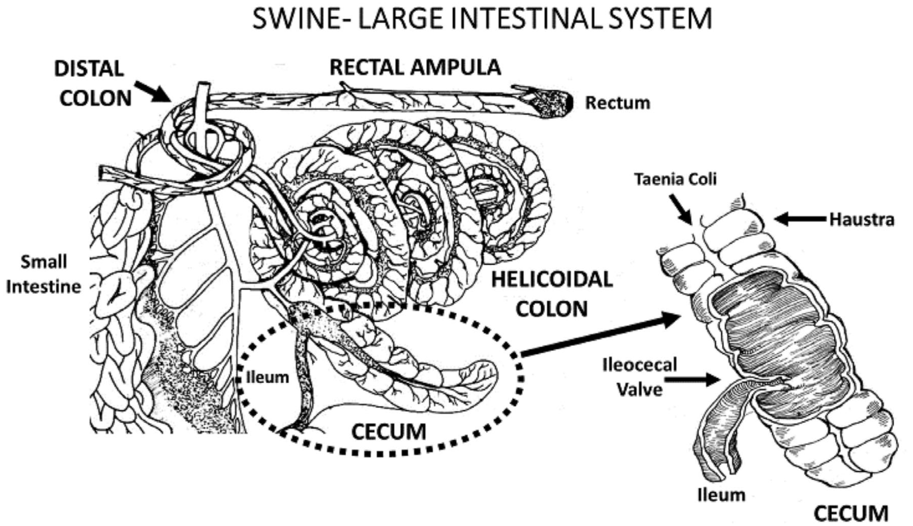
The schematic illustrates the spiral colon and caecum in pigs, highlighting the notably prominent and elongated caecum.
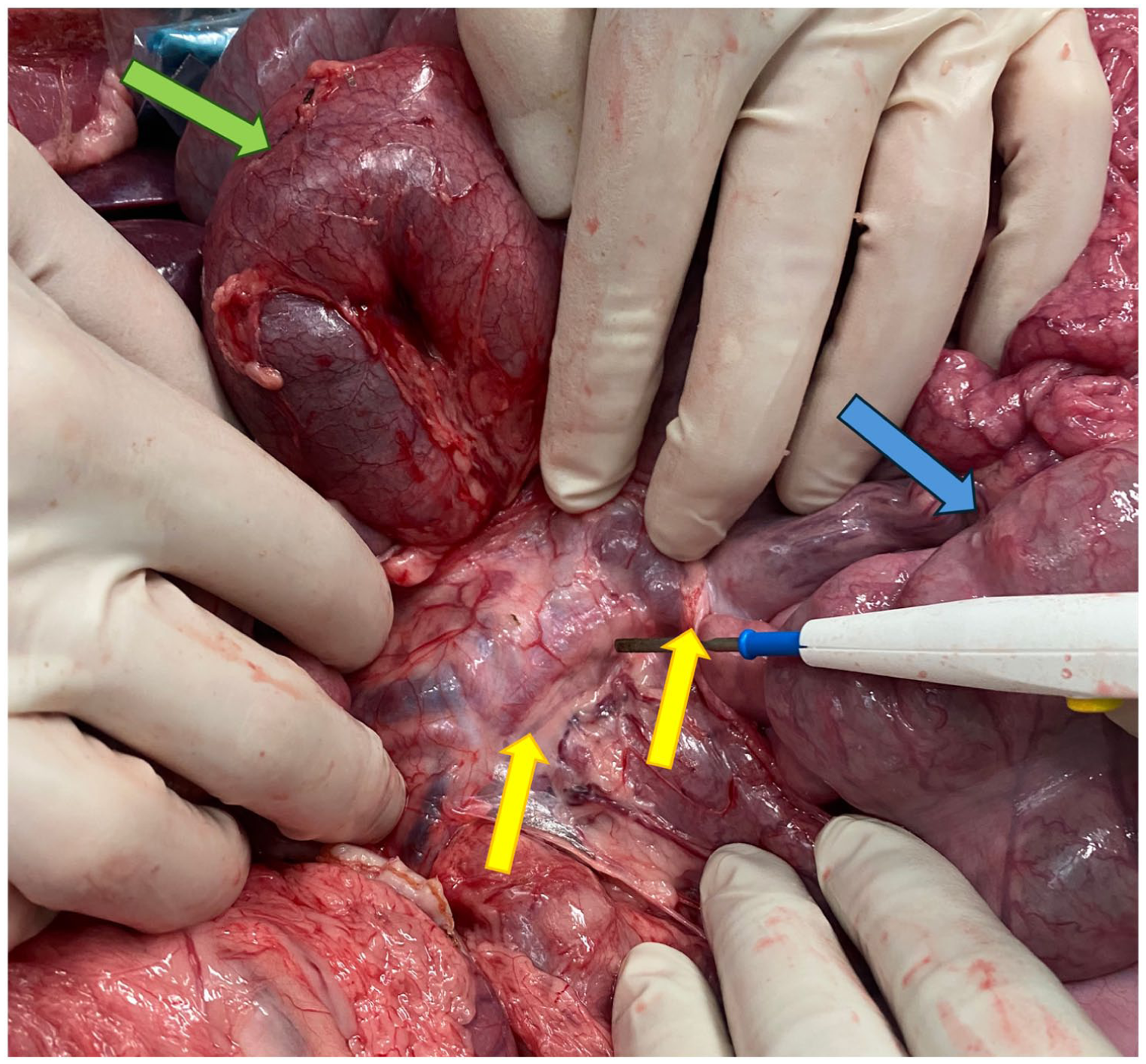
An intraoperative image illustrating the region of the retroperitoneum surrounding the duodenojejunal flexure in a porcine model. The green arrow indicates the reflected duodenum, and the blue arrow indicates the caecum. The yellow arrows highlight the prominent duodenocolic fold, which serves to anchor both structures together and must be meticulously divided to achieve complete colon mobilisation.
Hepatobiliary system
The liver, situated in the right upper quadrant of the abdomen, is the largest solid organ in humans and pigs. The human liver is characterized by two anatomical lobes and is wedge-shaped whereas the porcine liver is thinner, smaller overall by percentage volume and has four well-defined anatomical lobes giving it a clover-shaped appearance.17,29–31 Despite these differences in the gross morphology, the segmental anatomy of the liver is directly comparable to that described by Couinard in humans, with biliary and vascular radicals travelling together in a portal triad toward eight distinct segments25,30,32,33 (Figure 4). The retrohepatic inferior vena cava (IVC), however, is entirely intrahepatic and therefore procedures requiring caval clamping may require passive caval bypass to reduce haemodynamic instability as in an orthotopic liver transplant. 30 In humans, the arterial supply to each side of the liver is typically provided by the left and right hepatic arteries, which branch from the common hepatic artery at the liver’s hilum. However, variations are common, with accessory and replaced vessels frequently observed. 34 In pigs the hepatic artery branching occurs well before the hilum. Additionally, the right branch bifurcates into two and the left into three in the pig. The portal vein exhibits a different relationship, with division into left and right branches at the hilum in humans and before the hilum in the pig. The portal vein is formed by the confluence of the splenic vein and the superior mesenteric vein in both species. However, in pigs, the caudal mesenteric vein also joins at this confluence, analogous to the inferior mesenteric vein in humans. The confluence of the portal vein in humans is located posterior to the neck of the pancreas. In contrast, in pigs, the anterior and posterior portions of the pancreatic body encircle the portal vein. 35 The hepatic artery supplies 25% of the liver’s blood, while the portal vein provides 75%, a distribution pattern analogous to that observed in humans. 30 In humans, the left, middle and right hepatic veins all drain into the IVC, with the middle and left veins often converging to form a single trunk just prior to their insertion into this vessel. 34 The porcine liver is drained by four hepatic veins into the IVC. These veins are characterized by their thin walls, rendering them susceptible to damage. 31 The anatomical structure of the pig’s gallbladder closely resembles that of humans, with the primary distinction being that the cystic artery originates from a branch of the left hepatic artery (ramus quadratus), as opposed to the right hepatic artery in humans.30,31 The intra- and extra-hepatic biliary ductal system closely resembles that observed in humans, with the exception of the distal relationship between the common bile duct and the pancreatic duct, which is elaborated upon below. 30

Diagrammatic depiction of the lobar structure of the porcine liver, characterized by a ‘clover-shaped’ configuration (left), and the human liver, exhibiting a ‘triangular prism-shaped’ form (right).
The porcine pancreas closely resembles the human in terms of size, position and weight.36,37 A notable anatomical difference between the two species is that in pigs, the dorsal and ventral anlages are discrete and well developed. The pancreas exhibits a distinctive morphology characterized first by a head that is in contact with the pylorus and proximal duodenum. This then extends to the left, the body of the pancreas divides into anterior and posterior sections that encircle the portal vein, thereby imparting a unique ‘ring-shaped’ appearance to the porcine pancreas. These sections subsequently converge at the tail, which extends caudolaterally towards the splenic hilum. 35 The human pancreas in comparison is L-shaped with the head within the concavity of the duodenum and the tail intimately associated with the splenic hilum 37 (Figure 5). Crucially, the porcine bile duct and pancreatic duct enter the duodenum separately, unlike in humans, where they converge into a common channel, the ampulla of Vater, prior to piercing the duodenal wall at its second part. 11 The porcine pancreatic duct exhibits a brief extra-parenchymal course, positioned within a membrane located between the duodenum and the pancreatic head. 38 The total β-cell mass between humans and pigs is highly comparable and both species share the same β-cell mass-to-body weight ratio of 10mg/kg. 9 Though the presence of islets in pigs of less than 200kg has been reported only following isolation and not on pancreas histology, we have in our experimental work demonstrated the presence of such islets on hematoxylin and eosin stained histology in 80kg pigs. 39 The structure and appearance of these closely mirror those in humans. The spleen occupies a more prominent position in the porcine abdominal cavity compared with its location in humans. Instead of being situated dorsolaterally beneath the ribcage, it occupies a substantial portion of the left upper quadrant, extending into the epigastric region. The anatomical separation from the pancreas is more pronounced compared with humans, facilitating the performance of a splenectomy without the necessity for a concurrent distal pancreatectomy, or vice versa.
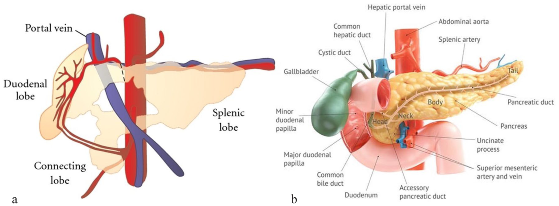
Diagram comparing gross morphology of porcine pancreas (a), which is ring-shaped, and human pancreas (b), which is L-shaped with the head and uncinate process embraced by the C-shaped curve of the duodenum. Also note the different relationship of the portal vein to pancreas between pigs and humans.
Genitourinary system
Pigs are particularly similar to humans in terms of the structure of the urinary system. A pig weighing 70kg possesses a kidney of a size comparable to a human’s 1 (Figure 6). The porcine kidney is lobulated and multi-papillate with true calices, analogous to that of a human.1,17 This makes it particularly attractive for translational research in renal transplantation. Typically, in both species, the renal artery arises directly from the abdominal aorta at a level between the L1-L2 vertebra. 40 The diameter of the main renal artery is also very similar between species (4.0–5.9 mm vs. 5.1–5.4 mm in humans and pigs respectively). 40 The arterial supply is less variable in pigs, with a single renal artery present in 93.4% of cases compared with 70% of humans.3,41 Once beyond the hilum, however, both have interlobar and segmental arteries supplying the parenchyma. 41 The left renal artery often originates slightly more cranially than the right. 23 Additionally, the renal arteries of pigs are also particularly prone to spasm when handled.3,15 Regarding venous anatomy, the left renal vein divides in two just beyond the lateral border of the aorta, which should be considered during dissection of the kidney off the retroperitoneum. Anomalies in venous anatomy in pigs, however, are rare, occurring in only 2.13% compared with 15–30% of humans. 40 The ureter is narrow in pigs, with a fragile mucosa that is prone to oedema, and gentle handling is required if allotransplantation is to be performed. 4 In pigs, the ureter traverses over the external iliac artery, as opposed to the bifurcation of the common iliac vessels observed in humans, due to the absence of this bifurcation in pigs. 40 The bladder is similar in position, volume and structure in both species. 40 The wall, however, is thinner in pigs and more prone to rupture when distended. 26 One other notable difference is that the entire bladder of the pig is an intraperitoneal structure instead of retroperitoneal as in humans. 1 Consequently, it is highly mobile and can easily be eviscerated to improve access to the pelvis without extensive dissection. 42 The anatomical and physiological similarities between the urinary systems of pigs and humans facilitate the performance of heterotopic renal transplantation in pigs in a manner identical to that in humans. This makes pigs an ideal large animal model for studying this procedure.

The green arrow indicates the right kidney, with the overlying retroperitoneum divided. Notably, there is a distinct absence of perinephric fat surrounding the kidneys of lean pigs whereas in humans the kidneys are encased by peri-nephric fat within Gerota’s fascia. The blue arrow illustrates the drainage of the right renal vein into the inferior vena cava, as observed in humans.
The anatomy of the genital tract of pigs has become more relevant to the field of abdominal transplantation in recent years since the description of the first live birth following uterine transplant in 2014. 43 Porcine models describing uterine auto-transplantation were followed by heterotopic allotransplantation, playing a crucial role in making uterine transplantation a clinical reality.42,44 Although the porcine uterus is recognized for its utility as a translational research model, it exhibits significant gross morphological differences compared with the human uterus. It is bicornuate in structure and adapted for large litters, with each cornu being tortuous and measuring approximately 26 cm in length.26,45 The cornu on each side is anatomically connected to the uterine tubes cranially and converge caudally at the unpaired uterine body. 46 The human uterus, in contrast, is pear-shaped and specifically adapted for single pregnancies. In both species, the uterine vessels are branches of the internal iliac system, with additional collateral supply provided through the uterine branch of the ovarian artery.42,47 In pigs, the primary arterial inflow is facilitated by a single uterine artery, while venous return occurs through both a superficial uterine vein and a deep uterine vein. The superficial uterine vein is located in the upper region of the pelvic floor and runs parallel to the uterine artery. 45 The deep uterine vein is situated at the base of the pelvic floor and lacks an accompanying artery. It is susceptible to damage, which can lead to catastrophic haemorrhage if injured during its trajectory along the pelvic floor. 45 The technique of anastomosing the donor aorta and IVC or internal iliac vessels with the recipient’s external iliac vessels has been described in uterine transplantation models in pigs.42,45
Vascular system
While the orientation of major abdominal organs is largely consistent across species, the fact that pigs are quadrupeds rather than bipeds results in differences in the orientation, branching and course of their vessels. 23 The abdominal aorta in pigs weighing 40–60 kg is smaller than that in humans, measuring only 7–10 mm in diameter compared with 20–25 mm in humans.23,48 Notably, in the context of heterotopic renal transplant procedures, the porcine abdominal aorta terminates in a trifurcation, with both the internal and external iliac arteries branching directly from the aorta, a significant divergence from the human anatomy.23,48,49 The internal iliac artery initially emerges as a common trunk from the aorta before bifurcating almost immediately after its origin. However, the external and internal iliac veins do converge into a common iliac vein in both pigs and humans. 49 The external iliac artery is quite small in pigs and this needs to be kept in mind when performing heterotopic transplantation procedures (Figure 7). The external iliac artery usually measures around 5mm in diameter in the pig and the internal trunk 5–8mm.48,49 The internal artery is therefore often preferred for heterotopic implants. Pigs possess a coeliac trunk and a cranial mesenteric artery, which is analogous to the superior mesenteric artery in humans. The coeliac trunk is notably short, measuring only 15 mm in length, with the splenic artery arising very proximally, near the abdominal aorta 23 (Figure 8). Unlike humans, pigs lack an inferior mesenteric artery; instead, they have a small posterior mesenteric artery that supplies only the distal colon and rectum.23,49 As previously mentioned, the terminal arteries supplying the small bowel do not form anastomotic vascular arcades in the mesentery but do so only in the subserosa. 25
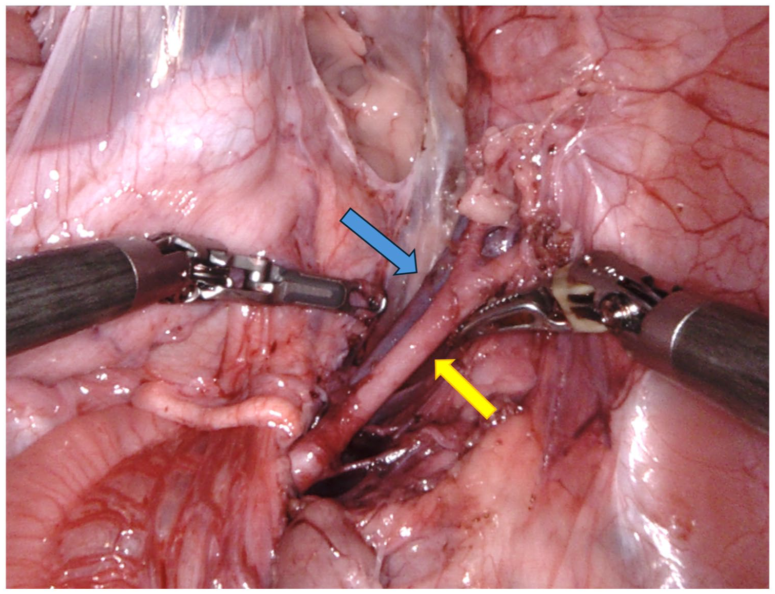
View into the porcine pelvis from a robotic heterotopic renal transplant demonstrating the narrow diameter of the external iliac artery (yellow arrow) and vein (blue arrow). For reference, the length of the daVinci robotic Maryland grasper on the right is 20mm, the artery is less than half this size.
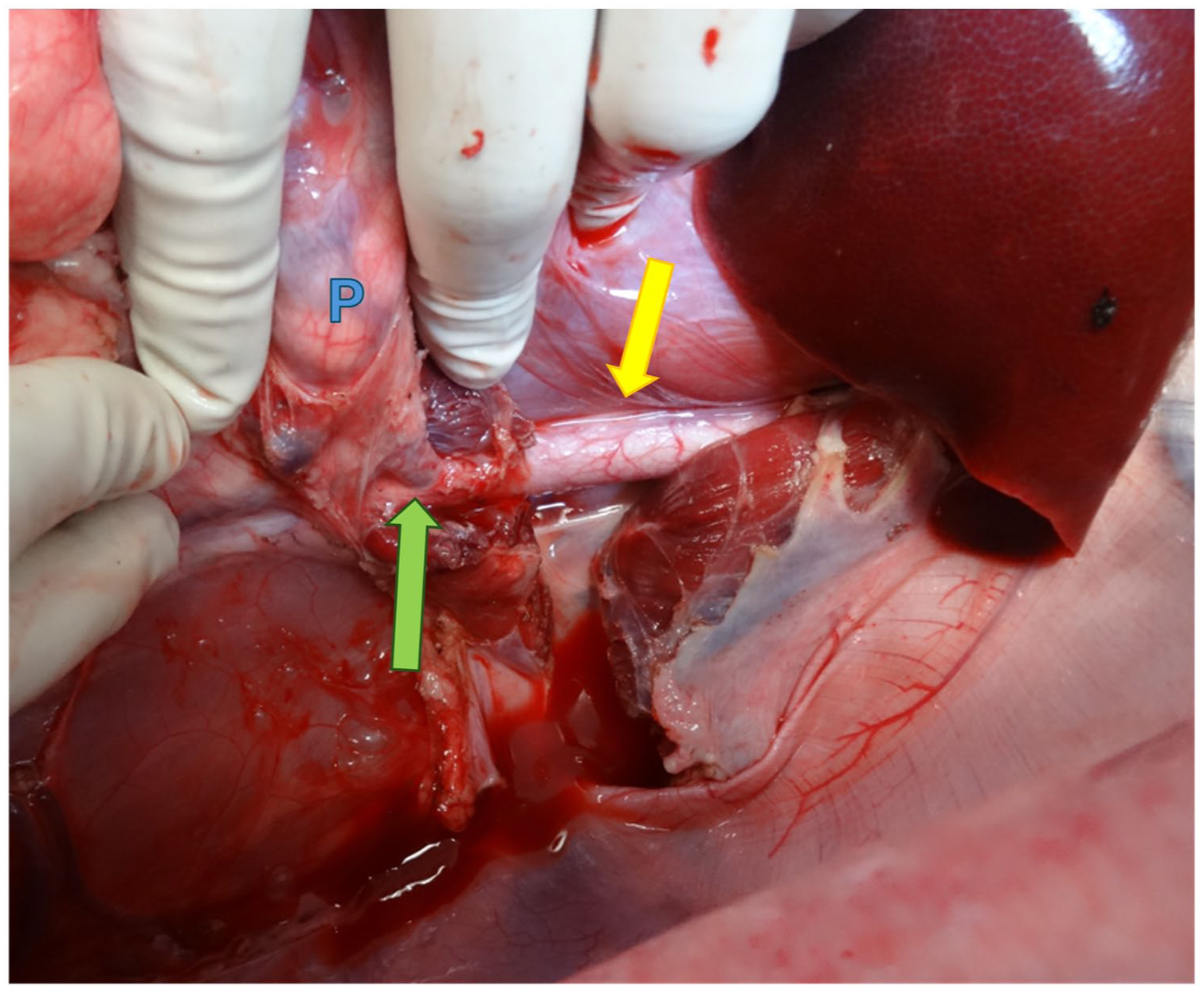
The yellow arrow indicates the abdominal aorta, with the diaphragmatic crus above it divided. The green arrow indicates the origin of the splenic artery, which arises almost directly from the abdominal aorta after a very short coeliac trunk, as observed in humans. This artery is seen extending cranially over the pancreas (P), which has been retracted medially by the surgeon’s hand.
Non-operative challenges with the use of pigs as translational models
Although their physiological and anatomical similarities make pigs a compelling model for transplantation research, several potential challenges necessitate careful consideration. Commercial pigs exhibit rapid and exponential growth as expected for an animal bred for food production.11,25 At birth they weigh approximately 1.5kg, which rapidly increases to 35kg at three months and then to 200kg by one year of age. 41 To support this typical weight gain, each individual pig requires roughly 1kg of feed per day, which adds considerable expense. Because of their rapid weight gain commercial pig breeds become increasingly challenging to handle, with complex husbandry demands, posing a manual handling risk for researchers from a relatively young age due to their size. 4 To address these issues, several minipig breeds, such as Yucatan, Hanford, Göttingen and Sinclair, are available for research purposes. These breeds reach sexual maturity at 4–6 months of age and weigh only 25–35 kg. 17 These breeds are anatomically and physiological identical to larger commercial breeds, differing only in size.9,41
Certain conditions are more frequently observed in pigs and can result in the premature demise of these animals. There is an increased incidence of midgut volvulus seen in pigs compared with humans based on the anatomical position of the superior mesenteric artery running between the small bowel and the large bowel in these animals. 50 Pigs, especially the Landrace breed, are prone to porcine malignant hyperthermia, which can occur during administration of volatile anaesthetics but can also be triggered by stress in the absence of anaesthesia.3,8,51 Additionally, pigs are also prone to the development of peptic stress ulcers and small bowel obstruction. 3 The myocardium of pigs is also susceptible to the development of possibly fatal ventricular arrhythmias in the setting of hypovolaemia, hypotension and shock as can occur during periods of intra-operative instability. 5 Given their pronounced sensitivity to stress, it is advisable to allow a period of 1–2 weeks for acclimatization to a new environment before initiating any experimental procedures. 8
Finally, while the popularity of pigs as a large animal model has grown rapidly, porcine-specific antibodies and kits for biological marker detection are expensive and not as common or exhaustive currently as for murine models. 41
Discussion
Considering the anatomical features described, it is pertinent to examine the critical factors to consider when conducting major abdominal and transplantation surgery in pigs. The relationship between the colon and duodenum is of paramount importance in preventing enteric content leakage. The peritoneal duodenocolic fold, which connects these two structures, is a substantial serous fold that must be severed to fully mobilize the colon from the abdominal cavity. It is also essential to note that the porcine colon is relatively thin-walled and susceptible to perforation from minimal trauma.
The considerable length of the small intestine and the space it occupies within the abdominal cavity can impede access to retroperitoneal structures, making it advantageous to reduce the small intestine to the minimum practical length. 52 When dividing sections of the intestine, it is crucial to avoid damaging proximal mesenteric branches of segments that will not be resected, owing to the absence of collateral arcades between branches in the mesentery. Given that the majority of nutrients are absorbed in the jejunum and ileum, preserving an adequate portion of the jejunum is advisable to maintain absorptive capacity for experimental purposes. 53
Early branching of the splenic artery from the coeliac trunk must also be acknowledged when performing procedures involving the abdominal aorta, medial mobilization of retroperitoneal structures, or the pancreas.
Conclusion
In conclusion, the growing use of porcine models in translational transplantation research necessitates that researchers in this domain possess a comprehensive understanding of comparative anatomy when conducting complex surgical procedures. The anatomical and physiological resemblances between pigs and humans, the successful application of advanced gene-editing technologies to surmount immunological challenges, the dependability of ex vivo perfusion systems, and the practical benefits of cost-effectiveness and availability will ensure the continued prevalence of porcine models in the future. To optimize the utilization of this valuable resource and minimize the risk of animal loss without benefit, it is crucial for surgical researchers to grasp the practical aspects of comparative anatomy to enable safe surgical interventions.
Footnotes

Author contributions
D. Barnett: concept, manuscript writing, figure preparation; R. Bhattacharjya: manuscript writing, figure preparation; S. Bhattacharjya: concept, manuscript editing.
Funding
The authors received no financial support for the research, authorship, and/or publication of this article.
Declaration of conflicting interests
The authors declared no potential conflicts of interest with respect to the research, authorship, and/or publication of this article.
Data availability statement
Data sharing not applicable to this article as no datasets were generated or analysed during the current study.