
Abstract
Estimation of age from microscopic examination of human bone utilizes bone remodeling. This allows 2 regression equation to be determined in a specific population based on the variation in osteon turnover in different populations. The aim of this study was to provide age estimation for Malaysian males. Ground undecalcified cross sections were prepared from long limb bones of 50 deceased males aged between 21 and 78 years. Ten microstructural parameters were measured and subjected to multivariate regression analysis. Results showed that osteon count had the highest correlation with age (R = 0.43), and age was estimated to be within 10.94 years of the true value in 98% of males. Cross validation of the equation on 50 individuals showed close correspondence of true ages with estimated ages. Further studies are needed to validate and expand these results.
Introduction
Morphologic skeletal features are used widely to perform age at death estimation in human remains, particularly useful for people aged less than 50 years, 1 while a histological approach has been advocated as the method of choice for age estimation mainly, for people aged over 50 years. 2 Bone structure changes with age by turnover, remodeling and modeling, and growth, which alters the morphology and histology of the bone throughout an individual’s life. 3 Age is an important factor, in which there is an increase in bone porosity with increasing age as remodeling results in increased bone resorption and less bone formation. 3
Combined approaches by using both gross and micro-morphological parameters have been used to offer better results,4,5 including cortical thickness (CT) and histologic parameters. Kerley’s method, 6 which requires a complete bone cross section, is widely cited as the most accurate of all histologic methods. Kerley 6 utilized various histologic parameters such as secondary osteons, osteon fragments, non-Haversian canals, and percentage of lamellar bone in specific regions on cross sections from femorae, tibiae, and fibulae.
There is a distinct variation in the remodeling pattern in various parts of the bone. Patterns of microstructural parameters differ in different cross sections of the same bone and in different locations of one cross section. 7 Any one part of the bone may not be representative of the entire cross section of the bone. Hence, the measurements from the entire cross section will be the best method to use. 7 The mid-diaphyseal part of a long bone was used to determine age in many studies,1,8 as this part of the bone tends to be more robust and last through exposure to weathering changes and predator or scavenger damage.9,10 Cortical bones of fibula, humerus, and ulna were used to estimate age in the population. 1 The anterior midshaft of femur was also widely used to study age prediction histologically.11,12
Various factors such as genetic, biomechanic, and environmental factors are known to have a major contribution to the morphology and microstructure of bone in the population. 13 The type of physical activity and metabolic and biochemical needs of the body in response to bone formation could lead to a distinctive bone microstructure and bone mass. 14 Individuals have been under-aged by 29.2 years due to poor nutrition resulting in significant retardation of osteonal growth in ribs.15,16 Other related factors such as life history, 17 disease, 18 physical activity, 17 diet and length of daylight,19,20 and nutritional stress 21 have some influence in determining the rate of skeletal ageing in an individual, which finally lead to variability in bone remodeling. 22
Applications of histological age estimation have been based largely on Western populations, and therefore continued to be used as a reference for age estimation of other populations.19,23–25 However, individual human skeletons have been shown to demonstrate age-related changes and progresses at different rates leading to differences in bone microstructure19,26,20 and bone mass.
27
Hence, the method used widely for
Methods
The study materials comprised specimens from 50 males, aged 21–78 years (mean 41.68, standard deviation (SD) 11.85) collected from the mortuaries of Forensic unit, Hospital Universiti Kebangsaan Malaysia and Hospital Kuala Lumpur. The procedure for legal handling of human bones is stated in the Criminal Procedure Code, Section 331 (part 2) of the Malaysian Law, which stated that the postmortem examination of the human body may be extended to dissection, and analysis of any portion of the body may be retained for further investigation and research purposes. 29
Samples were taken from humerii, ulnae, radii, femorae, fibulae, and tibiae, and only specimens confirmed to be free from any pathology following gross and microscopic examination were used. While other studies relied exclusively on histologic variables, this study included the morphologic variables such as CT and medullary cavity diameter (MCD), which were measured on the bone by using digital calipers. CT and MCD were determined to the nearest 0.01 mm after the bone marrow was removed.
A complete circumferential mid-diaphyseal 2 × 2 cm fragment was removed in a plane that was transverse to the longitudinal axis of the long bone. Bone fragments were defatted with diethyl ether in soxhlet and embedded in Buehler Epothin™ epoxy resin mixture using the manufacturer’s specifications. Thin sections of 30 µm were produced with a saw-microtome (Leica SP 1600), finished with a grinder/polisher (Phoenix Beta Buehler) and mounted with a cover slip on a microscopic slide using Histomount.
Microscopic analysis was performed under transmitted light at 100 × magnification with an Olympus BX 51 microscope. Measurements were made on four subperiosteal fields: anteromedial, anterolateral, posteromedial, and posterolateral quadrants, thus avoiding the linea aspera. Each field was subdivided into two subfields (Figure 1) separated by the width of one square grid measuring 10 × 10 µm (100 µm2), which was calibrated by an objective micrometer (AX0001, Olympus). Histologic parameters, namely osteon count (OC), osteon diameter (OD), Haversian canal diameter (HCD), osteon area (OA), Haversian canal area (HCA), osteon perimeter (OP), Haversian canal perimeter (HCP), and Haversian lamellae count (HLC) were measured using a commercially available image analysis program (SIS Soft Imaging System 3.2 Software Package). The morphologic and histologic variables are collectively known as microstructural parameters.
Bone thin cross-section: Eight fields for histological measurements. Note that histological measurements were made on eight fields: two areas of anteromedial, anterolateral, posteromedial, and posterolateral.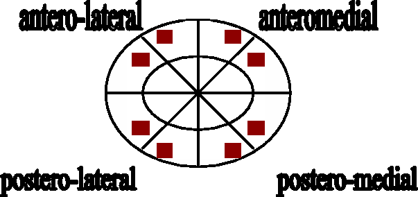
Measurements of osteons followed the criteria as described in Wachter et al. 30 with some modifications. Measurement of osteons, Haversian canals, and lamellae was made on intact osteons and canals. OC comprised the aggregate number of osteons and osteon fragments. In previous criteria, osteons and osteon fragments were counted separately. Osteons were counted as such that half or more than half of its Haversian canal was present in the visual field, and osteon fragments at the periphery of the field were also included, even if it was only partly within the field.
The complete slides of 10 individuals were tested for interobserver analysis with respect to osteon counting. Three independent observers, namely a forensic pathologist with a few years experience in bone histology, an experienced forensic anthropologist, and a forensic archaeologist were involved in the observational analysis. The osteon counting was done without any knowledge of the sample’s age, origin, or type of bone for blinding purposes. The analysis was repeated after one-week interval.
Statistical analysis
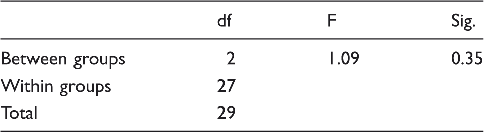
Correlations between microstructural parameters and age were tested by Pearson’s (R) correlation, and age regression equations were established using multivariate regression analysis in SPSS version 15.0. 31 All 10 parameters were subjected to stepwise multivariate regression analysis with age as dependent parameter so that only equation with the lowest standard error of estimates (SEE) was chosen. Cross-validation of the equation by independent sample t-test was performed on 50 deceased persons to determine the applicability of the method. The difference in osteon counting in each observer was tested by using paired t-test, and the difference in osteon counting among all observers was tested by using one-way analysis of variance (ANOVA).
Results and discussion
Descriptive statistics of OC 1 and OC 2 for all observers.
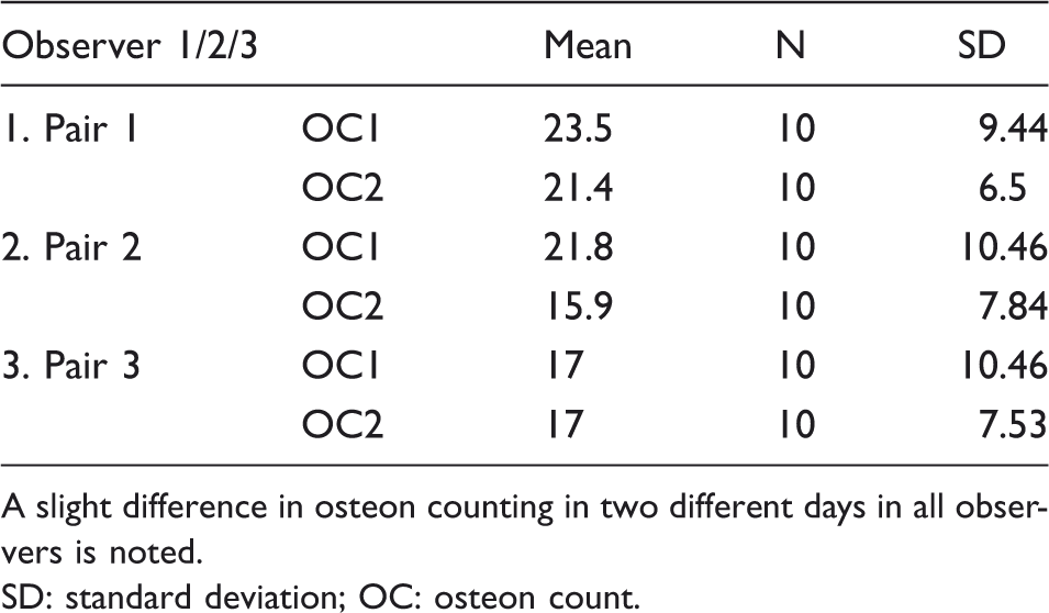
A slight difference in osteon counting in two different days in all observers is noted.
SD: standard deviation; OC: osteon count.
Paired sample t-test between the observers in osteon counting.
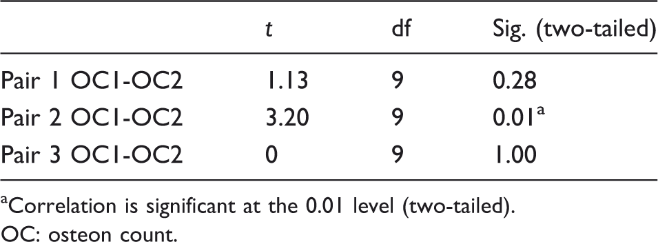
Correlation is significant at the 0.01 level (two-tailed).
OC: osteon count.
One-way analysis of variance (ANOVA) of the mean OC between the three observers.
Correlation analysis for the relation between age and microstructural parameters.
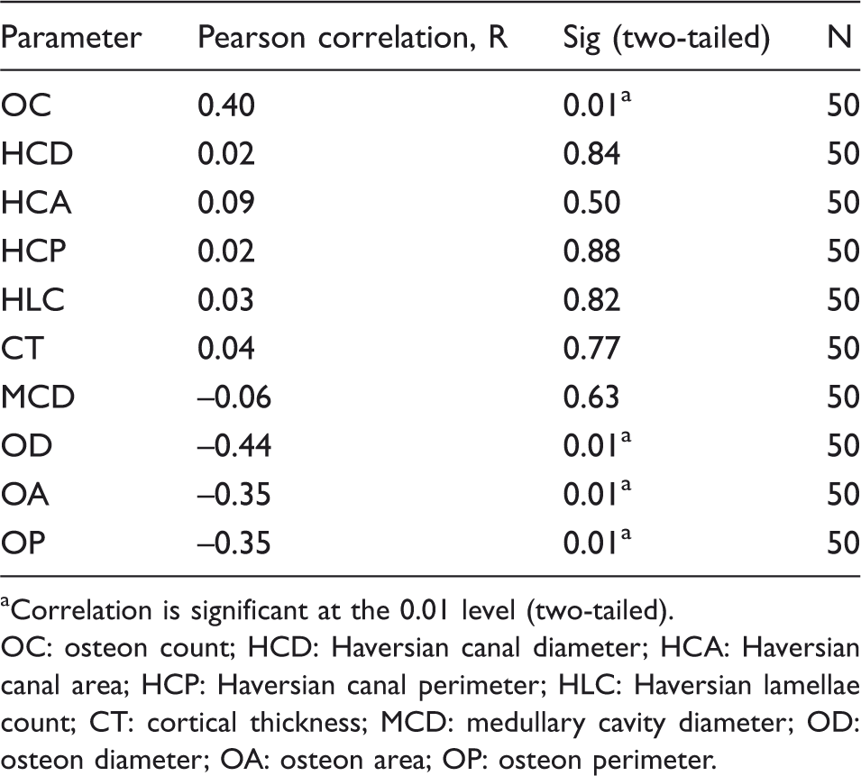
Correlation is significant at the 0.01 level (two-tailed).
OC: osteon count; HCD: Haversian canal diameter; HCA: Haversian canal area; HCP: Haversian canal perimeter; HLC: Haversian lamellae count; CT: cortical thickness; MCD: medullary cavity diameter; OD: osteon diameter; OA: osteon area; OP: osteon perimeter.
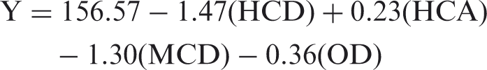
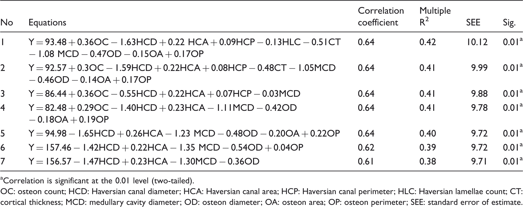
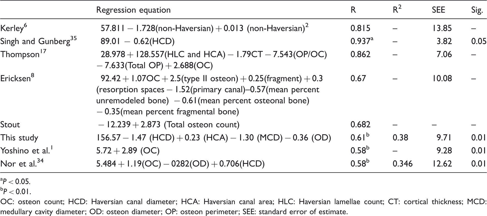
Age estimating equation derived from stepwise regression analysis.
Correlation is significant at the 0.01 level (two-tailed).
OC: osteon count; HCD: Haversian canal diameter; HCA: Haversian canal area; HCP: Haversian canal perimeter; HLC: Haversian lamellae count; CT: cortical thickness; MCD: medullary cavity diameter; OD: osteon diameter; OA: osteon area; OP: osteon perimeter; SEE: standard error of estimate.
Coefficients derived from regression analysis.
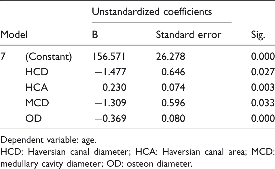
Dependent variable: age.
HCD: Haversian canal diameter; HCA: Haversian canal area; MCD: medullary cavity diameter; OD: osteon diameter.
It is pertinent to mention that this equation is applicable to all long limb bones, whether it is from upper or lower limb.
Comparisons of known age and estimated age for all males.
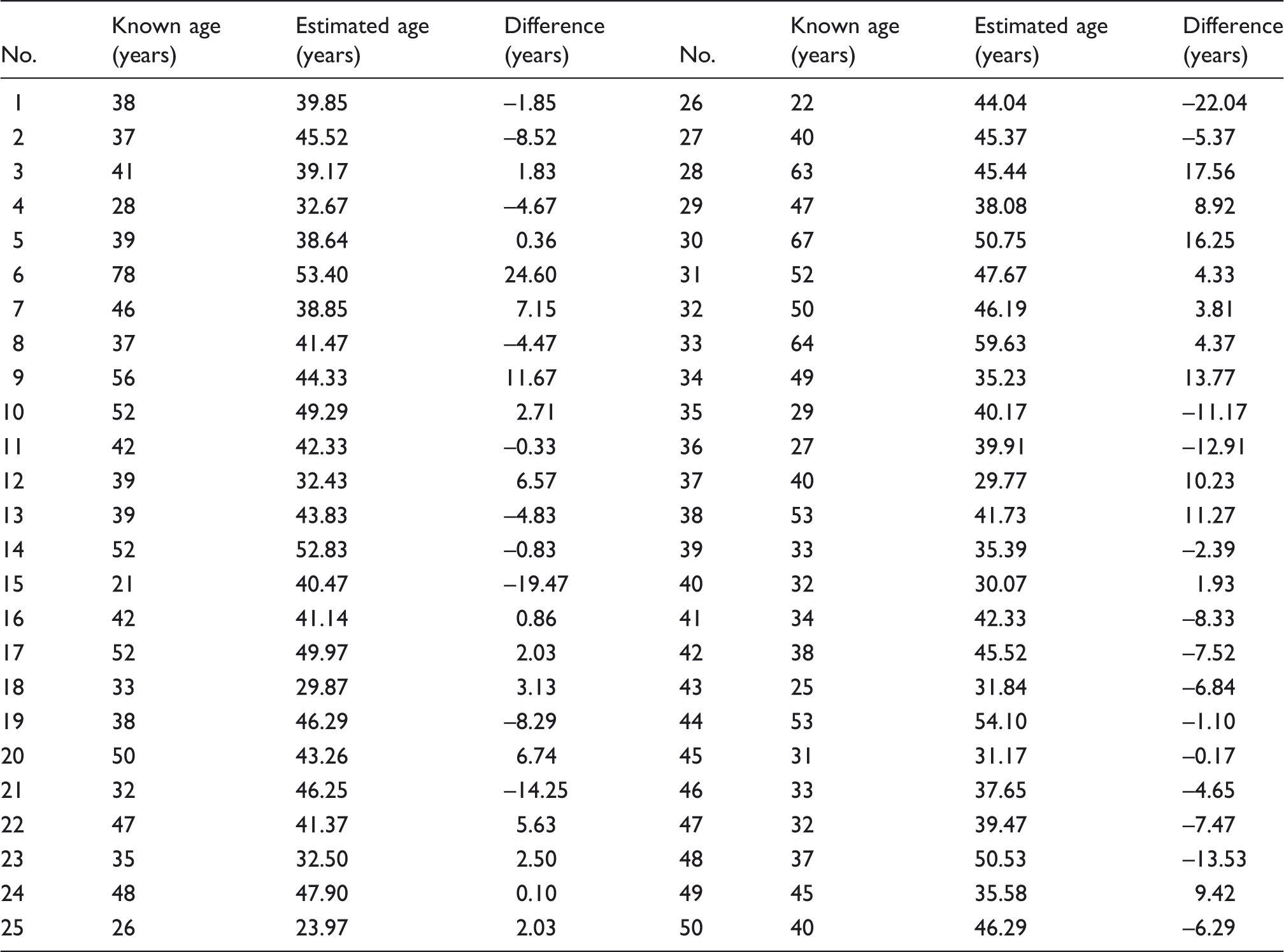
Paired sample t-test for known age and estimated age.
Age: known age; Age1: estimated age; SD: standard deviation.
P < 0.05.
P < 0.01.
OC: osteon count; HCD: Haversian canal diameter; HCA: Haversian canal area; HLC: Haversian lamellae count; CT: cortical thickness; MCD: medullary cavity diameter; OD: osteon diameter; OP: osteon perimeter; SEE: standard error of estimate.
Conclusion
For the Malaysian male population, a quantitative histological method of age estimation was analyzed by collecting the specimens and analyzing 10 microstructural parameters in the mid-diaphyseal cortex of upper and lower long limb bones. In microscopic transverse sections, quantitative assessments were taken on four subperiosteal locations, namely anteromedial, anterolateral, posteromedial, and posterolateral on the bone thin section. Interobserver analysis showed reasonable agreement between experienced and inexperienced examiners in the assessment of osteon counting. Despite the generally encouraging outcome, it is recommended that this study be used as a preliminary step to a more extensive research in future, which would include confounding factors, namely different types of bones, ancestry affiliation, dietary intake, disease, and environment in the estimation of age.
Footnotes
Declaration of conflicting interests
The authors declare that there is no conflict of interest.
Funding
The authors thank the Malaysian Government for funding the research project.
Contributorship
Faridah Mohd Nor – planned the study, compiled the data, and wrote the paper. Robert F Pastor and Holger Schutkowski – supervised the work and edited the paper.
Acknowledgements
The authors thank the Universiti Kebangsaan Malaysia Medical Centre and Hospital Kuala Lumpur for having allowed access to the human bone samples.