
Abstract
The ability to obtain an autosomal short tandem repeat (STR) profile of the semen donor from the reproductive tract of a living victim rapidly diminishes as the post-coital interval increases. This is of concern where victims of sexual assault provide vaginal samples several days after the incident. In order to overcome the technological impediments inherent in autosomal DNA typing with extended interval samples, we previously employed the use of Y chromosome STR profiling which, by targeting only male DNA, can eliminate masking of the male profile (by the victim’s alleles) or critical polymerase chain reaction reagent titration (due to excessive female DNA). Thus employing Y-STR profiling and additional enhancement strategies, we reported the ability to recover Y-STR profiles from samples collected 5 to 6 days after intercourse. However, the reproductive biology literature indicates that spermatozoa are found in the human cervix up to 7 to 10 days post coitus. Thus, even with improved extraction and profiling techniques, we failed to routinely recover profiles from samples collected ≥6 days after intercourse. The aim of the present work was to develop additional strategies to permit the recovery of male donor DNA profiles from ≥6 post-coital samples. Using nested polymerase chain reaction and DNA concentration procedures that together maximize the recovery and targeting of male DNA, we demonstrate the ability to obtain semen donor Y-STR profiles in extended interval post-coital samples collected 6 to 9 days after intercourse. This approaches the recognized time limits of sperm residence in the cervico-vaginal canal as described in the clinical literature.
Keywords
Introduction
DNA typing has revolutionized forensic biology by providing the ability to routinely obtain highly probative identification evidence from crimes involving the transfer of biological material. There are, however, still some situations whereby it is extremely challenging to recover a standard autosomal short tandem repeat (STR) DNA profile from a sample that contains human cellular material. One particular challenge can be “extended interval” post-coital vaginal tract samples recovered from rape victims who do not report the sexual assault until several days (e.g. >3 days) after the incident. Loss of a significant proportion of the original semen deposit in such extended interval post-coital samples arises due to vaginal drainage/lavage, menstruation and the normal intra-cervico-vaginal sperm degradative changes that occur over time.1,2 In these instances, the use of autosomal STR analysis would likely result in a failure to obtain a genetic profile of the male donor due to the overwhelming amount of female DNA present in the sample (i.e. “masking” of the male profile due to the titration of critical PCR reagents by the female DNA).3–6 A differential extraction is normally employed when sperm cells are present to separate the sperm (“male”) and non-sperm (“female”) fractions. However, in the case of extended interval post-coital samples, the sperm may be structurally fragile or damaged resulting in the premature lysis into the non-sperm (or primarily female epithelial cell) fraction. The use of Y chromosome STR analysis is a useful alternative to standard autosomal STR analysis in some of these case situations since it specifically targets the male DNA present in an admixed sample even amongst an overwhelming amount of female DNA.3–12 The reported high sensitivity of Y-STRs and the demonstrated ability to obtain genetic profiles of the male donors has resulted in the increasing use of these markers in sexual assault cases.3–6,13–15 Employing Y-STR profiling and additional enhancement strategies, we reported the ability to recover Y-STR profiles from samples collected 5 to 6 days after intercourse. 2
Despite the improvement in semen donor profile recovery in sexual assault evidence using Y-STR analysis, the analytical detection limit of the individual typing system or kit used impacts the success rate. Since extended interval post-coital samples contain minute numbers of sperm cells and/or male epithelial cells (if any at all) such samples will often fail to yield the male donor profile. The reproductive biology literature indicates that spermatozoa (albeit a small number) may be present in the human cervix up to 7 to 10 days post coitus.16–20 It is therefore pertinent to develop and test ultra-sensitive profiling methods for the analysis of the few sperm recoverable from such extended interval samples. We hypothesized that a viable approach to surpassing current analytical detection limits would be to pre-amplify segments of the male donor’s Y chromosome prior to standard Y-STR profiling.
In contrast to whole genome amplification (WGA) strategies that pre-amplify the entire genome (or large portions thereof), we report here the development of a targeted genome pre-amplification system that specifically amplifies multiple STR loci on the Y chromosome. This “nested” PCR-based pre-amplification reaction (or Y-chromosome targeted pre-amplification, Y-TPA) utilizes Y-chromosome specific primers in the first round amplification that are located in the flanking regions of 17 commonly used Y-STR loci. This results in the production of increased amounts of starting templates prior to subsequent Y-STR analysis. The Y-TPA primers are designed to be compatible with the primer sets used by commercial Y-STR amplification kits. Y-TPA, in combination with other DNA typing enhancement strategies including DNA concentration and purification, permit the recovery of potentially probative Y-STR profiles from cervico-vaginal samples collected up to and including 9 days after intercourse. This represents a significant improvement in the post-coital time frame during which DNA profiles from the semen donor might be recovered from sexual assault evidence.
Materials and methods
Body fluid sample preparation
All body fluid samples were collected in accordance with procedures approved by the University’s Institutional Review Board. Buccal swabs were collected from male and female volunteers by swabbing the inside of the cheek. Post-coital cervico-vaginal swabs (x2) were collected by each of four female volunteers who recovered the samples after separate acts of sexual intercourse at various time points (6, 7, 8, and 9 days) using sterile cotton tipped applicators (Lynn Peavey, Lenexa, KS). The volunteers were instructed to take the samples from the cervix by swabbing multiple times for 20–30 s at each specific time interval. All volunteers were asked to abstain from intercourse at least 2 to 3 days after sample collection to ensure a sufficient period between collections (i.e. 8–12 days in between separate acts of sexual intercourse). Only one pair of swabs per time period was taken subsequent to each act of sexual intercourse to preclude a progressive and unnatural loss of semen due to the sampling process itself. In some cases, a pre-coital cervico-vaginal swab was also obtained before coitus commenced to check for the presence of male DNA from previous sexual activity. All samples were dried overnight and then stored at −20℃ until analysis.
DNA isolation, purification, and quantitation
DNA was extracted from the samples using a standard non-differential organic extraction as previously described, 5 with a minor modification involving re-solubilization in 75 µL of TE−4 rather than 100 µL. The entire extract (75 µL) was subsequently purified and concentrated using the MinElute PCR Purification kit on the QIACube (QIAGEN, Germantown, MD). 21 The samples were eluted in 12 µL of nuclease-free water. DNA from purified post-coital samples (2 µL) was quantitated using the Quantifiler® Y Male DNA Quantification kit (Applied Biosystems (AB) by Life Technologies, Foster City, CA) in accordance with the manufacturer’s instructions. Buccal swab (reference) extracts were quantitated using the Quantifiler® Human DNA Quantification kit (AB). An extraction blank was included with each extraction as a negative control. This extraction blank was included in subsequent Y chromosome targeted pre-amplification and Y-STR amplification reactions.
Polymerase chain reaction
Y chromosome targeted pre-amplification (Y-TPA). Purified post-coital extracts (5 µL or ∼42% extract volume) were “pre-amplified” using a Y chromosome specific nested PCR multiplex. The 25 µL pre-amplification reaction mix employed the Type-It Microsatellite kit (QIAGEN) and consisted of the following: 1X Type-It Multiplex PCR master mix, 0.5X Q-solution, and 2.5 µL of primer mix (15 primer sets to amplify 17 Y-STR loci: DYS19, DYS385 a/b, DYS389I, DYS389II, DYS390, DYS391, DYS392, DYS393, DYS437, DYS438, DYS439, DYS448, DYS456, DYS458, DYS635, Y-GATA-H4) (Supplementary Table S1; see msl.sagepub.com/supplemental). The cycling conditions for the pre-amplification were: 95℃ 15 min; 15 cycles 95℃ 30 s, 60℃ 90 s, 72℃ 60 s; 68℃ 10 min (final extension). An amplification positive (male DNA, 15pg) and negative control (amplification blank, sterile Millipore water) were included with each amplification set. All pre-amplification products were purified with the MinElute PCR Purification kit on the QIACube (QIAGEN). All samples were eluted into 25 µL nuclease-free water.
Y-STR Amplification. Amplification with the AmpFlSTR® Yfiler® PCR Amplification kit (AB by Life Technologies) was performed in accordance with the manufacturer’s instructions, with a minor modification consisting of the use of a 12.5 µL reaction volume (half of the standard 25 µL reaction volume). A 0.5 µL aliquot of the purified Y-TPA product was used for amplification. An amplification positive (007 male DNA included in kit, 1pg) and negative control (amplification blank, sterile Millipore water) were included with each amplification set. As a control to ascertain the efficacy of the method, the remaining 5 µL (∼42%) of the original purified post-coital DNA extract was also amplified with Y-Filer without undergoing a targeted pre-amplification step (Y-TPA).
PCR product detection – Capillary electrophoresis
Amplified fragments were detected with the ABI Prism 3130 Genetic Analyzer capillary electrophoresis system (AB by Life Technologies). A 1.0 µL aliquot of the amplified product was added to 9.7 µL of Hi-Di™ formamide (AB by Life Technologies) and 0.3 µL of GeneScan™ 500 LIZ® size standard (AB by Life Technologies) using the following electrophoresis conditions: 16 s injection time, 1.2 kV injection voltage, 15 kV run voltage, 60℃, 20 min run time, dye set G5. All samples were analyzed with GeneMapper® Software v4.0 (peak detection thresholds of 25 RFUs). The accuracy of all obtained genotypes was confirmed by comparison to reference profiles.
Results
Post-coital sample processing
Four donor couples (1 through 4) were recruited for this study. Post-coital cervico-vaginal swabs (x2) were recovered by each of the four females at specified intervals after sexual intercourse (6 through 9 days). Each time point sample was collected after a separate act of sexual intercourse. Donor couples were asked to abstain from sexual intercourse for an additional 2 to 3 days after previous sample collection in order to provide an overall 8- to 12-day period in between sexual intercourse in order to reduce the potential for residual semen to be present before starting the collection process for the next time interval. Three of the four donor couples (couples 2, 3, 4) collected a pre-coital swab prior to coitus for each sampling as a control to demonstrate the presence, if any, of residual semen from a previous sexual act. No male alleles were detected in the pre-coital swabs for couples 2 and 3 (6- and 7-day time points) (data not shown). A small number of alleles (≤3) of low signal intensity matching the profile of the male participant were detected in the pre-coital swabs from couple 4 for the 7-, 8-, and 9-day samples (3, 2, and 1 allele respectively) after the enhanced profiling techniques described below (data not shown). We reasoned that the small number and intensity of the trace alleles in the pre-coital swabs of this donor couple (after abstention from sexual intercourse for at least 8 days prior to the acts of sexual intercourse being sampled) did not preclude the use of the post-coital samples from this couple in our studies. Indeed, as will be demonstrated, the high quality profiles recovered from couple 4 at the specified time periods could not be due to the presence of prior trace alleles.
A summary of the analysis schema used for all post-coital samples is provided in Figure 1. DNA from one of the two swabs collected at each time point was isolated using a non-differential organic extraction. The remaining swab from each time point was stored for possible further analysis, with the exception of the 9-day samples for couples 1 and 4 in which both swabs were extracted for comparison (described below). The extracted DNA was re-solubilized in 75 µL of TE−4. This large sample volume was not ideal for use with the post-coital samples as only a small aliquot could be used in subsequent amplifications due to PCR reaction volume limits. Since only a small amount of male DNA is expected to be present in these extended interval post-coital samples, ideally it would be best to amplify the entire extract so as to maximize the capture of the male DNA present in the extract. However, it is difficult to decrease the extract volume, as lower amounts of TE−4 result in failure of the DNA pellet to be completely re-solubilized due to the significant amount of female DNA present. We therefore employed the use of the MinElute PCR purification kit (QIAGEN) to purify and concentrate the sample extracts. In this study, samples were eluted into 12 µL of nuclease-free water to use for quantitation (2 µL), nested PCR pre-amplification (Y-TPA) (5 µL), and a non-pre-amplification control (5 µL). All samples were quantitated using the Quantifiler® Y Human Male DNA quantification kit (AB by Life Technologies) but, in all samples tested, undetectable quantities of male DNA were obtained. The 5µL aliquots of purified and concentrated extract were used in a subsequent Y-chromosome specific nested PCR pre-amplification (Y-TPA) (25 µL reaction volume). The Y-TPA products were then purified using the MinElute PCR purification kit (QIAGEN) with an elution volume of 25 µL (nuclease-free water) and a 0.5 µL aliquot for amplification using the AmpFlSTR® Yfiler® PCR amplification kit (AB by Life Technologies) (12.5 µL reaction volume). Five microliter aliquots of the non-Y-TPA treated extracts were also amplified with Yfiler® for comparison.
Experimental schema for enhanced profiling of the semen donor in extended interval post-coital samples.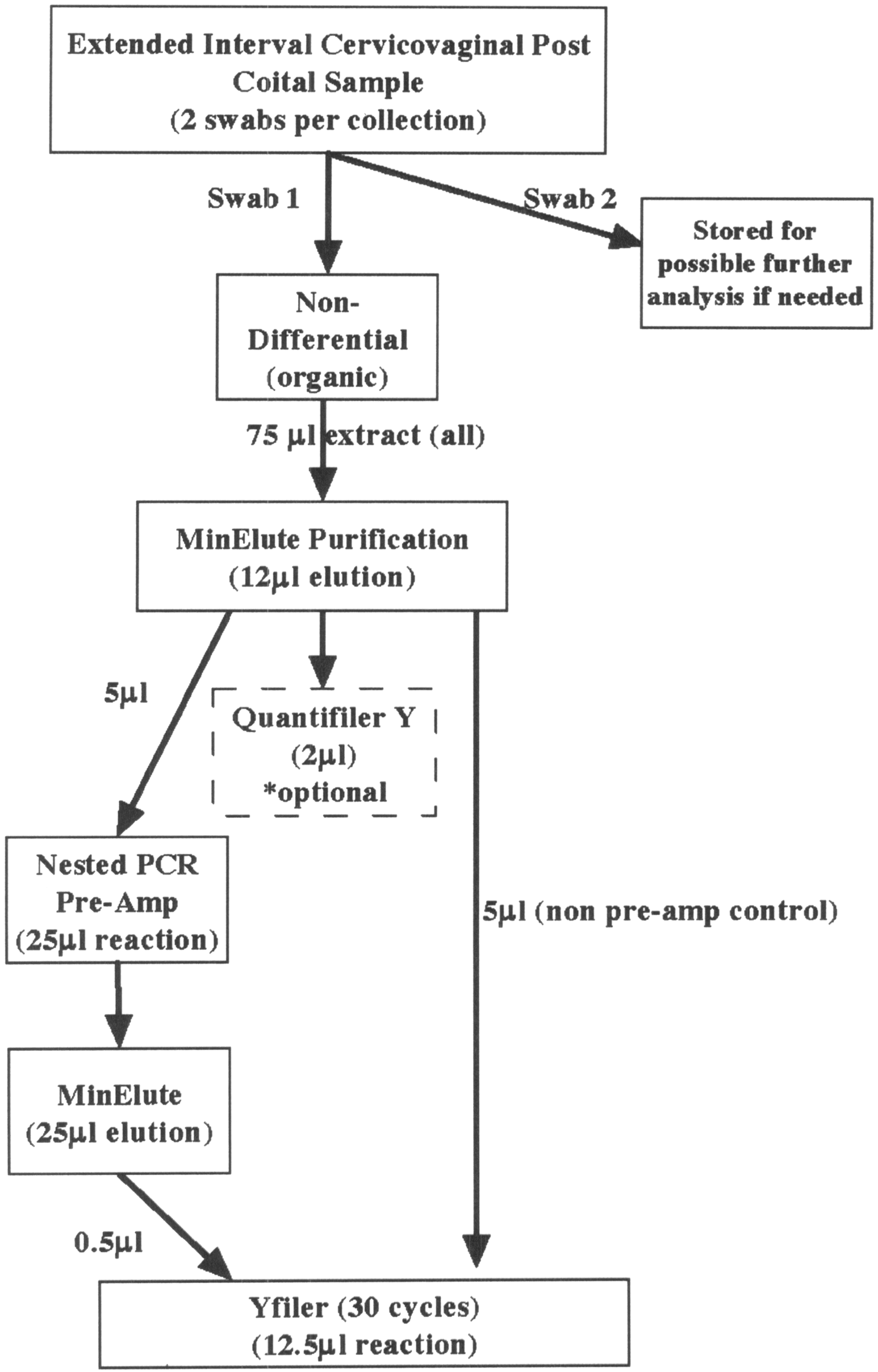
DNA profile recovery from extended interval (≥6 days) post-coital samples
The typing results of the 6 to 9 days post-coital samples subjected to a non-differential extraction, followed by extract purification and concentration, are summarized in Table 1. Representative electropherograms from 8 - and 9-day post-coital samples from couple 4 are shown in Figures 2 and 3, respectively (see p. 5; Figures 2 and 3 are also available as full-sized images at msl.sagepub.com/supplemental). For each sample, two electropherograms are shown: (A) without pre-amplification and (B) with pre-amplification (Y-TPA). For a majority of samples no alleles were detected without the use of Y-TPA. However, it was interesting to note that a small number of alleles were present in a few non-pre-amplified samples (couple 1: 6 days (1 allele), couple 2: 7 days (4 alleles), and couple 4: 8 days (4 alleles)) (Table 1, Figure 2(a)). In preliminary experiments prior to the development of the enhanced profiling strategies reported here, no DNA profiles (i.e. no alleles) were obtained from the 6 to 9 days collection time points using standard DNA extraction protocols (data not shown). These protocols used small aliquots from a larger extract volume (5–10 µL from a 50–100 µL extract). While this sample purification or concentration is not by itself novel, we demonstrate here its beneficial use in the analysis of extended interval post-coital samples as a simple additional procedural step that can, on its own without other enhanced detection methods, permit the recovery of some otherwise not observed male donor alleles.
Improved DNA profile recovery using enhanced Y-STR analysis – 8 days after intercourse. Y-STR profiles of the semen donor (from couple 4) in a 8-day post-coital sample without (a) and with (b) pre-amplification. Allele designations are indicated below each locus. Improved DNA profile recovery using enhanced Y-STR analysis – 9 days after intercourse. Y-STR profiles of the semen donor (from couple 4) in a 9-day post-coital sample without (a) and with (b) pre-amplification. Allele designations are indicated below each locus. DNA profile recovery from extended interval cervico-vaginal samples (6–9 days after intercourse). NT = not tested; NA = not available. The number of alleles recovered from one of the two swabs collected per time interval (6, 7, 8, and 9 days). The shading indicates the average RFU value of all alleles within the profile (white – not detected; light grey <500 RFUs; dark grey >1000 RFUs).


As can be seen from Table 1, the use of the Y-chromosome specific nested PCR pre-amplification resulted in a significant increase in the number of alleles detected, with the recovery of ≥70% of the male donor alleles (with the exception of couple 3) (Table 1). Remarkably, complete or nearly complete profiles were also obtained from samples collected 8 and 9 days after intercourse (couples 1 and 4) (Table 1, Figures 2 and 3). To our knowledge no other study employing carefully controlled and monitored sexual intercourse between subjects has demonstrated the recovery of male donor DNA profiles from the cervico-vaginal tract so long after sexual intercourse. Our own previous work demonstrated the facile recovery of male donor profiles from post-coital samples collected up to 5 days, with only limited partial profiles obtained for 6 days and a single 7-day sample. 2 The use of Y-TPA, therefore, significantly increased the time interval after which the profile of the semen donor can be obtained.
An additional 9-day post-coital sample from couple 1 was obtained after a separate act of sexual intercourse but was contaminated with menstrual blood (the female donor was in day 2 of menses when the sample was collected). Surprisingly, menses did not affect the ability of the Y-TPA method to recover the male donor DNA profile (Table 1). Further studies would have to be conducted to ascertain whether this is a genuine phenomenon or specific to this particular sample.
Comparison of DNA profile recovery from “duplicate” cervico-vaginal swabs
Comparison of profile recovery in multiple cervico-vaginal samplings 9 days after intercourse.

A “+” or “−” designation represents the presence or absence of an allele at a given locus, respectively. Two designations are provided for DYS385 since it is a bi-local locus and each donor is heterozygous. Couple 1 indicated which swab was collected 1st in the set of two swabs collected 9 days after intercourse. Couple 4 did not make this designation and therefore the samples are labeled swab 1 and 2 (not 1st and 2nd). The shading indicates the RFU value (white – not detected; light grey <500 RFUs; medium grey 501-999 RFUs; dark grey >1000 RFUs).
Effects of increased sample input
In the previous section, it was demonstrated that an increased number of alleles could be recovered by using the additional second swab available at each time point. An alternative method for increasing the number of alleles recovered would be to utilize more of the original sample extract from one swab in the subsequent Y-STR amplification. As described previously, only a 0.5 µL aliquot of the 25 µL (2%) purified pre-amplification product is used in the subsequent Y-STR amplification (AmpFlSTR® Yfiler®, 12.5 µL reaction volume). We therefore compared the use of a 5-fold increase in the volume of pre-amplification product used in the Y-STR amplification (increase from 0.5 µL to 2.5 µL) again using the 9-day samples from couples 1 and 4 (both swabs). There were no instances in which the use of the increased volume of pre-amplification product resulted in the recovery of additional alleles (data not shown). All Yfiler® amplifications up to this point were performed using the recommended 30 amplification cycles. We repeated the amplification of 0.5 µL and 2.5 µL aliquots using a low template DNA (LTDNA) approach with increased cycle numbers (32 and 36 cycles). For couple 1, one additional allele (77 RFUs) was observed using 0.5 µL of pre-amplification product and 32 amplification cycles (data not shown). While this additional allele matched the male donor reference profile, it was not observed with 36 amplification cycles or using 2.5 µL of pre-amplification product (data not shown). For couple 4, one additional allele was observed using 2.5 µL of pre-amplification product and 32 and 36 amplification cycles (269 and 88 RFUs, respectively) (data not shown). While an additional allele was recovered for each sample using the increased sample input and cycle number, a significant increase in the number of alleles recovered was not observed. Since the signal intensities of most recovered alleles in the Y-STR profile after pre-amplification are high (>500 RFU), the use of the increased sample and cycle number also led to saturation of these alleles and therefore made the analysis and interpretation quite challenging (data not shown). Therefore, after Y-TPA, there appears to be no significant advantage gained in allele detection using increased cycle number with the Y-Filer STR commercial kit used in this study. Accordingly we recommend using the manufacturer’s suggested cycling conditions (Figure 1).
Discussion
Using a combination of (i) DNA extract purification and concentration, and (ii) a novel Y-chromosome specific multiplex nested PCR pre-amplification (Y-TPA) method, we have demonstrated the ability to obtain potentially probative Y-STR profiles from the semen donor in cervico-vaginal samples collected up to 9 days after sexual intercourse. This is a significant improvement in the time frame after which male DNA profiles can be obtained and is similar to the limits of detection of semen in the cervico-vaginal canal reported in the clinical literature.
There may be somewhat of a reluctance by forensic personnel (sexual assault nurses and/or operational forensic casework laboratories) to agree to collect cervico-vaginal samples up to 9 days after intercourse due to a perception that this will significantly increase the backlog of DNA evidence waiting to be processed. There may even be concerns regarding the need for additional storage space for more sexual assault kits. However, it is likely that the number of cases involving an extended post-coital time interval between assault and sample collection (≥6 days) would be relatively small. For example, Morgan et al. report that in 30 months of sexual assault evidence analyzed by the Canton-Stark Country Crime Laboratory (Ohio, USA), only 4 of 83 (∼5%) of cases involved samples in which there were more than 73 h between assault and arrival at the medical center. 22 This is in broad agreement with an estimate of the number of sexual assaults from Orange County (Florida, USA) that are reported >72 h after the incident (∼4%). 23 Notwithstanding the small percentage of sexual cases that would be pertinent to the application of enhanced typing methods such as described herein, it is the authors contention that victims and individuals falsely accused of sexual assaults deserve that best efforts be made to recover genetic information from the semen donor. This should occur irrespective of when the victim reports the crime.
In addition to concerns regarding an increased volume of sexual assault evidence that will need to be processed, there is likely to be some concern regarding an increased risk of intra-laboratory contamination due to an increase in cycle number and the need for sample manipulation during purification and secondary amplification steps. While there is always a potential risk for additional contamination when a larger number of amplifications cycles are used,24,25 significant contamination issues (i.e. contamination greater than the normal sporadic contamination events seen by all DNA laboratories) were not observed throughout the course of this study. Extraction blanks were subjected to the same analysis as the post-coital samples. This included extract concentration, pre-amplification, purification and subsequent Y-STR amplification and detection. Contamination was not observed in any of the extraction blanks and drop-in alleles (not originating from the sample donor) were rarely observed. Therefore, it should be possible, with proper controls and procedures to employ these methods without significant contamination issues. Once the pre-amplification reaction has been performed, further processing of the samples (purification and Y-STR amplification) should occur in a post-amplification room. It is possible that small separate bench-top PCR workstations could be designated for purification set-up as well as secondary amplifications in order to isolate these reactions from other areas of the post-amplification environment and minimize contamination. Other standard practices of minimizing the time and frequency of tubes containing amplified product being open, sterilization of pipets and work spaces, and use of sterile consumables and reagents should also reduce the risk of potential contamination.
While we obtained results for a majority of samples in this study it is expected that, due to biological variation, not all post-coital samples collected within the same time frame would necessarily yield a DNA profile. A variety of factors will influence the amount of sperm remaining in the cervico-vaginal canal of the victim including: (1) physical activity level of the victim after the assault; (2) victim showering, bathing or douching after assault; (3) occurrence of the assault during the victim’s menstruation cycle; (4) sperm count of the perpetrator; (5) volume of semen ejaculated during the assault; and (6) number of times of ejaculation during the assault. Apart from the amount of semen remaining in the cervico-vaginal canal of the victim, there might also be variation in the efficacy of the sampling process itself. The potential differential success in profile recovery from samples collected from different individuals within the same time interval is evident even in this study with only four donor couples, with significantly less alleles recovered for couple 3 (6 days – 24% of alleles; 7 days – 18% of alleles) (Table 1). Also, the samples in this study were self-collected and it is possible that inter-individual variation in sampling might have affected the amount of semen recovered.
Finally, we have demonstrated the ability to obtain full and partial DNA profiles from cervico-vaginal samples recovered 6 to 9 days after intercourse using enhanced profiling strategies. Given the success and quality of the 9-day DNA profiles, it seems worthwhile to attempt to obtain profiles beyond 9 days despite the limits described in the medical literature. Future work will therefore include an evaluation of >9-day samples. Since only four donor couples were utilized in this study it will be necessary to expand the analysis to include a much larger sample set. This would permit a more accurate estimation of the variation in profile recovery between donors. We are currently involved in a much larger inter-institutional study to investigate the correlation between male DNA profile recovery in post-coital samples and various biological variables including the reproductive health of the victim. Preliminary DNA typing results confirm our herein described observations that use of enhanced DNA typing methods, particularly Y-TPA, permit the recovery of the male donor profile in post-coital samples taken up to 9 days after intercourse.
Footnotes
Acknowledgements
The authors would like to acknowledge all donors who provided samples for this study. The authors would also like to acknowledge Dr Holger Engel, Dr Francesca Di Pasquale, and Dr Sascha Strauss for their work on the development of the Y chromosome specific nested PCR pre-amplification multiplex.
Funding
This work was supported by the State of Florida through the National Center for Forensic Science at the University of Central Florida, and the National Institute of Justice, Office of Justice Programs, U.S. Department of Justice [award numbers 2007-DN-BX-K147 and 2007-DN-BX-K148]. The opinions, findings, and conclusions or recommendations expressed in this publication are those of the authors and do not necessarily reflect those of the Department of Justice.
Declaration of conflicting interests
The authors declare that there is no conflict of interest.
References
Supplementary Material
Please find the following supplemental material available below.
For Open Access articles published under a Creative Commons License, all supplemental material carries the same license as the article it is associated with.
For non-Open Access articles published, all supplemental material carries a non-exclusive license, and permission requests for re-use of supplemental material or any part of supplemental material shall be sent directly to the copyright owner as specified in the copyright notice associated with the article.