
Abstract
Age estimation occupies a prominent niche in the identification process. In cases where skeletal remains present for examination, age is often estimated from markers distributed throughout the skeletal framework. Within the pelvis, the pubic symphysis constitutes one of the more commonly utilized skeletal markers for age estimation, with the Suchey–Brooks method comprising one of the more commonly employed methods for pubic symphyseal age estimation. The present study was targeted towards assessing the applicability of the Suchey–Brooks method for pubic symphyseal age estimation, an aspect largely unreported for an Indian population. In order to do so, clinically undertaken pelvic computed tomography scans of individuals were evaluated using the Suchey–Brooks method, and the error associated with the method was established using Bayesian analysis and different machine learning regression models. Amongst different supervised machine learning models, support vector regression and random forest furnished lowest error computations in both sexes. Using both Bayesian analysis and machine learning, lower error computations were observed in females, suggesting that the method demonstrates greater applicability for this sex. Inaccuracy and root mean square error obtained with Bayesian analysis and machine learning illustrates that both statistical modalities furnish comparable error computations for pubic symphyseal age estimation using the Suchey–Brooks method. However, given the numerous advantages associated with machine learning, it is recommended to use the same within medicolegal settings. Error computations obtained with the Suchey–Brooks method, regardless of the statistical modality utilized, indicate that the method should be used in amalgamation with additional markers to garner accurate estimates of age.
Keywords
Introduction
Age of an individual, along with other parameters of sex and stature, plays a crucial role in human identification. 1 In cases of medicolegal significance where skeletal remains present for examination, age is often estimated from different bony markers. Such an investigation exploits the underlying association between the chronological age of individuals and the age expressed by their skeletal framework. 2 Amongst the numerous age markers located throughout the bony framework, the pelvis presents as a durable and resilient age marker. As an added advantage, the pelvis exhibits both age-related developmental and degenerative morphological changes, rendering it applicable for age estimation across a broader cohort,3–6 as opposed to ossification and fusion changes which encompass limited age classes.7,8
Within the pelvis, the pubic symphysis constitutes one of the more commonly scrutinized markers for age estimation.9,10 The pubic symphysis undergoes, throughout the lifespan of an individual, a series of irreversible and distinctly identifiable morphological changes, rendering it a suitable marker for age estimation.11,12 Following Todd's preliminary observations pertaining to the role of the symphysis pubis in age estimation, 13 multiple methods for pubic symphyseal age estimation were devised.11,14–19 Amongst these methods, the Suchey–Brooks method, 16 derived from Todd's 10-phase method, is one of the more commonly employed methods for pubic symphyseal age estimation. 20 The method entails classifying remains into one of six distinct phases based on morphological changes transpiring within the pubic symphysis. Subsequently, an age range specific to each phase is assigned to the remains in question to help generate a profile. This modified six-phase approach devised by Suchey and Brooks was observed to be relatively more accurate when compared to Todd's original method. 21 However, in the years that followed, investigations undertaken with the Suchey–Brooks method using different population groups demonstrated varying applicability.9,22–72 Moreover, despite being one of the most commonly utilized age estimation methods, investigations evaluating the forensic suitability of the Suchey–Brooks method for ageing an Indian population are currently limited. 73
Validating previously derived age estimation methods warrants undertaking a thorough evaluation using contemporary, large-scale, population-specific skeletal repositories. Such population-specific investigations not only help rule out the influence of genetic and environmental factors which can often undermine or overmine a method 26 but also help derive population-specific age estimation models. However, the lack of large-scale contemporary, demographic specific skeletal repositories outside the USA often impedes evaluating the applicability of previously derived age estimation methods. A computed tomographic examination of the skeletal framework presents as a suitable alternative to this dilemma. Post-mortem or clinically available CT scans of the pelvis enable a non-invasive evaluation of morphological changes transpiring within the bony surface. As an added advantage, computed tomography helps circumvent issues arising due to interfering tissue remnants, which can often hamper a clear visualization of surface changes. Previously undertaken computed tomography (CT)-based investigations with the Suchey–Brooks method have demonstrated a satisfactory reproduction of ensuing morphological changes.31,33,39,55,56,60,62,64,66,67 Owing to the lack of a large-scale documented repository for an Indian study population, an alternative computed tomographic investigation was explored within this study.
The present study was targeted towards assessing the applicability of the Suchey–Brooks method for pubic symphyseal age estimation in an Indian population, through clinically undertaken computed tomographic examinations of the pelvis. Bayesian analysis and supervised machine learning models were utilized to establish the overall error associated with the method, whilst overcoming issues of age mimicry and eliminating any influence of human bias. Additionally, error computations obtained with these two statistical modalities were comparatively evaluated to establish the superior mathematical approach for forensic age estimation.
Materials and methods
Sample collection
The present study was carried out in the Department of Forensic Medicine and Toxicology and Department of Diagnostic and Interventional Radiology at the All India Institute of Medical Sciences, Jodhpur, India. Ethical clearance for this prospective cross-sectional investigation was obtained from the Institutional Ethics Committee vide Letter No. AIIMS/IEC/2019-20/1007 prior to study commencement. For the present investigation, participants aged 10 years and above, scheduled to undergo computed tomographic examinations of their pelvis/abdomen as per directions of their treating physicians at the Department of Diagnostic and Interventional Radiology, were approached. Such patients/their guardians were appraised about the study parameters in detail, and their consent for voluntary participation into the study was sought. Patients who consented to participate were included into the study after verifying their age through proper documentation. Consenting individuals who could not produce any valid proof of age documentation and/or those suffering from any known skeletal injuries/congenital anomalies capable of interfering with the analysis of interest were excluded from the study. Furthermore, CT scans with fractures, patient movement-induced artefacts or CT-specific artefacts within the region of interest, which became apparent during CT examination, were also excluded from the investigation.
A total of 465 CT scans, satisfying the inclusion criteria, were collected between January 2020 and January 2022 and incorporated into the study. Using the random number table, the total study group was arbitrarily divided into a training group comprising of 365 individuals and a test group of 100 individuals (50 males and 50 females). CT scans of all participants were coded to blind the investigators to patient information capable of inducing bias.
Scanning parameters
Dual-source CT – SOMATOM Definition FlashTM (Siemens Medical Solutions, Erlangen, Germany) – was employed to acquire CT scans of consenting participants. A slice thickness of 1.0 mm (imaging parameter standardized by the healthcare centre for treatment purposes) was utilized for evaluating each scan. Collected CT scans were then processed and analysed using 3D Slicer 4.11.20200930.74,75 The bone window for each scan was selected, and a 3D volume-rendered (VR) reconstruction of the pubic symphyseal region was generated. The obtained 3D VR representation was subsequently cropped to remove other interfering bony regions using the scissor tool. The generated pubic symphyseal image was scrutinized from different planes and rotations so as to permit a clearer visualization and appreciation of transpiring morphological changes.
Age estimation using the Suchey–Brooks method
Wink in 2014 described CT-specific diagnostic features for each Suchey–Brooks phase using clinically undertaken CT scans of living individuals. 60 Since the present study utilized a similar investigation with clinical scans, Wink's diagnostic features were relied on. CT-specific depictions for different phases provided by Wink 60 were utilized as reference images. On the basis of the observed changes, the pubic symphyses (both left and right) were classified into one of six phases. The characteristic description for each phase has been listed under Table 1. 3D CT representations of morphological features associated with all six phases are shown in Figure 1.

3D CT representation of morphological features associated with all six phases.
Pubic symphyseal age estimation method employed in the present study.

Statistical analysis
Statistical analysis was undertaken with IBM Statistical Package for Social Sciences (SPSS) v26 and RStudio (RStudio Team (2022). RStudio: integrated development environment for R (RStudio, PBC, Boston, MA URL http://www.rstudio.com/). R scripts and codes for relevant statistical tests were sourced from previously undertaken studies.76–80 A p-value < 0.05 was considered statistically significant for all computations.
Inter- and intra-observer error in the present study was assessed using Cohen's weighted κ. Fifty samples were evaluated by two independent observers (VW and RS) to compute the inter-observer error. The lead investigator (VW) re-evaluated the same 50 samples after a duration of 2 weeks from the time of initial assessment in order to establish the intra-observer error. Obtained weighted κ values were interpreted following the system devised by McHugh, 81 that is, κ < 0.20, no agreement; κ = 0.21–0.39, minimal agreement; κ = 0.40–0.59, weak agreement; κ = 0.60– 0.79, moderate agreement; and κ = 0.80–0.90, strong agreement; κ > 0.91, almost perfect agreement.
Bilateral differences in the scoring of the pubic symphysis were assessed using the Wilcoxon test. Sex differences in the expression of morphological changes within the pubic symphysis were evaluated with the Mann–Whitney U test. The correlation between observed morphological changes and documented age was evaluated using Spearman's rho. Each of these statistical evaluations was undertaken using the entire study group of 465 individuals.
Transition age parameters associated with the Suchey–Brooks phases were established using a log-age cumulative probit model. Computed transition age parameters were expressed in terms of mean age and standard error, both in the log-scale and the corresponding exponential scale.
Using Bayesian statistics, highest posterior density (HPD) and highest posterior density region (HPDR) were established for each phase. Bayesian statistics employs Bayes theorem to postulate the probability (also known as the posterior probability) of an individual being a particular age given that the morphological changes expressed within their skeletal framework (in this case, the pubic symphysis) corresponds to a specific phase.38,82–84 This posterior probability is computed as follows: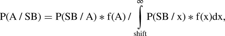

The aforementioned transition age parameters, Bayesian HPD and HPDR (95% confidence interval of HPD), were computed with the training group of 365 individuals. Obtained values were subsequently utilized for establishing the inaccuracy, bias and root mean square error (RMSE) associated with the Suchey–Brooks method.
Inaccuracy, that is, the absolute error associated with age estimation, was determined as follows:


Different machine learning models of support vector regression (SVR), gradient boosting regression (GBR), decision tree regression (DTR) and random forest (RF) were employed to estimate the age of individuals. The training group was utilized to train these models, and the inaccuracy, bias and RMSE associated with each of these supervised machine learning approaches were computed using the test group (N = 100).
Error computations obtained with Bayesian analysis and different supervised machine learning approaches were comparatively evaluated to establish the superior statistical approach for age estimation, as well as to demonstrate the lowest error computations attainable with the Suchey–Brooks method for an Indian population.
Results
The entire study group comprised of 198 females with a mean age and standard deviation of 48.68 ± 16.181 years and 267 males with mean age ± SD of 46.30 ± 18.033 years. Age and sex distribution of the study group is shown in Table 2. Owing to the relatively lower representation of individuals in the 70–79 and 80–89 years age cohorts, they were clubbed together to create a truncated age category of ≥70 years.
Age and sex distribution of the study group (N = 465).

Obtained weighted κ value of 0.933 indicates an almost perfect agreement between the two sets of phase allotment to the pubic symphysis through CT scans, by a single observer. When two independent observers evaluated the pubic symphysis through 3D CT images, a weighted κ value of 0.763 was obtained, indicating a moderate agreement in the scoring of age-related changes. No statistically significant bilateral differences were observed in the scoring of pubic symphyseal changes (p = 0.128, Z-score = −1.523). Additionally, a partial correlation analysis between the two pubic symphysis, whilst controlling for age, yielded a high correlation of 0.946 substantiating the observed bilateral symmetry. Bilateral asymmetry of a single-phase category was observed in 14.62% scans, with the left pubic symphysis displaying characteristics of the higher phase in 58.80% of such cases. A statistically significant sexual dimorphism was obtained during the grading of the right pubic symphysis (p = 0.008, Z-score = −2.636) and left pubic symphysis (p = 0.013, Z-score = −2.477). Owing to the overall bilateral symmetry observed within the study, only one-half of the pelvis was utilized for further statistical reporting. In keeping with Overbury et al., 45 the pubic symphysis displaying characteristics of the higher phase was utilized for statistical analysis. Bayesian analysis and machine learning were undertaken separately for both sexes due to the significant sexual dimorphism observed with the method. An overall statistically significant correlation of 0.605 was obtained between phases allotted to the pubic symphysis and the documented age of individuals in the combined study population (N = 465). Females garnered higher correlation values of 0.617 with age, in comparison to males (0.595).
The training group comprised of 148 females with a mean age ± SD of 50.48 ± 15.427 years and 217 males with a mean age ± SD of 46.82 ± 17.427 years and was utilized to establish transition age parameters and posterior density estimates. Transition age parameters (mean age and standard error), depicting the uni-directional transition from phase I to VI, are shown in Table 3. The mean age for transition between consecutive phases was higher in females, when compared to males. For the transition from phase V to VI, however, males garnered a higher mean transition age value. The standard error values associated with consecutive transitions were also higher in females, barring the standard error for the transition from phase V to VI, wherein a higher error was observed in males. Higher HPD values were obtained in females for phases I–IV. On the other hand, for phases V and VI, higher HPD values were observed in males (Table 4).
Transition age parameters computed in the present study (N = 365).

TA: mean transition age in log-scale; SE: standard error in log-scale; Exp (TA ± SE): exponential value of transition age and associated standard error.
HPD and highest posterior density region (HPDR/95% confidence interval of HPD) for each phase (N = 365).

HPD: highest posterior density; CI: confidence interval.
The test group comprised of an equal number of male and female participants with a mean age ± SD of 44.08 ± 20.502 years and 43.46 ± 17.318 years, respectively. Inaccuracy, bias and RMSE values computed for the test group using Bayesian analysis are shown in Table 5. Lower inaccuracy and RMSE values were observed in females. However, lower bias values were observed in males, in comparison to females. Obtained bias values indicate an overall tendency to under-age using the Suchey–Brooks method, in both sexes.
Error computations for pubic symphyseal age estimation obtained with Bayesian analysis and machine learning approaches (N = 100).

RMSE: root mean square error.
Error computations obtained with different supervised machine learning models for the test group are also shown in Table 5. Between the four machine learning approaches utilized herein, SVR furnished the lowest inaccuracy (9.26 years) and bias values (−2.11 years) in females, whilst RF garnered the lowest RMSE (11.23 years) in females. RF yielded the lowest inaccuracy value (11.67 years) in males, whereas SVR garnered the lowest RMSE (14.20 years) and bias values (−0.86 years) for this sex. A negative bias was obtained with all four machine learning approaches in both sexes, indicating the overall tendency of this six-phase method to under-age individuals of an Indian population.
Inaccuracy, bias and RMSE obtained with Bayesian analysis and machine learning indicate that all machine learning approaches yield lower RMSE values in comparison to Bayesian analysis, in females. However, only SVR furnished lower inaccuracy values when compared to Bayesian analysis. GBR, DTR and RF yielded marginally higher inaccuracy values in comparison to the 9.55 years obtained with Bayesian analysis. Bias values obtained with machine learning were higher than those obtained with Bayesian inference, in females. Similarly, in males, machine learning furnished lower RMSE values in comparison to Bayesian analysis. Inaccuracy values obtained with Bayesian analysis were lower than those obtained with machine learning in males. With SVR and RF, however, this difference was marginal. Bias values computed for males using machine learning approaches were higher than those obtained with Bayesian inference.
Discussion
The pubic symphysis constitutes a resilient, non-synovial, amphiarthrodial structure with limited mobility and displays age-related changes in a fairly predictable pattern, 40 rendering it an ideal age marker. Whilst Todd's preliminary observations aided in establishing the pubic symphysis as an efficient age marker, 13 his 10-phase pubic symphyseal age estimation method suffered from certain pertinent drawbacks. Suchey and Brooks, in 1990, coalesced Todd's 10-phase method 13 into six distinct phases using stepwise linear regression, with the aim of overcoming these limitations and devising a more robust age estimation method. 16 Whilst devising their modified method, Suchey–Brooks utilized a large sample comprising of 1225 male and female contemporary remains of diverse socio-economic and demographic backgrounds, effectively deriving a method which would be appropriate for both sexes, as well as different population groups. Previously undertaken systematic reviews and meta-analyses with the Suchey–Brooks method validate this hypothesis.86,87 However, targeted research studies investigating the applicability of the Suchey–Brooks method for ageing an Indian population are unreported. It is this lacuna that the present study purports to fill.
No statistically significant bilateral differences were observed in the present study, indicating that age estimation models derived using the Suchey–Brooks method can be applied to either half of the pelvis with equal vigour. A high partial correlation between the two pubic symphysis, whilst controlling for age, further substantiates the lack of any significant bilateral differences. Our findings pertaining to bilateral symmetry are in agreement with previously reported findings32,33,61 and in contrast to certain other studies.31,39,45,57 It is prudent to mention that a significant share of research using the Suchey–Brooks method utilized one-half of the pelvis alone and did not explore the facet of bilateral symmetry/asymmetry. For the majority of cases which presented with bilateral differences in the present study, the left pubic symphysis displayed characteristics of the higher phase, a finding corroborated in part by Lottering et al. 39 On the other hand, Overbury et al. reported this directional asymmetry towards the right pubic symphysis. 45 This observed directionality within the pubic symphyses could be attributed to different biomechanical and physiological influences. However, further research is wanting to corroborate our findings and better elucidate this phenomenon. For the observed 14.62% cases of bilateral asymmetry in the present study, the pubic symphysis displaying characteristics of the higher phase was utilized for age estimation, as opposed to the usual norm of employing either the left or right half of the pelvis. This was done in accordance with Overbury et al. 45 and Passalacqua, 88 with the aim of garnering more accurate estimates of age.
A significant sexual dimorphism was observed on applying the Suchey–Brooks method to an Indian population. Our findings pertaining to sexual dimorphism substantiate those reported previously.24,72 However, sex differences observed with the Suchey–Brooks method contradict the original hypothesis of Suchey and Brooks, who incorporated features corresponding to both sexes with the aim of developing an age estimation method which would encompass both sexes. 16 Nevertheless, these observations validate the findings of Gilbert-McKern 15 and Berg 11 who recommend the use of separate/revised methods for pubic symphyseal age estimation in females. This observed sexual dimorphism could be attributed to the influence of different intrinsic factors, such as parity in women. Furthermore, females were relatively, albeit marginally, underrepresented within our study. A similar underrepresentation of females exists within the original study by Suchey and Brooks. 16 Further investigations should be undertaken with equally represented male and female participants of different population groups to validate/dispute our findings of sexual dimorphism.
A moderate positive correlation89,90 was observed between estimated age and documented age in the present study, which substantiates the findings of previous investigations.9,22,29,31,32,35,39,41,42,56,61 Certain other studies, however, reported relatively higher correlations with age.27–29,33,50,55,64
A major share of investigations with the Suchey–Brooks method utilized descriptives reported within the original study to establish the accuracy and precision associated with the method.9,22–26,28,29,31–33,35,40–43,45,47–57,59–61 However, the application of such fixed descriptive parameters to demographically variant population groups can often induce bias, namely, age mimicry and population bias amongst others. A characteristic result of this age mimicry bias is the over-ageing observed with younger individuals and under-ageing observed with older individuals. 91 Alternate statistical approaches such Bayesian analysis offer an improvement over this commonly employed simple descriptive evaluation of data. Bayesian inference, coupled with transition analysis, is believed to yield more accurate and realistic estimates of age through its use of probability density functions. As an added advantage, Bayesian analysis circumvents issues of age mimicry,82,92 a common artefact in age estimation studies, characteristically observed even with simple regression analysis. 93 Bayesian statistics in amalgamation with transition analysis has previously been utilized for pubic symphyseal age estimation.11,30,33,38,39,92 Machine learning presents as another equally efficient statistical modality for assessing the applicability of any age estimation method. In addition to eliminating the component of human bias, machine learning, too, does not suffer from issues of age mimicry.94–96 Furthermore, supervised machine learning works equally well with missing data, has fewer assumptions to satisfy and can largely render the measurement-statistics controversy irrelevant. 97 The use of supervised machine learning for assessing the applicability of the Suchey–Brooks method is currently unreported. Thus, by extension, a comparative evaluation of the performance of Bayesian analysis and machine learning for age estimation using the Suchey–Brooks method is also presently lacking.
Transition age parameters computed with the training group indicate an earlier transition between consecutive phases in males. For phases V–VI transition, however, females yielded a lower mean age-at-transition. The associated standard error for consecutive phase transitions was also higher in females, barring phases V–VI, wherein males furnished a higher standard error. This delay in transition along with the higher error observed in females demonstrates the higher variability associated with ageing this sex. This could further be attributed to the influence of hormones and parity-induced trauma, amongst other intrinsic factors. The difference between mean age-at-transition values for consecutive phase transitions was observed to increase with progressing transitions, that is, whilst the transition ages for phases I–II and phases II–III displayed a gap of about 3–5 years, transition ages for phases IV–V and phases V–VI exhibited a gap spanning 10–30 years. Similar findings have been reported in previously undertaken studies.30,38,39 The standard error for consecutive phase transitions was also observed to increase with progressing transitions in both sexes, corroborating the higher error rates associated with ageing older individuals.9,23–26,28,29,31–33,35,41,48,50–52,54–57,61 Mean transition age values obtained with males of an Indian population are lower than those reported by Kimmerle et al. 38 for a Balkan population. However, the mean transition age obtained herein for phases V and VI in males is higher than those reported by Lottering et al. 39 for an Australian population and Hisham et al. 33 for a Malaysian population. Mean transition ages computed for females of an Indian population are lower than those reported by Hisham et al. 33 for Malaysians. However, mean transition age values for phases IV and V, and phases V and VI in Indian females are higher than those reported by Lottering et al. 39 for Australian females. Both sexes in the present study displayed a higher standard error for each phase transition, in comparison to previously undertaken investigations with different population groups.
Higher HPD values were obtained with Bayesian analysis in females for phases I– IV. Phases V and VI, however, furnished higher HPD values in males. Higher HPD obtained in females suggests a delayed onset and progression of different pubic symphyseal changes within this sex. HPD values computed for males of an Indian population are lower than those reported by Lottering et al. 39 for an Australian population. HPD reported by Kimmerle et al. 38 for phases V and VI in a Balkan population are higher than that obtained with Indian males. Females exhibited comparable HPD values to those reported by Lottering et al. 39 and Kimmerle et al. 38 for phases I–V.
Error obtained with Bayesian analysis in Indian males is lower than what has been reported previously through a simple descriptive evaluation of data in different populations.9,31,48,52,54,55,57,64 With females too, Bayesian analysis garnered lower error computations, in comparison to simple descriptive evaluation.9,31,34,40,49,52,54,56,57,61,69,70 However, caution is recommended during such comparisons owing to the methodical differences across these studies, that is, gross morphological examination of historical osteological collections versus prospective CT-based examinations. A comparative evaluation of inaccuracy and bias values obtained herein, against those reported by Lottering et al. 39 who utilized CT images for the evaluation of different age-related changes and Bayesian analysis for age estimation, indicates higher error computations in an Indian population. Females furnished lower inaccuracy values in comparison to males in the present study, contradictory to the findings of Lottering et al. 39 for an Australian population. These observed differences can be ascribed to the demographic variability between these two study groups.
Amongst the different supervised machine learning models utilized in the present study, SVR furnished the lowest inaccuracy and bias values in females. RF on the other hand, yielded lowest RMSE values in females. With males, RF garnered lowest inaccuracy values, whereas SVR furnished lowest RMSE and bias values. The greater precision observed with RF could be attributed to its use of a bagging technique to circumvent the problem of overfitting, as well as its ability to work well with non-linear data. Just as with RF, SVR too is quite adept at handling non-linear data. 98 Additionally, SVR minimizes noise through its use of a maximum marginal hyperplane, thus garnering more accurate estimates of age. 98 With all four machine leaning models, females garnered lower inaccuracy and RMSE in comparison to males. Our findings pertaining to error computations obtained for the Suchey–Brooks method through machine learning models could not be compared across studies due to a lack of similar data.
The primary objective of the present investigation was to ascertain the applicability of the Suchey–Brooks method for pubic symphyseal age estimation in an Indian population, an aspect largely unreported. In order to do so, statistical modalities of Bayesian analysis and machine learning were utilized with the aim of illustrating the most accurate estimates of age attainable with the method for the population in question, whilst circumventing age mimicry bias, an issue commonly encountered with descriptive and regression analysis.91,93 Inaccuracy and RMSE values obtained with both statistical approaches indicate that the overall error associated with the Suchey–Brooks is quite high, more so in comparison to alternate markers within the pelvis such as the acetabulum. 99 Our findings, thus, substantiate the original observations of Suchey and Brooks, who recommend employing their method in amalgamation with other age markers to garner more accurate estimates of age. 16 Applying pubic symphyseal age estimation, alongside the popular Lovejoy et al. auricular age estimation method, 100 and recently devised San-Millán–Rissech component-based acetabular age estimation method 101 might help further augment the accuracy and precision associated with forensic age estimation and human identification. Previous investigations utilizing a systematized combination of pubic symphyseal and auricular changes, amongst other markers, have furnished improved estimates of age 102 and should be incorporated routinely into the age estimation practice. Lower inaccuracy and RMSE values, along with the stronger correlation observed in females, suggest that the method demonstrates greater applicability for this sex. Similar findings were reported by other investigations, albeit through a descriptive analysis of data.9,31,48,51,54,69,70 The overall accuracy and precision associated with the Suchey–Brooks method may further be augmented through the use of convolutional neural networks (CNN). Previously undertaken CNN-based investigations with alternate age markers within the pelvis have yielded improved estimates of age, more so in comparison to the traditional approaches, 103 and thus should be explored with the pubic symphysis as well.
The second objective of this study was centred around comparatively evaluating the performance of these two statistical approaches, that is, Bayesian analysis and machine learning for pubic symphyseal age estimation. Inaccuracy values obtained with SVR were lower than those garnered with Bayesian inference in females. The remaining three models furnished marginally higher inaccuracy values in comparison to Bayesian analysis. With males, all four machine learning approaches yielded higher inaccuracy values in comparison to Bayesian statistics, albeit marginally. RMSE values obtained with machine learning were lower than those obtained with Bayesian analysis, in both males and females. The overall bias obtained with machine learning, however, was greater in comparison to Bayesian analysis, for both sexes. Inaccuracy and RMSE values obtained with Bayesian analysis and machine learning in the present study indicate that both statistical modalities furnish comparable error computations for pubic symphyseal age estimation using the Suchey–Brooks method. However, given the ability of supervised machine learning to eliminate human bias, handle multidimensional data and effectively work around the measurement-statistics controversy, it would be beneficial to rely on machine learning for age estimation.
The present study also indicates that the use of CT diagnostic features of this popular six-phase method given by Wink, 60 permits a simple and quick evaluation of transpiring changes, significantly reducing the error commonly associated with phase-based age estimation. 104 Nevertheless, the use of CT as an age diagnostic procedure alone is not recommended in living individuals. Alternately, clinically undertaken CT scans for therapeutic purposes can be employed for age estimation (as done within our study). Post-mortem CT investigations may also be undertaken for non-invasive age estimation in unidentified remains. Lastly, the present study incorporated participants as young as 10 years old with the aim of establishing the youngest age at which age-related pubic symphyseal changes can be appreciated in an Indian population. Findings of our study illustrate that 16-year-old males and 22-year-old females first display morphological changes characteristic of phase II described within the method.
Conclusion
CT-specific diagnostic features of the Suchey–Brooks method help simplify the process of pubic symphyseal age estimation. In the present study, no significant bilateral differences were observed in the scoring of the pubic symphysis, indicating that either half of the pelvis can be utilized for age estimation with equal vigour. However, statistically significant sex differences were observed with the method. Transition age parameters and HPD values suggest an initially delayed onset and progression of age-related pubic symphyseal changes in females, followed by an earlier progression for the latter phases. Lower error computations obtained with Bayesian analysis and machine learning, along with the stronger correlation observed in females, suggest that the method displays greater applicability for ageing this sex. Error computations obtained with the Suchey–Brooks method, irrespective of the statistical modality utilized, indicate that the pubic symphysis should be used in amalgamation with additional markers to furnish accurate estimates of age. Despite the comparable error computations garnered with Bayesian analysis and machine learning, given the numerous advantages conferred by the latter, machine learning should be preferred for forensic age estimation.
Footnotes
Acknowledgements
This research article is a part of an ongoing doctoral research being conducted by one of the authors (VW) in the Department of Forensic Medicine and Toxicology, All India Institute of Medical Sciences, Jodhpur, India
Declaration of conflicting interests
The author(s) declared no potential conflicts of interest with respect to the research, authorship and/or publication of this article.
Funding
The author(s) received no financial support for the research, authorship and/or publication of this article.