
Abstract
Venous disease, including varicose veins and chronic venous insufficiency, is one of the most common chronic medical conditions. Haemorrhage from rupture of varicose veins in the legs is rare and can lead to sudden death. Nevertheless, this condition is not included among the causes of sudden death. In this case, an 88-year-old man was found dead in a large pool of blood at home. Initially, investigators mistakenly assumed that it was a case of homicide. Bloodstain pattern analysis was performed. An external examination of the victim showed an ulcer on the left foot and evident varicose veins untreated on the lower limbs. The aim of this study is to emphasize the role of treatment of varicose veins in the prevention of adverse events such as sudden death from acute haemorrhage.
Introduction
About 15–50% of the population is affected by varicose veins. It is also a substantial source of morbidity in the United States and the Western world.
Rarely, the presence of varicose veins in the legs can be complicated by venous haemorrhage. This happens spontaneously or as consequence of even a mild trauma. 1 Generally, chronic venous insufficiency is not fatal; however, if the rupture of varicose veins is followed by massive venous haemorrhage and by inadequate bandaging, it can lead to death. One predisposing factor to breakage is the localization of varicose veins above a protuberance, particularly in the malleolar region and lower leg. Also, the skin is more vulnerable if altered by chronic venous stasis, or if crossed by vessels with sclerotic walls. In these cases, a trivial trauma may cause rupture.
Compliance with medical treatment is important to facilitate healing and avoid the risk of recurrence. Due to the rare nature of this event, fatal haemorrhagic death by rupture of a varicose vein may confuse the forensic pathologist in the process of identifying the manner of death. The analysis of this uncommon case seeks to demonstrate the causes of rupture of varicose vein and, in addition, to emphasize the importance of investigative accuracy in such uncertain “crime” scenes.
Case history
This paper describes the case of an 88-year-old man found dead, in a pool of blood, in the bedroom of his apartment (Figure 1). The police and prosecutor were called by first responders who accessed the house. Traces of blood were found in the whole room. The deceased was lying on his back on the bed. His legs were out of the bed and the soles of the feet on the floor (Figure 2). He was not wearing socks, which were about 80 cm away from the left foot. The right foot wore a slipper, whereas the left foot was bare and showed a wound.
Crime scene. Varicose formations of the lower limbs.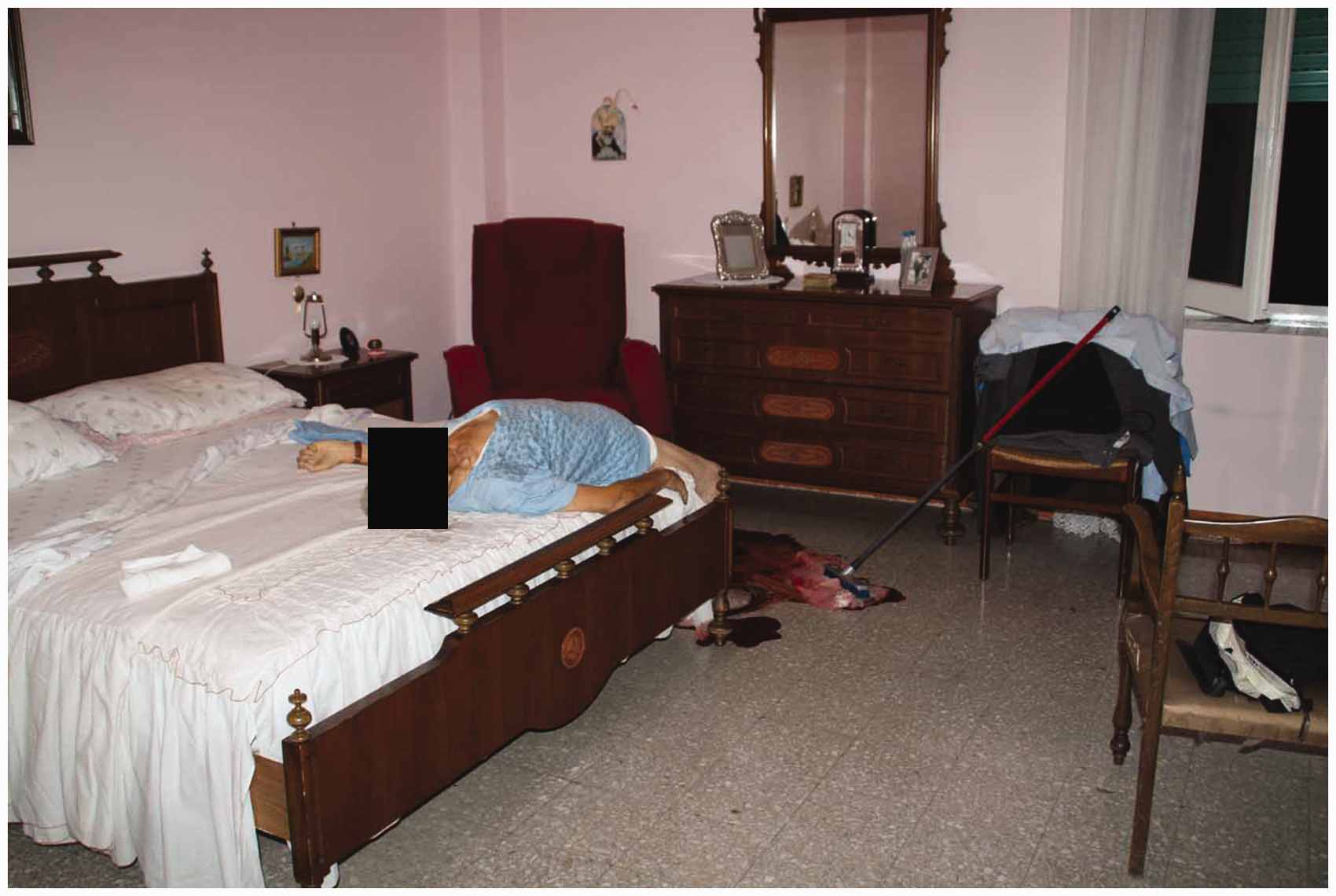

Materials and methods
Bloodstain pattern analysis
Blood pattern analysis was performed in order to analyze all the traces of blood at the scene, and specifically, to evaluate the source of the bleeding.
Autopsy and histological analysis
Autopsy was performed including sampling and analysis of the organs.
The organs were analyzed by histopathological investigations, through the use of paraffin and staining in hematoxylin-eosin. A microscopic analysis of the slides was carried out.
Analysis of published forensic studies
A literature search was conducted on the search engine PubMed NCBI, by entering the following keywords: “fatal”, “varicose veins”, “rupture”.
Results
Circumstantial data
The victim’s feet were surrounded by a large pool of blood. The puddle measured 120 × 100 cm. The presence of small blood spatters on the back of the left foot was observed. The ulcer was bordered by small blood splatters: they appeared as though they were spurted from an arterial source. Blood stain pattern analysis (BPA) was performed in order to understand the origin and the location of venous and/or arterial bleeding. Vector analysis of BPA established that the origin of the bleeding was compatible with a localized ulcer on the victim’s left leg.
Autopsy findings
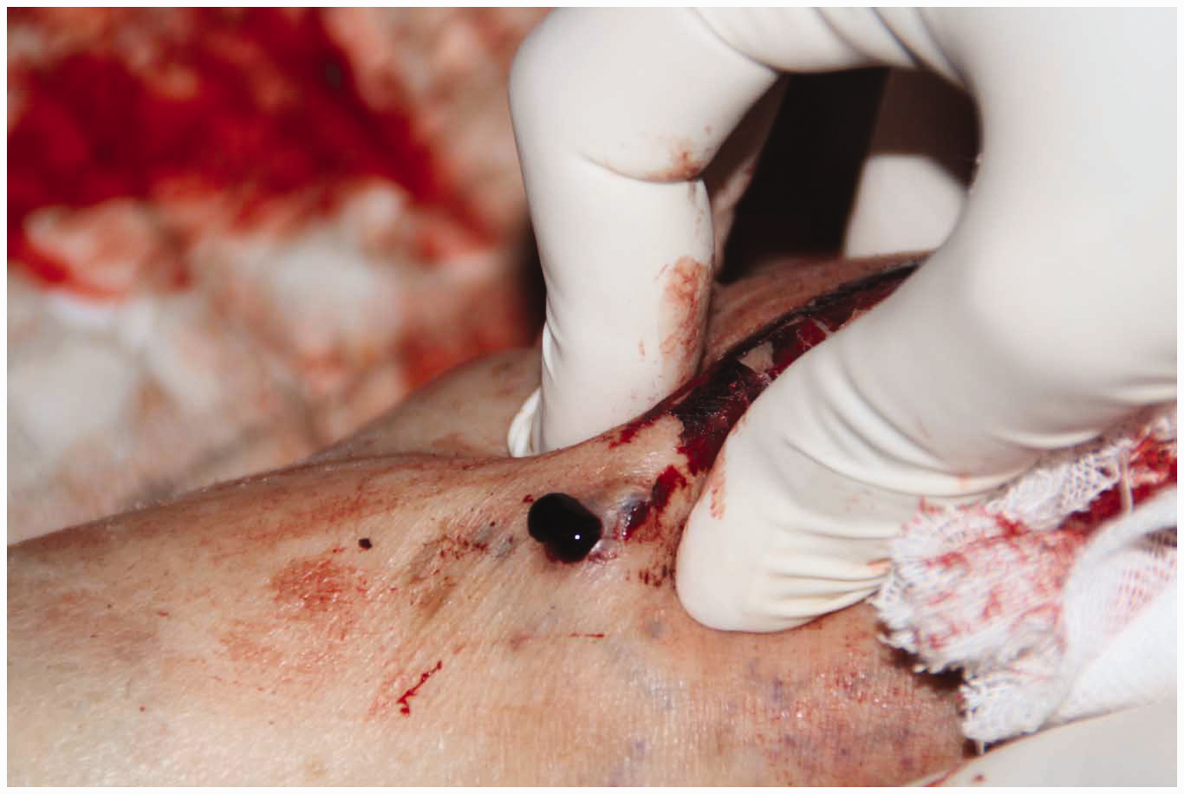
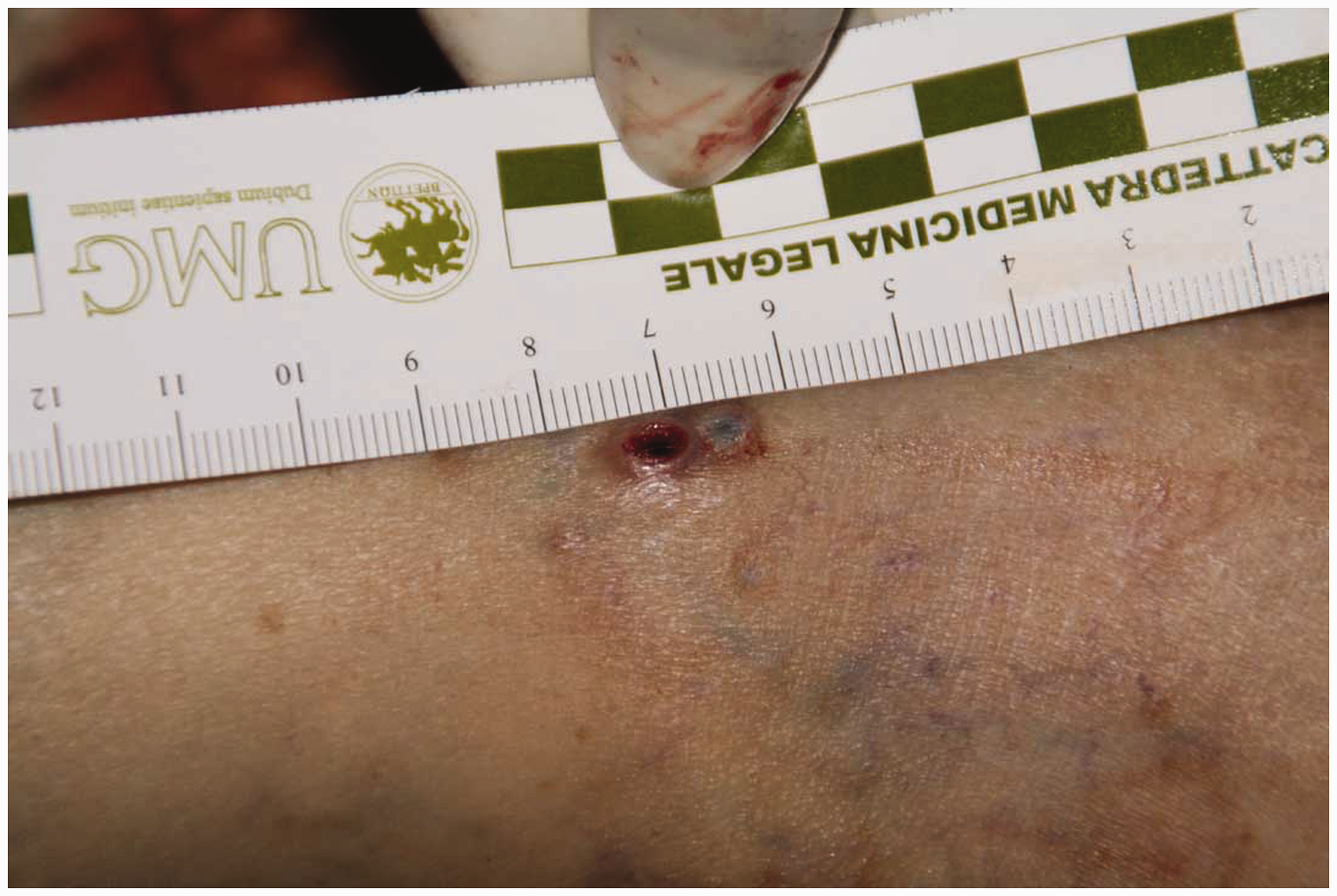
The body showed typical signs of death due to exsanguination. At external examination of the lower limbs showed diffuse skin discolorations with varicose formations of bluish colour (Figure 2). A circular lesion linked to subcutaneous arteriovenous anastomoses was observed. The largest ulcer measured 0.5 cm, with sharp margins and loss of haematic material from the inside (Figure 3). The analysis of the path of the ulcer showed an arteriovenous fistula between the dorsal venous arch and the arcuate artery (Figure 4). Further examination disclosed the presence of two small wounds of circular shape (Figure 5). There was no other traumatic lesion. The right hand had patches of blood, attributable to the attempt to staunch the bleeding. Significantly, macroscopic analysis of the inside of the left sock showed a circular blood crust that corresponded to the diameter of the skin ulcer of the (bare) left foot (Figure 6).
Ulcer with arteriovenous fistula. Loss of haematic material. Small wounds of circular shape. Circular blood crust on the left sock.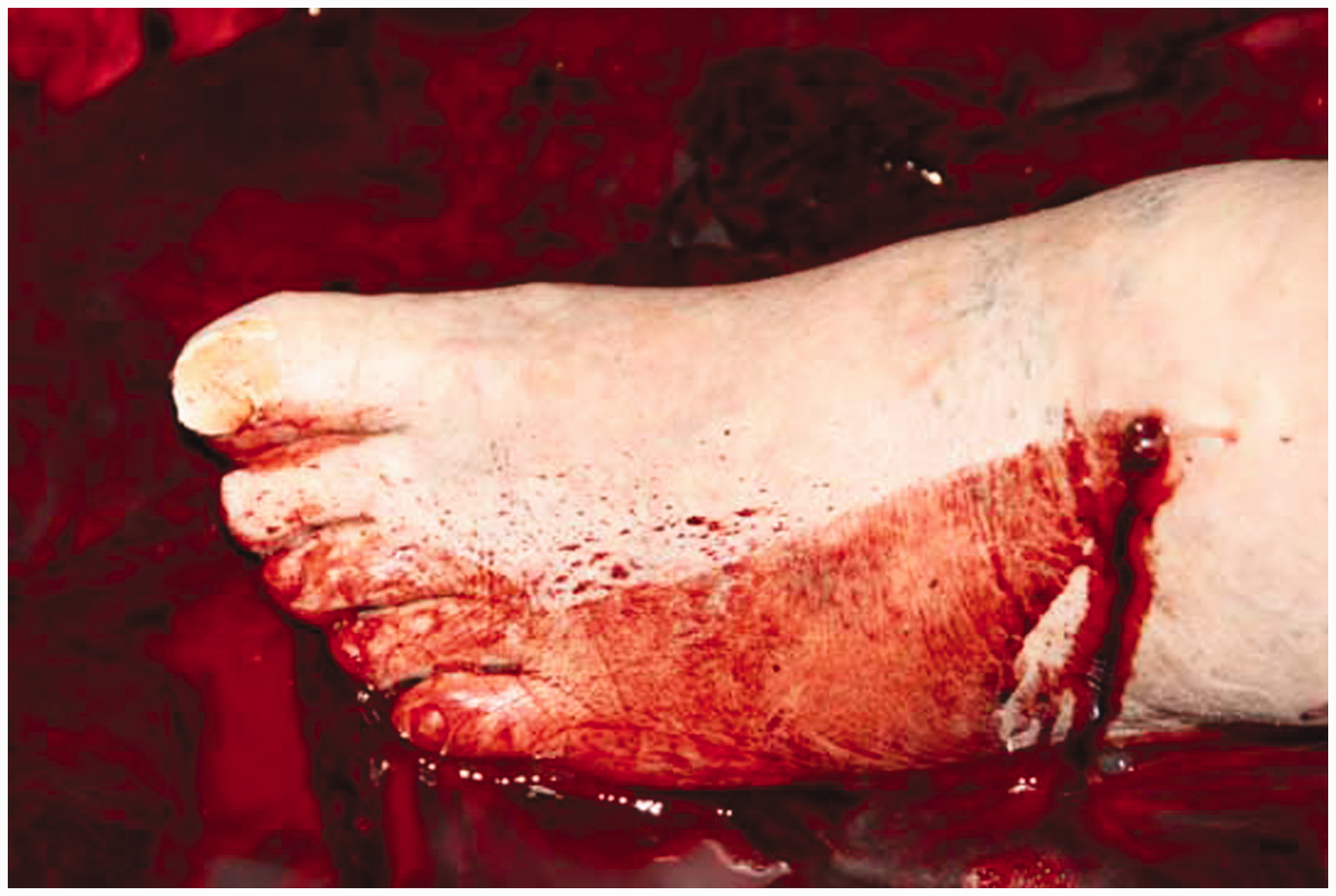

Discussion
Analysis of literature confirmed the utmost importance of the methodical inspection, the external examination or autopsy in achieving a possible explanation for the deaths.1–7 It is notable that in almost all cases, the victims’ death occurred in their own homes. Usually, people affected are aged over fifty. There is no correlation between this fatal event and gender. Aetiology of this fatal outcome is thus related to the pathology of varicose veins disease, usually untreated with proper therapies and medical devices, in patients who have no other risk factors. Some patients (like the one in our study) were treated with anticoagulants for other diseases. The source of bleeding is often a connection between skin ulcer and varicose vein of the limbs or arteriovenous anastomoses. This is illustrated in this case by the analysis of blood stains, which showed evidence of arterial origin at the top of the left foot.
This study aims to emphasize the importance of treating this disease which is considered, especially for women, to be only an aesthetic problem. This case demonstrates how varicose veins and their complications can become a much bigger problem in terms of morbidity and mortality and should be included among the risk factors for sudden and unexpected natural death.
Sudden and unexpected death is described as an event which occurs in a time interval of less than 6 h after the onset of the warning signs. 6 According to estimates, 12% of natural deaths are sudden and 88% of these are due to cardiac disease. 8
With regard to deaths due to rupture of varicose veins in the lower limbs, the term “sudden” illustrates how quickly death occurs. In this case, death took place 5–7 min after the start of bleeding. It must be emphasized that in the treatment of varicose vein disease, both thrombotic and haemorrhagic complications need proper treatment to avoid/prevent bleeding that could lead to death. Therefore, the correct use of anticoagulants, careful monitoring of coagulation parameters by INR and the search of other coagulation factors related to patients with varicose vein disease are necessary.
Finally, from the forensic point of view, this study shows the importance of judicial inspection and external examination of the body. The “crime scene” plays a fundamental role in the solution of suspected cases. In particular, the bloodstains may indicate, for example, whether they were caused by a physical struggle, violence or a murder. BPA refers to the collection, categorization and interpretation of the shape and distribution of bloodstains connected with a crime. 7 The BPA is based on a systematic appraisal of the crime scene, good photographic documentation, the study of autopsy findings and declarations of the author or the victim. Through careful analysis, it is possible to work out the sequence of events. Here, the first responders who accessed the victim’s bedroom were confused by the large pool of blood and warned the competent authorities.
At first sight, fatal haemorrhage, with massive loss of blood, could look like a non-natural death due to a crime. In this case, the deceased's sock presented a circular blood crust that had served as a “cap” on the skin lesion. When he removed his sock, the old man also removed this “cap” from the lesion, inevitably provoking the fatal haemorrhage. During his life, he took anticoagulants to treat other illnesses.
Several cases of fatal bleeding from rupture of varicose veins documented by literature are very similar to “suspicious crime scenes”, in which death may be due to traumatic causes. As in this case, large quantities of blood observed by first responders on the spot can be misinterpreted and deflect attention from the real cause of death. To distinguish a death from a possible criminal assault, from a natural death caused by a natural event, it is extremely important to carry out:
the analysis of blood stains; the search for the source of the bleeding; the evaluation of external injuries found on the victim's body.
Footnotes
Acknowledgments
Preliminary analyses were accepted and presented at 22nd IALM Congress 2012 in Istanbul and at AAFS 65th Annual Scientific Meeting 2013 in Washington DC.
Declaration of conflicting interests
The author(s) declared no potential conflicts of interest with respect to the research, authorship, and/or publication of this article.
Funding
The author(s) received no financial support for the research, authorship, and/or publication of this article.