



Abstract
Research indicates that women are more susceptible to pain than men, but the reason for this difference is unclear. While estrogen and progesterone have been implicated, testosterone has not received adequate consideration in the literature. Additionally, incorporating behavioral expressions, or exaggerations, of pain as an important aspect of pain perception is receiving increasing attention. The current study examined the role of testosterone in female pain expression and perception via the cold pressor test. Following all participant exclusions, 46 healthy participants (32 women) provided saliva samples for testosterone analysis using enzyme-linked immunosorbent assay before and after rating their pain during the cold pressor test. Participants used a visual analog scale to indicate how the 2℃ water was perceived, ranging from “worst pain imaginable” to “no pain.” The researcher also noted whether a participant displayed overt behavioral expressions of pain such as jumping and cursing. The results revealed that men reported lower visual analog scale scores than women, indicating less perceived pain. A subgroup of women who displayed overt behavioral responses to pain seemed to be driving this sex/gender difference. It was expected that this subgroup of females would have corresponding changes in testosterone that would further explain the observed sex/gender differences, but this was not supported. Collectively, these data add to the previous literature investigating sex/gender differences in pain perception and highlight the importance of studying overt behavioral expressions of pain. Testosterone may alter this behavior and subsequent pain perception, but the contributions of testosterone are likely subtle and were not detected in this study.
Introduction
Of the many psychological constructs experienced by humans, pain is arguably one of the most motivating and complex. Millions of people are affected by pain on a daily basis, and it is one of the primary reasons people seek medical attention. It contributes to an annual cost of more than $600 billion in health-care costs and loss of productivity (Rutchick & Slepian, 2013) and can significantly impact one’s quality of life. Due to the fact that pain is a highly individualistic experience, studying the influencing factors on pain can be challenging. A biopsychosocial model provides a theoretical framework for better understanding the many mechanisms that predict experiences of pain (Gatchel, Peng, Peters, Fuchs, & Turk, 2007). Studying these various factors provides valuable insight into the management and treatment of pain.
One interesting intersection of biological, psychological, and social contributions to pain perception is observed when comparing the pain response of males and females. An abundance of research indicates prevalent sex/gender differences, intimating that women are more susceptible to both acute and chronic forms of pain (Etherton, Lawson, & Graham, 2014; Hellström & Lundberg, 2000). This effect is seen consistently in women reporting higher pain intensity and displaying lower levels of tolerance and threshold (Ruau, Liu, Clark, Angst, & Butte, 2012), as well as having an increased willingness to report pain and even take analgesic medications (Wise, Price, Myers, Heft, & Robinson, 2002). Though the reasons for these differences are not definitively clear, two complimentary explanations surface regularly in the body of literature surrounding this topic. First, there are specific physiological mechanisms, namely sex hormones, which differ between biological sexes and may affect pain perception (Musey et al., 2014; Wiesenfeld-Hallin, 2007). Second, psychosocial factors shaped by society and gender-role expectations contribute to one’s personal experience of pain (Hellström & Lundberg, 2000; Wise et al., 2002).
In considering the biological contributions to these sex/gender differences, steroid hormones offer a plausible explanation. Indeed, both estrogen and progesterone are implicated in female pain responding. High progesterone levels correlate to lower pain thresholds (Stening et al., 2007), while dynamic estrogen levels throughout the menstrual cycle are consistently recorded as a modulator of nociception (Bartley et al., 2015; Lu et al., 2012; Mogil, 2012; Rezaii, Hirschberg, Carlström, & Ernberg, 2012). Interestingly, however, reports of whether this modulation is pro- or anti-nociceptive are inconsistent and many studies do not fully consider other potential mediating factors, such as testosterone. Testosterone has not received adequate consideration in investigating sex/gender differences in pain perception, despite the fact that women have significantly lower average baseline levels than men.
It has been suggested by previous findings that testosterone may serve a role in decreasing pain. For example, one study found that greater testosterone levels in normally menstruating women corresponded to decreased pain intensity during experimentally induced pain (Bartley et al., 2015). Testosterone therapy utilizing transdermal gel administration decreased symptoms of pain and fatigue in chronic pain patients with minimal apparent risk (White et al., 2015; White & Robinson, 2015). Another line of research revealed an analgesic effect of testosterone metabolites for diabetes-related neuropathy (Calabrese et al., 2014). However, this body of literature is scarce and requires further replication and exploration.
In conjunction with such biological factors, psychological factors influencing behavioral expression of pain are frequently tied to differences in pain perception. Various gender-related factors have been explored and found to be important determinants of pain perception in females. For example, there is a relationship between sex/gender-role beliefs and pain expression. Females are consistently shown to overtly express pain more readily than males, and men who rate high in masculinity are significantly less likely to report their pain (Fillingim, 2000). This is interesting given that behavioral expressions of pain are associated with enhanced pain intensity, especially in women (Keefe et al., 2003; Nayak, Shiflett, Eshun, & Levine, 2000; Roditi, Tobinson, & Litwins, 2009). Currently, the relationship between behavioral expression of pain in females and testosterone has not been explored. Given that each variable has been suggested to alter pain perception, studying the combined impact of these variables may provide new and needed insights into previously observed gender differences.
The present study aimed to expand previous findings on sex/gender differences by specifically investigating the testosterone levels of women in relation to subjective pain scale ratings both before and after experimentally induced pain. It was expected that changes in testosterone in females would correspond to alterations in reported pain. Specifically, based on previous literature, females with higher baseline testosterone levels or more notable testosterone increases while experiencing pain should display less pain. Further, overt behavioral expressions of pain would be associated with increased pain ratings and lower levels of testosterone.
Method
Participants
Fifty-six individuals were recruited from an undergraduate university (38 women, 16 men, and 2 other gender) using flyers and campus-distributed emails. Participants were offered the opportunity to enter a raffle for a $25 Visa gift card as compensation. Individuals were screened online for eligibility and not permitted to participate if they had history of hand or wrist pain, arthritis, cardiovascular problems, diabetes, chronic pain conditions (such as fibromyalgia and neuropathy), Raynaud’s disease, or were pregnant or breastfeeding. These factors were chosen as eligibility requirements both to maximize participant safety and to minimize the impact of pre-existing pain conditions on the data.
Of the 56 participants from whom data were collected, eight (six women, two men) were excluded for medication use or medical conditions known to affect hormone levels (e.g., Lipitor; PCOS), with the exception of hormonal contraceptives (used by 19 women). Two more participants were excluded because they did not identify as male or female in the questionnaire, and the given sample was not large enough to account for a third gender variable. All exclusions yielded a total sample size of n = 46 (32 women, 14 men). Participants ranged in age from 18 to 35 (M = 21.61, standard error of the mean (SEM) = 0.55); 41% of the participants identified themselves as White/Caucasian, 34% as Hispanic/Latino, 9% as Black/African American, and 16% as multi-racial.
Materials
Participants completed a health and background questionnaire (van Anders, Goldey, & Bell, 2014), providing information about factors that may affect either pain perception or testosterone levels, including prescription medication, days since the last period, and contraceptive use. The questionnaire was administered in the lab via Qualtrics.
The cold pressor test (CPT) is a commonly used procedure for experimentally inducing pain or discomfort (Etherton et al., 2014; Rutchick & Slepian, 2013). For the present study, the test involved placing the non-dominant hand, up to the wrist, in a container filled with water at room temperature (22℃) for 2 minutes. After this time, the participant moved the hand directly into a container of water maintained at 2℃ until either his/her pain tolerance level was reached, or 5 minutes had passed, whichever came first. Level of pain intensity was reported using a visual analog scale (VAS)—a 10-cm vertical line on a piece of paper with the words “worst imaginable pain” across the top and “no pain” at the bottom. Participants simply marked the spot on the line that corresponded to their pain level on a new VAS for each separate indication. The non-dominant hand was chosen for the CPT to allow use of the dominant hand in marking the VAS. The time it took to withdraw the hand was recorded for each participant. In addition to VAS scores, the researcher took note of individuals who displayed outward behavioral expressions of discomfort including jumping at the cold sensation, shrieking, cursing, grimacing, yelling/moaning/whining noises, or explicit statements that the cold water was painful. Every instance of behavior was not indicated within a single individual and behaviors were not tallied; therefore, this was a yes (overt expression was observed) /no (no overt behavior observed) variable. The individuals who collected data were similar in age to one another and trained to avoid displaying emotion or engaging in conversation during the CPT. They remained in the room with the participant in order to administer the CPT.
To measure testosterone levels pre- and post-CPT, participants provided two 2 to 4 mL samples of passive drool into polystyrene collection tubes, once just before and once 10 minutes after undergoing the test. This method and the timing were chosen based on previous work showing that testosterone changes can be detected in saliva 10 minutes following various manipulations to mood and arousal (van Anders et al., 2014). Saliva was stored at −20℃ until analyzed. At the close of the data collection period, all samples were assayed in duplicate simultaneously to measure testosterone concentration, using commercially available enzyme-linked immunosorbent assay kits from Salimetrics. The sensitivity of the kits was reported by the Salimetrics company to be <1.0 pg/mL. Coefficients of variation for inter-assay testosterone were 6.88% for high and 10.05% for low; intra-assay coefficient of variation was 6.03%. Percent change in testosterone from pre- to post-CPT was used in some analyses due to its sensitivity to individual differences in baseline measures and its normative use in testosterone research (van Anders et al., 2014).
Procedure
The study design and protocol were granted approval by the university’s institutional review board before data collection began. All data were collected between 11 am and 7 pm in order to account for diurnal fluctuations in testosterone (van Anders et al., 2014). Sessions were initiated by the researcher with a detailed description of the procedure. After obtaining informed consent, the researchers exited the room and the initial saliva sample was collected while participants simultaneously completed the health and background questionnaire. Prior to beginning the CPT, participants were reminded that they could end the test at any time by removing their hand from the water, but were asked to attempt to reach their point of tolerance (i.e., they could no longer tolerate the pain). The researcher stood nearby with a stopwatch, noting the length of time until the withdrawal of the hand. A VAS score was taken within the first 30 seconds as an indication of the participant’s threshold for pain, and a final VAS score was collected when the participant withdrew their hand from the CPT as a measure of the participant’s tolerance level. If a participant remained in the CPT for the maximum 5 minutes, the test was ended and a final VAS score was recorded.
After CPT exposure, all participants were shown a 10-minute emotionally neutral travel documentary, previously determined not to affect testosterone (Goldey & van Anders, 2015). During this time filler, they were asked to refrain from using cell phones or speaking to the researcher. A second saliva sample was collected following the video. Finally, just prior to ending the session, participants reported their pain level on the VAS one last time to ensure that they were not experiencing lingering pain at the end of the experimental session.
Results
Sex/gender differences in pain perception
To investigate differences in pain perception between the male and female participants, separate independent samples t-tests were conducted for the average VAS scores reported at the time of threshold and at the time of reported tolerance. Analyzing threshold and tolerance with separate t-tests is commonly employed in the CPT literature (e.g., Compton, Ling, & Torrington, 2008). Although there were no significant differences between male and female VAS scores at threshold, t(44) = 0.73, p = 0.468, significant differences were observed at tolerance, t(44) = 2.13, p = 0.039. Males had significantly lower VAS scores (M = 51.43, SEM = 9.41) than females (M = 71.03, SEM = 3.53), indicating that males reported less pain/higher tolerance to the CPT-induced pain than females (see Figure 1).
Average VAS scores for males (n = 14) and females (n = 32) at the time of threshold and tolerance in the CPT. No differences were observed at threshold (p > 0.05), but females reported significantly more pain at tolerance (p = 0.039). Error bars indicate ±SEM.
Behavioral indication of pain and pain perception
In support of a biopsychosocial perspective on pain, recent literature suggests that exaggerated pain responding can be an important determinant of overall pain intensity (Beneciuk, Bishop, & George, 2010; Parr et al., 2012). As the purpose of the study was to investigate influences on pain perception, the researcher noted whether a participant displayed any overt behavioral responses during the CPT, including physical gesturing and vocal expressions of their discomfort (e.g., using profanity, shrieking, jumping, and yelling). Interestingly, no males displayed these behaviors, while the majority of females did. Of the 32 females included in this study, 23 (or 71.9%) outwardly expressed their pain. To better investigate the differences in pain perception between females who engaged in these behaviors and those who did not, separate independent samples t-tests were conducted for pain reported at threshold and at tolerance. Unlike the original sex/gender differences observed, here there were significant differences between females who did and did not outwardly express pain at both threshold, t(30) = 2.35, p = 0.026, and tolerance, t(30) = 2.53, p = 0.017. Females who overtly displayed their pain reported significantly higher VAS scores, indicating more perceived pain at threshold (M = 65.04, SEM = 4.09) and tolerance (M = 77.65, SEM = 3.46) than females who did not display these behaviors (threshold: M = 46.67, SEM = 6.93; tolerance: M = 54.11, SEM = 12.20) (see Figure 2). Additionally, none of females who outwardly expressed their pain were able to withstand the cold pain for longer than half the maximum allotted time, or 2 minutes and 3 seconds, which was not the case for females who did not display expressions of pain (data not shown).
Average VAS scores at threshold and tolerance for females who displayed behaviors indicating pain versus those that did not display such behaviors. Females who displayed their pain behaviorally had significantly higher reported pain at threshold (p = 0.026) and tolerance (p = 0.017). Error bars indicate ±SEM.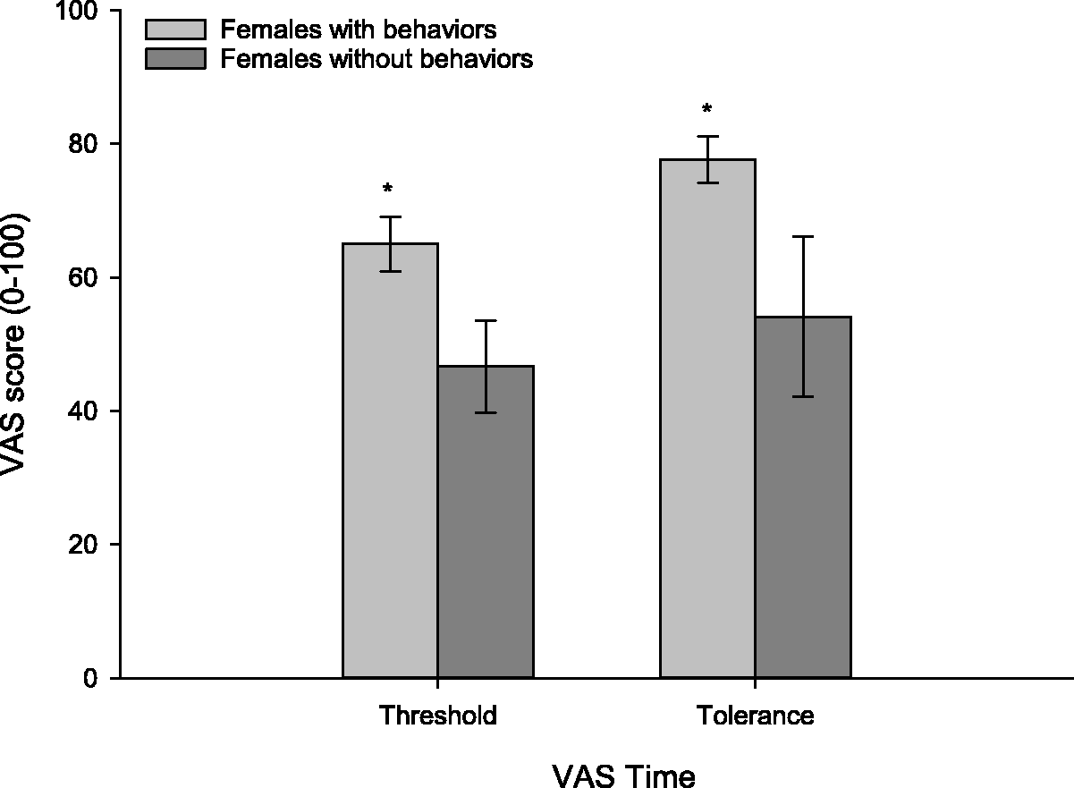
Testosterone differences and pain perception
Based on our hypothesis that female pain responding is related to changes in testosterone, we anticipated that testosterone levels would be significantly different between these two female subgroups. Independent samples t-tests were conducted to investigate the relationship between testosterone levels and behavioral indications of pain in females. No significant effects were found for baseline levels of testosterone, posttest levels of testosterone, or percent change in testosterone (for all p > 0.05) (see Figure 3).
Testosterone levels (pg/mL) and percent change in testosterone for females who did and did not display behaviors to indicate pain during the CPT. No significant differences were observed at baseline, after the CPT, or as a function of the percent change in testosterone (p > 0.05). Error bars indicate ±SEM.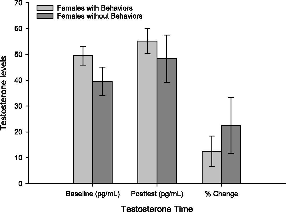
To investigate the differences between males and females, we initially confirmed that males did in fact have higher testosterone levels than females at baseline, t(44) = 8.83, p = 0.0001 (males: M = 121.92, SEM = 10.86; females: M = 46.72, SEM = 3.11). This difference was still present following the CPT, t(44) = 7.54, p = 0.0001 (males: M = 133.84, SEM = 13.07; females: 53.29, SEM = 4.25) (see Figure 4(a)).
Testosterone levels in males versus females. (a) Testosterone levels for males and females during the baseline and post CPT data collection time points. Males had significantly higher levels of testosterone (p = 0.0001). (b) The percent change in testosterone from baseline to the post CPT time point for males and females. Although the average percent change in testosterone was greater for females, this was not a significant finding (p > 0.05). Error bars indicate ±SEM.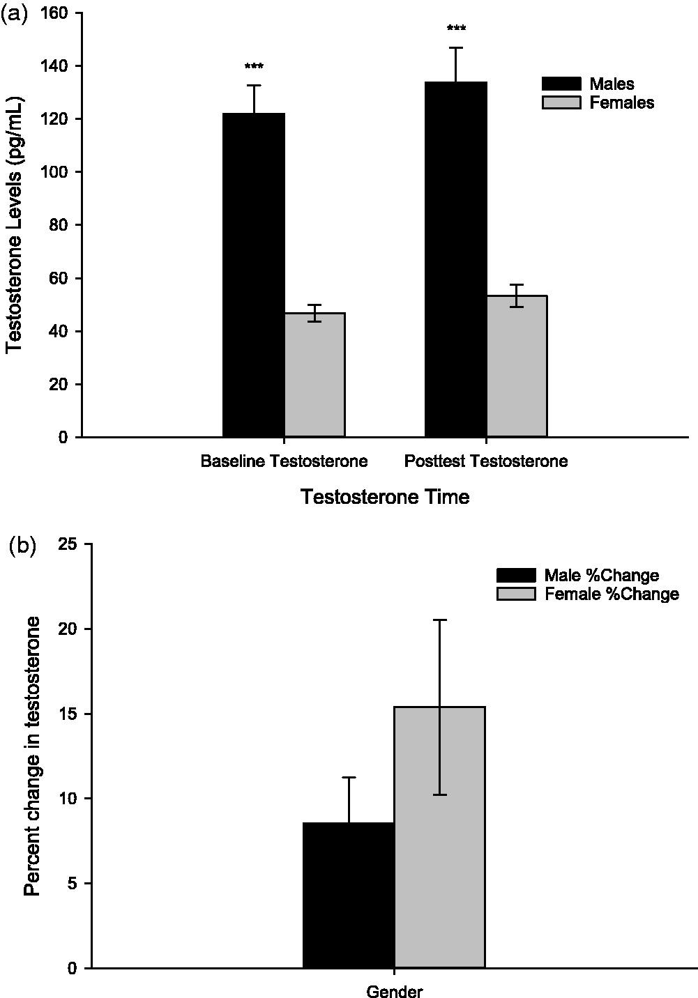
An analysis of the sex/gender differences in the testosterone change from baseline to post-CPT measurements was conducted. Although women experienced a greater change in testosterone following the CPT than men (males: M = 8.54, SEM = 2.69; females: 15.38, SEM = 5.16), a t-test revealed that this effect was not significant, t(44) = .85, p = 0.399 (see Figure 4(b)).
Discussion
The findings from this study support previous literature on sex/gender differences in pain perception; women showed lower tolerance (i.e., heightened pain perception) in the CPT when compared to men. Such differences were not observed during the initial exposure to the CPT-induced pain (threshold). Instead, it was only when taking into account the finding that some women were more expressive of their pain than others that differences in pain perception were found at the point of threshold. It was expected that variations in testosterone levels would explain the differences between the females displaying behaviors associated with their pain and those that did not, but this was not supported. Further, we did not find that sex/gender differences in pain perception could be explained by differences in testosterone. Collectively, these results indicate that behavioral expression of pain is related to pain intensity, but the contributions of testosterone to this relationship, if any, are likely very subtle. Rather than a hormonal mechanism serving as the primary determinant of pain perception, the findings presented here were better explained by accounting for overt expressions of pain. Indeed, females who outwardly expressed their pain through gestures and vocalizations were those who seemed to be driving the observed sex/gender differences.
Psychological expectations regarding pain can produce a catastrophizing response to painful stimuli, and this is increasingly recognized as an important predictor of pain (Beneciuk et al., 2010; Parr et al., 2012), as well as a potentially significant contributor to sex/gender differences in pain threshold and tolerance (Edwards, Haythornwaite, Sullivan, & Fillingim, 2004). Women tend to experience psychological exaggerations more often than men, corresponding with increased perception of pain intensity (Sullivan et al., 2001). This is evidenced by physical manifestations of increased overt displays in response to the painful experience. The degree to which one outwardly expresses their pain may even contribute to chronic conditions or disability caused by pain (Etherton et al., 2014). Given the observed behavioral differences between the female subgroups, and their correspondence with heightened subjective pain intensity scores, the results of the present study support these previous conclusions. Further, as the females with no fervent expressions were able to withstand the cold pain for the longest duration, it would appear that these intense outward expressions do more to enhance the perception of pain than cope with it, supporting prior research (Keefe et al., 2003; Nayak et al., 2000; Roditi et al., 2009).
Overt expression of pain in this study was indicated as either present or absent, and any observable instance of pain was considered “overt.” There are issues with this approach, including the fact that experimenter bias could have occurred. We contend that this is unlikely, however, due to the fact that all researchers collecting data followed a script when administering the CPT and had no reason to expect that particular females (or males) would display their pain over others. Another issue with the approach used was that subtle differences in pain perception related to behavioral expressions of pain were likely missed since participants were coded as either showing expressions of pain or not. The use of a more standardized scale, such as the Pain Catastrophizing Scale (Parr et al., 2012), would have provided the opportunity to more fully address the contributions of pain behavior to pain perception.
The directional relationship (i.e., the causal factors) between increased likelihood to report pain and enhanced pain perception is not abundantly clear, indicating a need for more work in this area. Some studies do suggest however that testosterone offers a viable means of better understanding this relationship. For instance, higher levels of endogenous testosterone appear to serve an analgesic role in females, even when accounting for menstrual phase (Bartley et al., 2015). That study further reported that sensitivity to painful stimuli was greatly diminished when testosterone levels were higher (Bartley et al., 2015). In another recent study, men with lower testosterone levels reported higher levels of pain and anxiety related to pain in an experimental setting (Choi et al., 2017). Given these results, testosterone appears to serve a function in pain perception and response.
It is therefore unexpected that the results presented herein do not conclusively implicate this hormone in female versus male pain perception. There are various explanations for our lack of finding that testosterone modulated the pain response. A small body of research points to an interaction between estrogen and testosterone in women’s responses to pain (Vincent et al., 2013), which was not accounted for in this study. It is quite possible that the two sex hormones work in tandem, having an interdependent effect on the experience of pain. This can be seen through hormone therapy in gender reassignment. Individuals taking estrogen supplements have experienced increased pain sensitivity and even some chronic pain, while females transitioning to males and supplementing testosterone reported less general pain and decreased symptoms of existing chronic pain (Aloisi et al., 2007; Musey et al., 2014). As estrogen was not directly measured in the current study, its influence cannot be fully accounted for. Female participants did indicate the use of hormonal contraception and the days since their last period. Regression analyses were conducted to investigate the influence of these factors on testosterone levels and pain reporting but were not found to be significant (data not shown). While there are published accounts of pain sensitivity changing across the menstrual cycle, others have reported that estrogen fluctuations do not significantly alter pain in females (Stening et al., 2012). It is also possible that our small sample size made it difficult to detect the influence of testosterone on pain. Bifurcating our sample into groups to investigate the sex/gender differences meant that the power to detect testosterone effects was significantly lowered, and it is therefore possible that testosterone did have more of an influence on pain than is captured in these data.
Although hormone analysis through saliva has been verified in many studies (e.g., van Anders et al., 2014), the ideal method of hormonal assay is through the use of serum. Saliva was utilized for this study because it does capture testosterone changes and was deemed to offer a methodological advantage by avoiding the stress that can be induced by blood-draw methods. However, this method may not fully capture blood-level changes in testosterone. That idea, combined with varying recommendations of time between exposure to pain and final saliva sample collection, may have contributed to a minimal captured change in testosterone. However, the timing used here (10 minutes post manipulation) is relatively standard in research investigating testosterone in response to mood and affect manipulations, and it is therefore unlikely that our findings are simply due to a lack of capturing the pain-induced changes in testosterone in this study. And again, the use of saliva analysis to quantify testosterone changes following experimental manipulations have been previously validated (van Anders et al., 2014).
While the gender of the researcher can influence the results in the CPT (Vigil, Rowell, Alcock, & Maestes, 2014), those collecting data in the current study were females of a similar age. Therefore, any influence exerted by the gender of the researchers would have been consistent for all participants. It is also the case that having the researcher in the room while participants rated their pain could significantly impact pain ratings (Vigil et al., 2014), but this was also consistent for all participants, and the researchers were trained to avoid displaying emotional expression or engaging in conversation during the CPT in an attempt to minimize experimenter-expectancy effects as much as was possible.
Further, the present study was limited in part by the use of healthy volunteers and an acute, controllable pain stimulus, and therefore does not capture the biological and psychological changes that would occur during chronic pain. Future studies would be benefitted by incorporating individuals with chronic pain conditions to explore testosterone’s influence on pain. Although the cold pressor test is not an apt representation of chronic pain experienced by patients, it is a reliable way to gather physiological data in response to pain in a laboratory setting that poses minimal risk to human subjects. Similarly, generalizability was lessened by the use of young college students, and future research should incorporate a more diverse (age, education, etc.) background. Although this study does contain methodological limitations, the methods used are a reliable and valid means to induce and study pain.
In conclusion, it would appear that pain perception can be predicted largely by psychological components and the way in which one responds to pain, specifically by overtly expressing their discomfort. While it is possible that testosterone plays a role in the modulation of pain perception, these effects were not captured in this study. Further research should be conducted keeping in mind the potential mediating effects of other biological factors. Likely, there is an interaction between the biological and psychological realms that should be more thoroughly explored in future studies to provide better understanding of pain processing and improvements in treatment efforts.
Footnotes
Acknowledgments
The authors would like to thank Sara Gardon and Chantal Neutzler for their assistance with data collection.