




Abstract
Gold nanostructures were synthesized by reduction of gold ions using aqueous extract of grapefruit pulp (Citrus paradisi). This eco-friendly bioreduction method allows the formation in solution and support of gold nanostructures on silk fibers. Bioreduction techniques involve biomolecules of grapefruit extract for reducing a gold precursor to obtain different kinds of nanostructures. Carbohydrates and organic acids, present in C. paradise, are believed to be responsible for the formation of nanoparticles. Analysis of gold–silk nanocomposites by electron microscopy shows gold nanostructures with quasi-spherical, hexagonal, and triangle shapes. The evolution of functional groups in the silk fibers before and after the bioreduction process was followed by infrared spectroscopy. Diffuse reflectance spectroscopy (DRS) and laser scanning confocal microscopy (LSCM) were used to probe surface plasmon resonance and fluorescent behavior in the silk–gold composite. This simple and novel methodology for obtaining these types of nanocomposite may have important applications in the development of functional fibers.
The search for novel synthetic methodologies of nanostructured materials has become an important topic of nanoscience and nanotechnology. 1 The study of metal nanostructured materials has attracted special interest in recent years because of the large number of applications in materials science and engineering. 2 In particular, metal nanoparticles have attracted interest because of their size-dependent optical, mechanical and magnetic properties. In addition, the shape control of metal nanostructures can be a strategy to adapt their physical and chemical properties for various applications such as biological labels, 3 pharmaceutical materials, 4 sensors, 5 catalysts, 6 and surface-enhanced Raman scattering (SERS) substrates. 7 Formation of nanomaterials can be carried out by biomolecules from biological systems readily available in nature. 8 Biosynthesis with biological systems such as plant biomass extracts is an alternative to physical and chemical methods to produce nanostructured metals. 9 Several methodologies have been developed to obtain nanoparticles of different metals. For example, silver nanoparticles with spherical shape and sizes between 16 and 40 nm were obtained by employing geranium leaves extract (Pelargonium graveolens). Gold nanotriangles and silver nanoparticles have also been obtained using Aloe vera extract.10,11 Our group has previously proposed a green method to synthesize gold nanoparticles with spherical shape and average particle size of around 40 nm using green tea leaves aqueous extract (Camellia sinensis). 12 Gold nanostructures have also been produced using microorganisms and human cells.13–18 A strain of Neurosporacrassa fungus with a mixture of HAuCl4 and AgNO3 aqueous solutions was used to produce mainly spherical monometallic and bimetallic nanoparticles, with average diameters of 11 nm for silver and 32 nm for gold. 19 When in contact with AgNO3, Plectonema boryanum, a cyanobacterium, generates spherical silver nanoparticles in solution. 20 Also, proteins from biological systems can be used for producing silver nanoparticles from a mushroom substrate. 21 However, in most cases nanoparticles must be supported on solid materials, such as natural or synthetic polymeric fibers, to make usable devices. 22 Haratifar et al. deposited gold on magnetite (Fe3O4) using an ethanol extract of Eucalyptus camaldulensis as a natural reducing agent. 23 In this contribution, we report on the synthesis of gold nanostructures embedded in silk fibers using grapefruit (Citrus paradisi) aqueous extract as a reducing reagent of metal ions.
Experimental details
Materials
Tetrachloroauric acid (HAuCl4) was obtained from Aldrich Chemicals Co. Silk fabric swatches were acquired from A.K. Exporters Ltd, Raksha-Impex Company, Bangalore, India. Materials were used without any further treatment. The silk swatches were probed using infrared (IR) spectroscopy. The spectrum obtained was compared with a published report on natural silk hence confirming the composition of the material. 24
Citrus paradisi extract
A total of 10 g of thoroughly washed C. paradisi (grapefruit) pulp was finely cut and stirred with 90 ml of de-ionized water. The mixture was boiled for 2 minutes and then cooled down to room temperature. The infusion was filtered with a linen cloth to retain fibrous residues and left at room temperature. The resulting extract was used for the bioreduction experiments.
Synthesis of gold nanoparticles in solution
To synthesize Au nanoparticles in solution, the grapefruit extract was mixed with HAuCl4 solution (10−3 M) in a ratio of 1:1 by volume (v/v). The bioreduction was carried out in darkness, at room temperature, for 24 hours. No further treatments were carried out. Transmission electron microscopy (TEM) samples were prepared by placing drops of the reacted mixture over carbon-coated copper grids and allowing the solvent to evaporate.
Silk–gold composite formation
The composite was prepared by adding grapefruit extract (8 ml) to a glass vial containing a silk swatch (2.54 cm × 2.54 cm), then pouring 2 ml of 10−2 M HAuCl4 de-ionized aqueous solution. This mixture was allowed to stand for 24 h at room temperature. The silk–gold composite fabric was removed and air-dried at ambient conditions.
In order to evaluate the performance of the method for anchoring gold on silk fibers, atomic absorption (AA) studies were carried out in the original 10−2 M HAuCl4 solution and also in the residual solution, after the silk fibers were soaked in it. The measurements were collected in an AAnalyst 200 Perkin-Elmer atomic absorption spectrophotometer, employing a Lumina N305-0107 Perkin-Elmer lamp with a wavelength of 242.8 nm.
Characterization
The morphology and size of the synthesized gold nanoparticles were determined in low-resolution mode with a JEOL 2010 transmission electron microscope with a LaB6 filament. Field emission scanning electron microscopy (FESEM) was performed on a Leo 1550 microscope instrument with a point resolution of 1 nm at 20 keV and 2.5 nm at 5 keV. A Bruker Angle X-ray Scattering Microanalysis was used for elemental analysis. UV–Vis diffuse reflectance spectra were recorded using a Cary 5 spectrophotometer (Varian) equipped with a deuterium lamp. The UV–Vis absorption signal was recorded over a range from 200 to 800 nm. Laser scanning confocal microscopy (LSCM) was carried out in a LSM-510 Meta, Carl Zeiss microscope, equipped with an argon ion laser for excitation at 488 nm and green fluorescent protein (GFP) filters for emission at 515–530 nm. FT- IR measurements were collected on Shimadzu Prestige 21 spectrometer with a spectral range 4000–500 cm−1.
Results and discussion
Synthesis of gold nanoparticles in solution
Using extract plants for obtaining gold nanoparticles can be advantageous over other biological processes, by eliminating the complicated process of maintaining cell cultures. If the biosynthesis of gold nanoparticles can compete with traditional chemical methods, there is a need to achieve faster synthesis rates. Grapefruit (C. paradisi), is a year-round accessible fruit in North America which contains biomolecules capable of reducing metal ions and stabilizing nanoscale materials. Water soluble compounds in C. paradisi are composed mainly of organic acids and sugars. In general, sugars such as sucrose, glucose and fructose are present at a concentration of 76–85.5 g/l and organic acids range from 22.5 to 26.8 g/l. 25 The main acids in C. paradisi are citric (12.99 g/l) and malic (0.871 g/l) acids with trace amounts of tartaric (0.237 g/l), ascorbic (0.41 g/L), and oxalic acids (0.268 g/l). 26 It is important to note that glucose, 27 ascorbic acid, 7 and citric acid 28 have been reported previously to be responsible of formation of metal nanostructures with controlled size.
It is well known that gold nanoparticles exhibit violet–purple color with these colors arising due to excitation of surface plasmon vibrations. When C. paradisi extract was added to 10−3 M aqueous HAuCl4 solution, the yellowish color appearance of the original solution changed to a violet color, indicating the formation of gold nanoparticles. The inset in Figure 1(b) shows pictures of aqueous [AuCl4]– ions samples before reduction (labeled: br) and after reduction (labeled: ar), respectively.
TEM studies of gold nanostructures obtained in solution. Au nanoparticles were formed after 24 hours of reaction with Citrus paradisi extract at 10−3 M HAuCl4. (a) Representative TEM micrograph of gold nanostructures. (b) Size distribution histogram of gold nanoparticles obtained in solution. The inset in Figure 1(d), shows the photographs of aqueous [AuCl4]− before (br) and after (ar) the bioreduction. (c) TEM image of planar nanotriangle and decahedral nanoparticles, the inset shows SAED pattern of the gold nanotriangle. (d) SAED pattern of gold nanoparticles, the diffraction rings correspond to 1(111), 2(200), 3(311), 4(420), and 5(422) lattice planes of FCC gold structure.
TEM micrographs of gold nanoparticles synthesized with C. paradisi extract reveal polydisperse morphology. Figure 1(a) corresponds to a TEM micrograph of gold nanoparticles solution produced with a 24-hour reaction. It can be observed that the morphology was mainly quasi-spherical with an average size of 30 nm (Figure 1(b)), but polyhedral structures could also be identified.
The TEM analysis in Figure 1(c) clearly reveals the formation of triangular nanoplates and decahedral gold nanostructures in low concentrations, in addition to quasi-spherical nanoparticles. The inset in Figure 1(c) shows the selected area electron diffraction (SAED) pattern of a gold nanotriangle with the hexagonal pattern reflecting its single crystalline nanostructure. The spots marked using triangles, squares and ovals correspond to reflections from the 1/3(422), (220), (311) lattice planes of the faced-centered cubic gold structure. Zone axis of the gold nanotriangle is [111], with the top surface normal to the electron beam.29, 30 The presence of the forbidden 1/3(422) reflection has been explained previously by Pileni and co-workers as a result of stacking faults in nanotriangles. 31
The SAED pattern in Figure 1(d) reveals that the diffraction rings with d-spacings of 2.355, 2.039, 1.23, 0.912, and 0.8325 Å that could be indexed as (111), (200), (311), (420), and (422) reflections corresponding to face-centered cubic gold.
The chemical activity of C. paradisi extract is attributed to its high content of reducing molecules, such as ascorbic acid, 7 glucose, 27 citric acid 28 and phenolic compounds. It has been established that extracts of different plant species produce distinct size and shapes of nanoparticles. This observable fact is due to the presence of functional groups such as hydroxyl (–OH), carboxyl (–COOH), amino (–NH2), and sulfhydryl (–SH), which also act as stabilizing agents. 32 Furthermore plant secondary metabolites including terpenoids and polyphenols such as flavonoids, anthocyanins, and isoflavones can also be involved in the reduction process of Au3+ ions. For this reason, the exact mechanism of reduction and stabilization for the nanoparticles synthesis assisted by the grapefruit extract is complex to elucidate and further work is required. 33
Silk–gold composite formation
After the addition of the HAuCl4 aqueous solution to the mixture of silk fibers and grapefruit extract, the fibers changed coloration from pale white to violet confirming the presence of gold nanoparticles. The atomic absorption analysis shows that 99.086% of the gold from the HAuCl4 solution is deposited on the silk fibers.
It is well known that Silk created by Bombyx mori, is formed by two main proteins, sericin and fibroin. Fibroin is the structural center of the silk, whereas sericin is the sticky material surrounding it. The primary structure of fibroin consists mainly of recurrent amino acid sequence (Gly–Ser–Gly–Ala–Gly–Ala) n . Some of these amino acids contain carboxylic functional groups besides amino groups. Both moieties show good affinity to metallic atoms and positive ions due to their high electronic density, furthermore amino groups also possess reductive properties. For this reason carboxylic and amino groups could be responsible for the stabilization and capping of the gold nanoparticles.
In addition, recently Zhang et al. 34 reported that during the in situ synthesis of silver nanoparticles on silk fabric with a polyamide network polymer, the amino groups are able to entrap silver ions as well as acting as reducers. This report found larger silver nanoparticles supported on silk fibers than silver nanoparticles obtained in aqueous solution. This can be attributable to the high surface energy of silver nanoparticles on the surface of silk fibers, which induce aggregation of silver nanoparticles. That behavior is also present in our system.
When the bioreduction occurs gold nanoparticles are electrostatically attracted by the functional groups, such as carboxylic and amino moieties, on the silk fibers and these same chemical moieties may help to stabilize the nanostructures and control the particle size. After 24 h of bioreduction the nanoparticles size increases to 32 nm due to nucleation of nanostructures.
24
In addition, we speculate that some of the biomolecules present in grapefruit extract may play the role of stabilizers, such as polyphenols do when green tea extract is used to obtain silver nanoparticles.
12
Figure 2(a) shows a FESEM image for gold nanoparticles supported on silk substrate (Figure 2(b)). Gold nanostructures with quasi-spherical shape and a size of about 100 nm are uniformly distributed on silk fibers. Gold hexagonal nanostructures and gold clusters are also observed in Figure 2(b). Analysis of silk–gold bionanocomposite by energy dispersive spectroscopy (EDS) confirmed the presence of Au, O, and C elements, and the absence of chloride (Figure 2(c)). Silk fibers also exhibit a very uniform violet color (Figure 2(d)).
FESEM micrograph of silk coated with gold nanostructures at 1.14 K X (a) and 23.90 K X (b), respectively. (c) EDS spectrum of silk fiber coated with gold nanoparticles. The presence of the Au peak indicates the effectiveness of electrostatic assembly of the particles on the surface of silk fibers. (d) Optical image of the composite exhibiting a violet color. The color can be associated with surface plasmon resonance absorption of the gold nanoparticles.
Analysis made by laser scanning confocal microscopy (LSCM) on the bionanocomposite reveals characteristic fluorescence signal of gold nanoparticles at 545 nm (Figure 3(a)). Individual silk composite fibers were analyzed to probe the distribution of gold nanostructures by fluorescence. Figure 3(b) shows a phase contrast microscopy image for gold–silk nanocomposite and the overlapping between phase contrast and fluorescence (Figure 3(c)). Silk fabric without gold nanoparticles is not fluorescent. Figure 3(d) shows the UV–Vis spectrum for a solid silk–gold composite sample, where a plasmon resonance band at 540 nm appears which is characteristic for gold nanoparticles. During preliminary tests the silk–gold nanocomposite fabric exhibited high resistance to loss of color, which means that gold nanostructures are well attached to the fibers.
Confocal microscopy images of silk–gold nanocomposite showing: gold nanostructures fluorescence in silk fibers (a), phase-contrast image of the nanocomposite (b), overlapping of (a) and (b) in image (c). UV–Vis DR absorption spectrum silk fibers decorated with gold nanoparticles (d).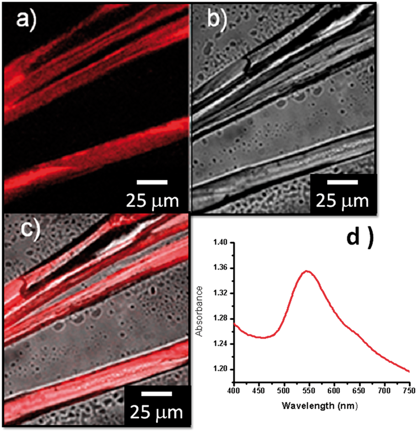
Silk fibers do not appear to influence the reduction process because there are no significant changes in infrared spectroscopy. Figure 4 shows a comparison between the silk substrate (spectrum 1) and gold nanocomposite (spectrum 2). The spectra have no significant differences and the functional groups are clearly representative of proteins. The peaks at 1621, 1512, and 1224 (connected at 1260) cm−1 are assigned to the amide I, amide II, and amide III bands of silk protein. The peak at 1444 cm−1 may be assigned to the carboxyl group in silk. Thus, biosynthesis of gold nanoparticles and their support on silk fibers appear to depend on the appropriate selection of the bioreducing agent and the concentration of the metal precursor.
FTIR studies of silk fibers (spectrum 1) and silk–gold nanocomposite (spectrum 2).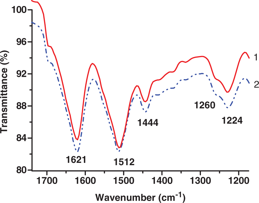
Conclusion
The green biosynthesis used in this work, using C. paradisi extract as a reducing agent, was shown to produce gold nanoparticles supported on silk fibers. Gold nanostructures supported on the silk fibers exhibit unique optical properties such as fluorescence and surface plasmon resonance. This work demonstrates that combining suitable biotemplates and bioreducing agents, the synthesis of organic–inorganic advanced nanomaterials can be easily achieved. Moreover, these novel nanomaterials may have a promising future in sustainable dyeing process in the textile industry as well as low-temperature catalysts.
Footnotes
Acknowledgements
We thank to Alicia Callejas, Ernestina Castro and Rosa Mouriño from Life Science Department-CICESE, Ensenada-Mexico for confocal microscopy technical support and advice. We also thank Francisco Ruiz from CNyN-UNAM for technical assistance with TEM measurements.
Funding
JPH acknowledges financial support from US Department of Agriculture (project USDA 2007-1065-01). Nolasco-Arizmendi is grateful to Mexico’s National Council for Science and Technology, CONACyT, for a Ph.D. scholarship. This work made use of the Microscopy facility of the Cornell Center for Materials Research (CCMR) with support from the National Science Foundation Materials Research Science and Engineering Centers (MRSEC) program (DMR 0520404).