
Abstract
The antibacterial activity of ZnO is reported by several authors. We present the preparation and application of inorganic–organic hybrid polymers modified/filled with ZnO nanoparticles of varying particle sizes. Inorganic–organic hybrid polymers employed here are based on 3-glycidyloxypropyltrimethoxysilane (GPTMS). ZnO is prepared by hydrolysis of zinc acetate in different solvents (methanol, ethanol or 2-propanol) using lithium hydroxide (LiOH ċ H2O). The hybrid materials prepared are applied to cotton (100%) and cotton/polyester (65/35%) fabrics. The antibacterial performance of these sol-gel derived hybrid materials is exemplarily investigated against Gram-negative bacterium Escherichia coli and Gram–positive Micrococcus luteus. Effects of particle size and concentration for the antibacterial performance are examined. Literature discusses various (active) species and processes responsible for the antibacterial action of ZnO. Therefore, particular attention is paid to investigate active species available in the described systems as well as to observe possible interaction between the nanoparticles and bacteria; the first results are presented.
The growth of bacteria on textile material during use or storage harmfully affects the textile or in case of clothes the person wearing the garment. By employing durable antimicrobial finishing of textiles using broad-spectrum biocides, on can limit at least the unpleasant and sometimes dangerous growth. Biocides or bacteriostatic agents employed can be divided into two types, organic or inorganic ones respectively. Organic biocides include organic acids or tertiary alkyl amines; a widely used bacteriostatic agent is triclosan, a polychloro phenoxy phenol.1,2 Although such compounds are widely used there are certain drawbacks as they are often less stable at higher temperatures and show a limited durability. Furthermore different organic biocides or bacteriostatic agents bear an allergic potential that should not be disregarded. Specifically, the organic compounds employed are discussed as being responsible for the spreading of resistant germs. Some organic biocides are pollutants and/or enrich in the environment. 1 Inorganic biocides are metals such as silver or copper and oxidic materials, e.g. ZnO, CuO, MgO or TiO2 3 . Silver is actually a widely employed antibacterial material not only used in textile finishing. 3
The inorganic materials are discussed as advantageous for several reasons. In many cases the mode of action is less specific which allows reducing a wide range of germs and prevents the development of resistances at least to a certain degree. Some argue that the increase of resistances is excluded for the antibacterial activity of silver. 4 The durability of metals or metal oxides is high compared with various organic alternatives and the harmful potential is often lower with metals or metal oxides. 3 Some of these metal oxides such as ZnO and TiO2 have been used in the production of cosmetics or personal care products, e.g. sun-creams. 4 Such materials are of interest for many further applications. As an example, ZnO, a semiconductor with a wide band-gap (3.36 eV), has achieved increased attention concerning possible electronic applications due to its unique optical, electrical and chemical properties. 5
Antibacterial properties of the inorganic materials are often explained by the release of ions interfering with bacteria 6 which has been reported in detail for silver ions. 4 In the case of titanium dioxide, the biocidic effect can be explained by the so-called photo-catalytic effect. The photo-catalytic effect is very complex; one of the main reasons for its antibacterial activity is in the fact that water or hydrogen ions respectively are reduced on the oxide’s surface producing hydroxyl radicals. 7 When these very reactive species interfere with organic material as well as with microorganisms they cause decomposition. Since these radicals are able to decompose nearly all organic molecules the development of bacterial resistances is highly improbable. The release of the active ions as well as the photo-catalytic effect is strongly dependent on the specific surface area of the materials used. The specific surface area is well known and is connected to the particle size of the materials; therefore, nanoparticles are of an enormous interest in this field. When the primary particle is smaller, the specific surface area is higher. 8
There are many studies investigating the antibacterial effect of ZnO nanoparticles. Sawai et al.,9–12, for example, reported that ZnO particles show antibacterial activity against both Gram-positive and Gram-negative bacteria. ZnO particles are known to be active even against spores that are high temperature and high pressure resistant. ZnO treatment influences the activity of the particles; high-temperature treatment of the oxide lowers the particle’s antibacterial activity.9–12 Yamamoto et al. 13 reported that the surface area and the concentration play an important role in the antibacterial activity, while the particle shape and crystalline structure have lower impact. Yamamoto et al. 13 also reported that the antibacterial activity increased as the particle size decreased (which necessarily leads to an increased surface area) 3 . Adams et al., 14 Brayner et al. 15 and Jeng et al. 16 reported that ZnO behaves in a different way towards microorganisms than towards other metal oxides such as SiO2, MgO, TiO2 and CaO.15–17 All observations reported by the researchers were explained by a number of mechanisms. These mechanisms include the formation of active oxygen species in the presence of the corresponding nanoparticles12,18,19 which damages the membrane cell wall because of the binding of the particles on the bacteria surface due to the electrostatic forces19,20 or the penetration of particles or ions through the cell membrane. 18
The study presented here is a part of an ongoing research project aimed at the development of a stable and highly durable multifunctional product for finishing textiles. We recently reported on the use of such inorganic–organic hybrid polymers filled with ZnO nanoparticles first focusing on a use as an ultraviolet protection finish for textile. 21 The investigations shown here prove that ZnO-containing hybrid materials are suitable to develop antibacterial textiles with excellent activities. This work describes the synthesis, characterization and application of appropriate ZnO nanoparticles. Furthermore, it specifies the investigation of the antibacterial activity as well as the experiments helping to understand the mechanisms responsible for the antibacterial action of our ZnO-based materials.
Experimental section
Materials
The coating experiments were carried out using plain-weaved fabrics made of scoured and bleached cotton (100%), with a mass per unit area of 250 g/m2, and blended fabrics made of polyester/cotton (65/35%), with a mass per unit area of 162 g/m2. Both materials were standard test materials supplied by WFK, Germany. Prior to application, the fabrics were purified by washing with warm water (40℃) and methanol. For the washing, 1 g/l of non-ionic detergent (Marlipal® O 80/13) was added. After washing, the fabric was rinsed with warm water and dried, acclimatized for 24 h (22℃, 65% rel. humidity) and weighted before using.
Chemicals
Zinc acetate dihydrate (ZnAc), lithium hydroxide monohydrate (LiOHċH2O, 98%), methanol, ethanol and 2-propanol were obtained from Merck. 3-Glycidyloxypropyltrimethoxysilane (GPTMS, 98%) was obtained from ABCR. Further chemicals used were fluorescein isothiocyanate (FITC, Aldrich, 90%), catalase from bovin liver, (activity ≥ 2000units/mg from Sigma-Aldrich, Germany) and 2,2-diphenyl-1-pikrylhydrazyl (DPPHċ, Aldrich, 98%). For catalyzing the cross-linking reaction of the epoxy group of the GPTMS 1-methylimidazole (97%, Fluka) was used. All chemicals were used without further purification. Lysogeny broth (LB broth) was obtained from Sigma-Aldrich, Germany.
Microorganisms
Escherichia coli DSMZ498 and Micrococcus luteus ATCC-No. 9341 were incubated overnight at 37℃ in a nutrient broth (LB broth medium: 20 g LB broth/liter water). The cultures obtained were diluted with autoclaved nutrient broth to obtain cell suspensions which were adjusted to a final working concentration.
Preparation of zinc oxide nanoparticles
The preparation procedure employed has been described in previous work 21 and is a variation of a preparation described by Spanhel et al. 22 A two-neck round bottom distillation flask was used to suspend 2.8 g of (ZnAcċ2H2O) in 100 ml of the particular solvent (methanol, ethanol or 2-propanol) by reflux heating for 3 h. 0.75 g of lithium hydroxide was dissolved in 100 ml of solvent at room temperature by magnetic stirring. The ZnAc suspension was cooled to 0℃ before the lithium hydroxide solution was added by drops under vigorous stirring. The mixture was treated in an ultrasonic bath (SONOREX TK 52H) at room temperature for about 2 h. The resulting sol theoretically contains 0.675 wt.% ZnO. Higher amounts of ZnAc and lithium hydroxides were used (using constant amounts of solvent) to prepare colloidal solutions of higher concentration.
Preparation of zinc oxide nanoparticles labeled with FITC
ZnO prepared as described above had been centrifuged and washed, before 0.1 g were resuspended in 100 ml 2-propanol. 20 mg of FITC were added to this solution before it had been stirred for 1 h. Subsequently 100 µl of the resulting ZnO/FITC solution was added to 50 ml of an aqueous solution containing E. coli cultures (108 colony forming units [CFU]/ml) which were grown overnight at 37℃ in a nutrient broth medium (LB broth medium). A control solution of the same concentration was prepared in parallel. FTIC is expected to link chemically to ZnO and is therefore used to label the nanoparticles. The dye FTIC shows a distinct green fluorescence which allows a visualization of the labeled nanoparticles in the fluorescence microscope. Furthermore, the attachment of the labeled particles to the bacteria can be visualized. The approach is employed to prove an occurring of the attachment of the ZnO nanoparticles to the bacteria surface. If it occurs, a decomposition of the bacteria cells by time can also be visualized. The stained bacteria were observed using a microscope (KEYENCE all-in-one-Type Fluorescence Microscope, BZ-Analyzer 8100E, Japan).
Preparation of the GPTMS-sols
10 ml of GPTMS were dissolved in 100 ml of solvent before hydrolyzation using 0.01 M hydrochloric acid. The resulting sol was stirred for at least 3 h to form the basis sol.
Preparation of the hybrid polymer
Before the finishing of the textiles, the zinc oxide dispersion and the GPTMS-sol were mixed in different ratios and finally 1-methylimidazol (0.5 ml/10 ml GPTMS) was added as a catalyst for the cross-linking reaction of the epoxy group of the GPTMS. The labeling used in the text gives the amount of ZnO in relation to GPTMS, therefore 10% ZnO means 1 g ZnO per 10 g GPTMS.
Characterization of the ZnO nanoparticles
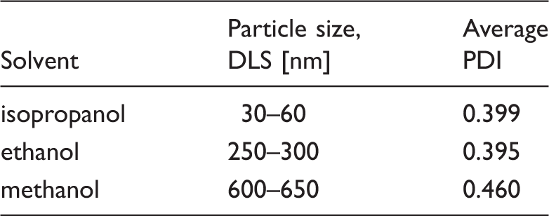
The size of the ZnO nanoparticles was measured by dynamic light scattering (DLS), using a Zetasizer, Nano-S, produced by Malvern. For all samples high polydisperity index (PDI) values in the range of 0.4–0.5 were calculated indicating broad dispersions. Samples were only analyzed and used for further investigations if the size quality report of the calculation showed no errors.
Coating process
The hybrid polymer sol was applied to the fabrics by a pad–cure–method. The coating was carried out by a padding process with a laboratory padder equipped with roles with a standard rubber hardness of 70° Shore (Mathis, Switzerland). The nip-pressure was adjusted to a value guaranteeing a wet pick up of 100%; the role speed was 4 m/min. After padding, the samples were dried in a labcoater (Mathis, Switzerland) at 130℃ for 30 min before they were washed to remove residual by-products.
Inductively coupled plasma–optical emission spectrometry
The amount of ZnO or zinc respectively was investigated by inductively coupled plasma–optical emission spectrometry (ICP–OES). Coated cotton samples were treated with concentrated nitric acid (65%) before the decomposition of the material was carried out at 180℃ in a microwave digester (MarsXpress, CEM, Kamp-Lintfort, Germany). The resulting solutions were completely transferred in volumetric flasks and diluted to a defined volume with distilled water. ICP–OES measurements were carried out with a Varian 720-ES.
Scanning electron microscopy
For qualitative assessment, the samples were studied by scanning electron microscopy (SEM). The micrographs in Figure 1 were prepared using a Topcon-Microscope (ATB-55) those in Figure 6 using a Hitachi S-3400 Type II (Tokyo, Japan).
SEM micrographs of a blank cotton fabric (upper micrograph) and a cotton fabric treated with a GPTMS-sol modified with 10 wt.% ZnO particles (size 30–60 nm) (lower micrograph).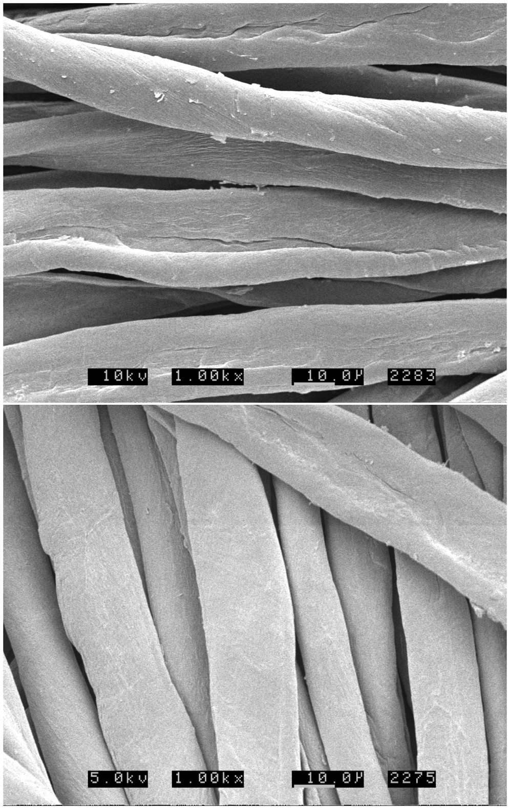
Antimicrobial testing
The antimicrobial activity of the coated fabrics was tested against the Gram-negative bacterium E. coli and the Gram–positive M. luteus using the following qualitative as well as quantitative tests:
zone of inhibition test (disc diffusion test); American Association of Textile Chemists and Colorists (AATCC) Test Method 100-2004; tetrazolium/formazan test (TTC).
The zone of inhibition method (disc diffusion test) has been employed as a method to evaluate the antibacterial activity of textiles. The test is carried out according to EN ISO 20645:2004. Sterilized agar plates are prepared before a textile sample to be investigated is placed onto the surface. Afterwards the agar plate is inoculated and placed in an incubator for 24 h at 37℃. The pure agar medium will be overgrown by the bacteria, if antimicrobial substances diffuse into the agar medium growth will be prevented which can be observed by a halo (zone of inhibition). The diameter of the halo can be used as a measure of the antibacterial activity.
Modified AATCC test method 100-2004: the purpose of the AATCC test method is to “provide a quantitative procedure for the evaluation of the degree of antibacterial activity” of finishes on textile materials.
12
The method is based on an inoculating test and was modified for the experiments presented; the procedure valid here is as follows. Textile samples (diameter 6 cm) are placed in a flask sealed with aluminum foil before sterilization. In the next step, 3 ml of inoculum (1-2*106 CFU/ml) are added. Samples are taken immediately after the addition and after 24 h. Samples were taken by the addition of 100 ml 0.9% NaCl before shaking vigorously for 1 minute, followed by extracting 1.5 ml of the solution, and applying 500 µl to agar plates in logarithmic dilutions 0, 1, 2 (except the blanks after 24 hours, which were plated with up to six logarithmic dilutions since stronger growth was expected). The plates were incubated at 37℃ for 48 hours, after which they were removed from the incubator and any formed colonies were counted. After that the number of bacteria was counted and the reduction of bacteria, R, was calculated as follows:
9
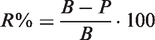
R = reduction in percentage
B = CFU/test sample in blank
P = CFU/test sample for treated samples
Tetrazolium/formazan test (TTC): in the presence of bacteria, TTC is reduced to red formazan. 23 The red formazan obtained therefore indicates the activity and viability of the cells. Therefore, the TTC test method is considered as a fast method for evaluating the antibacterial activity of fabrics. To complete this test, circular swatches of test fabrics of a diameter 3.8 ± 0.1 cm were cut into a small size. The number of swatches to be used was six. Both, the test and control swatches, were sterilized at 110℃ before incubation. All swatches were placed in a 40 ml nutrient broth medium containing 10 µl an E. coli solution (108 CFU\ml) as an inoculum volume. All flasks were incubated while shaking at 37℃/200 rpm for 3 h; then, 1 ml from each flask containing the fabrics to be tested was added to the sterilized test tubes containing 100 µl TTC solutions (0.5% w/v). All tubes were incubated at 37℃ for another 20 min. The resulting formazan was centrifuged at 4000 rpm for 3 min followed by decantation of the supernatants. The pellets obtained were dissolved in ethanol and centrifuged again. The red formazan solution obtained at the end which indicated the activity and viability of the cells was measured by a photometer at 480 nm.
High-performance anion exchange chromatography–pulsed amperometric detection measurements
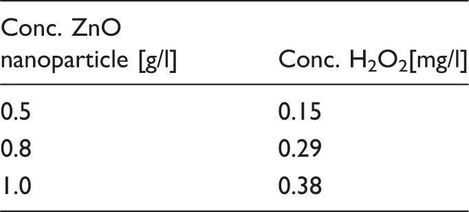
The generation of H2O2 from ZnO were detected using a high-performance anion exchange chromatography–pulsed amperometric detection (HPAEC–PAD) system from DIONEX (Idstein, Germany). The system was equipped with a Carbo Pac PA 1 column and 0.5 M NaOH acted as eluent. Calibration was carried out in a range from 0.2 down to 0.0012 g H2O2/l. The calibration curve is the average area of three repeated measurements. For measuring the amount of H2O2 formed in ZnO dispersions, different amounts (0.5, 0.8 and 1 g/l) of ZnO particles (30 nm) were suspended in water before the resulting dispersions were injected.
Laundering
A single laundering was performed in a laboratory washing machine (Linitest, ATLAS in accordance to DIN EN ISO 105-C06 using a non-ionic surfactant (Marlipal® O 80/13), 40℃). All textile samples were washed once before the different investigations were carried out.
Results and discussion
Particles size of ZnO nanoparticles prepared in different solvents measured by DLS
After preparation of ZnO nanoparticles, particles dispersed in the mentioned solvents were mixed with a freshly prepared GPTMS-sol. The resulting sol was applied to textile materials by a padding process. After drying and curing all samples were laundered before further experiments were carried out. To investigate the homogeneity of the coating and to exclude the occurrence of undesired agglomeration of the ZnO nanoparticles SEM investigations were carried out. The corresponding SEM micrographs are shown in Figure 1. Compared to the blank sample, the treated fiber sample shows a very smooth, less structured surface. Big agglomerates are not observed indicating a homogeneous distribution of ZnO in the coating layer.
Evaluation of antibacterial activity for ZnO-treated fabrics
As mentioned before, fabrics are excellent media for the growth of microorganisms when the basic requirements such as nutrients, moisture, oxygen and appropriate temperature are available. Natural fibers like cotton are more susceptible to microbial attack than synthetic fibers. Antibacterial activity of cotton and cotton/polyester blended fabrics coated with hybrid polymers containing ZnO nanoparticles were evaluated to assess the effectiveness of ZnO nanoparticle coating. Antibacterial activity is evaluated qualitatively and quantitatively.
Results for cellulosic cotton fabrics are shown as exemplarily for the evaluation of the antibacterial activity of textiles finished with hybrid polymer coatings prepared with ZnO-sols. The hybrid polymers were either prepared with ZnO-sols of various concentrations of particles of a given size, or with widely differing particle sizes of a given concentration. Antibacterial activity was tested against E. coli and M. luteus on agar plates by evaluating the resulting zone of inhibition (area of halo). The results of the zone of inhibition test for a sample coated with a hybrid polymer loaded with different amounts of ZnO particles of a given particle size are shown in Figure 2 (left photograph). The test is carried out against M. luteus and it can be observed that the blank sample (1) and the one coated with the hybrid polymer without ZnO (5) yields no antibacterial effect. Samples modified with ZnO yield a distinct activity which increases with the increasing amount of ZnO. We investigated the amount of zinc oxide deposited on corresponding samples by ICP-OES supplementary. The values measured for zinc are ranging from 8 g/kg fabric (for sample “10% ZnO”) to 28 g/kg fabric (for sample “50% ZnO”). The values increase linearly (R2 = 0.99642) increasing the amount of ZnO particles added to the GPTMS-sol. Figure 2 (right photograph) shows the results for the hybrid polymer coatings of the same amount of ZnO with varying particle sizes. As can be clearly seen, when the particle size is decreased, the antibacterial activity increases. These tests are carried out with E. coli.
Disc diffusion tests with M. luteus (left) and E. coli (right). Samples in the left dish were coated with the hybrid polymer loaded with ZnO particles of a given size distribution (30–157 nm) and various concentrations. (1) untreated, (2) 10% ZnO, (3) 30% ZnO, (4) 50% ZnO, (5) treated with GPTMS-sol only. Samples in the right dish were coated with the hybrid polymer loaded with ZnO particles of a given concentration (10% ZnO) and various size distributions, (1) untreated, (2) 600–650 nm, (3) 250–300 nm, (4) 30–60 nm. (The relative share of ZnO is always calculated as wt.% in relation to GPTMS in the sol.)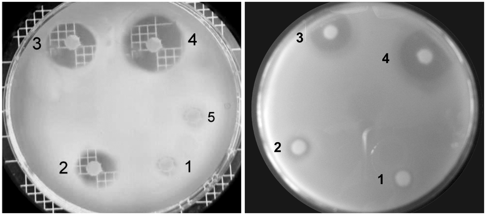
In general, from Figure 2 it can be concluded that the untreated fabrics as well as the fabrics treated with only GPTMS had no antibacterial activity. An antibacterial activity can be observed when fabrics are treated with the hybrid polymer modified with ZnO nanoparticles. Activity of treated fabrics increases with increasing nanoparticle concentration as well as with decreasing particle size. Both variations mean an increase of the active surface area of ZnO and therefore an increased biocidal effect.
For a given set of experiments, either by increasing the amount of particles of a given size or by decreasing the particle size at a given mass of particles the active of ZnO surfaces increase which seems to be the reason for an increased activity.
In further experiments, antibacterial activity of the coated fabrics was determined for E. coli and M. luteus according to the AATCC 100-2004 standard method. Following a slightly modified AATCC Test Method 100-2004 (see the experimental section)
12
the antimicrobial activity of the treated fabrics was evaluated in several test series. Qualitative results for the antibacterial activity of cellulosic fabrics coated with the ZnO-modified hybrid polymers are presented in Figures 3 and 4. As expected, no reduction of the bacteria E. coli and M. luteus was detected for the untreated fabrics. On the contrary, there was a decrease in the number of bacteria counted after 5 h incubation (compared with 0 h contact time). As shown in Figure 3, a cotton sample coated with a GPTMS-sol loaded with ZnO (10%, 30–60 nm) leads to a 98.8% reduction of E. coli after 5 h and a 97.3% reduction of M. luteus.
Reduction rate of E. coli and M. luteus versus time for cotton fabrics treated with a GPTMS-sol modified with ZnO nanoparticles (10% ZnO). Reduction rate of E. coli and M. luteus after 5 h in the presence of cotton/PET blended fabric (65/35%) treated with the GPTMS-based hybrid polymer loaded with 10% ZnO nanoparticles of different particle sizes.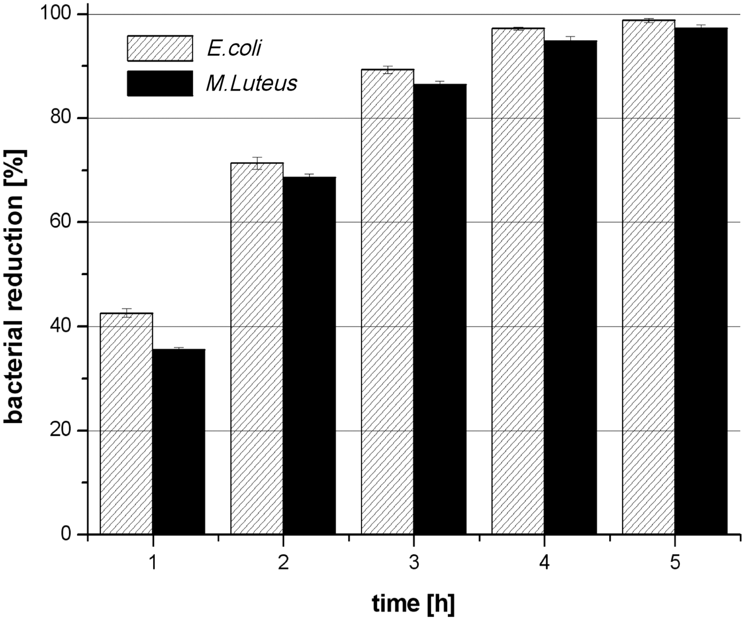
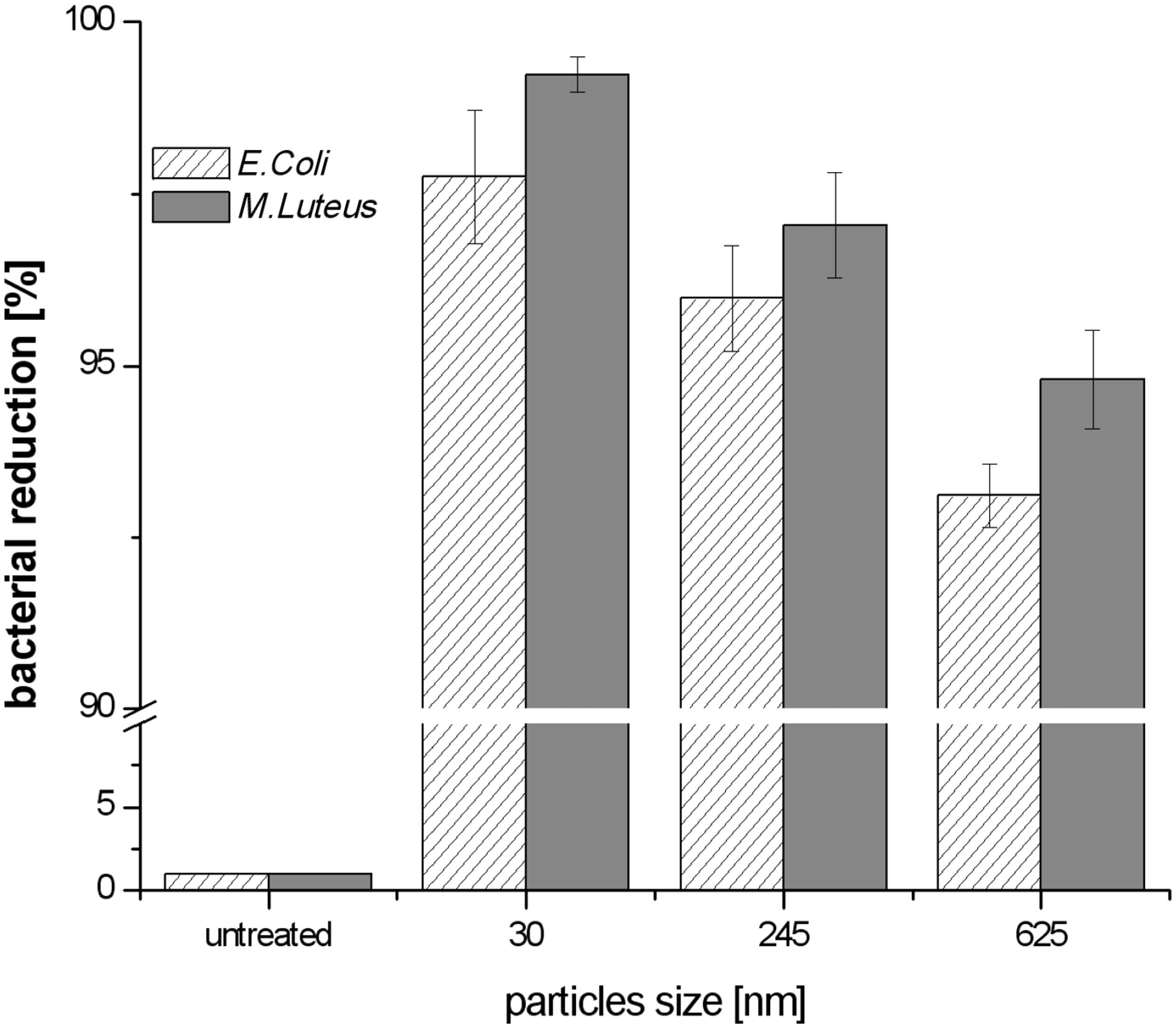
The same measurements were carried out to evaluate the antibacterial activity of cotton/polyester blended fabrics coated with the hybrid polymer modified with a constant mass of ZnO but with a varying particle size. Measurements were performed against E. coli and M. luteus. As shown in Figure 4, treatment of the bacteria for 5 h with the fabric coated with the hybrid polymer loaded with ZnO particles of the smaller particle size (30–60 nm) leads to a complete reduction of the E. coli and M. luteus populations, while fabrics coated with hybrid polymers loaded with bigger ZnO particles (600–650 nm) achieve a 93% and 97% reduction of the E. coli and M. luteus populations, respectively. The results for ZnO particles in the range of 245 nm (PDI = 0.456) are in between.
A further quantitative investigation of the antibacterial activity of fabrics coated with the hybrid polymer modified with different amounts of the active ZnO particles – with a given particles size in the range of 30–60 nm– has been carried out using the TTC test. Details about this method were described in the experimental part. This test serves as indicating the system for the determination of the viability of microorganisms. The absorbance of formazan that is formed in the presence of vital microorganisms is directly proportional to the amount of living bacteria. Corresponding data shown are in good agreement with the results shown before. We observe a strong reduction of the vital cells due to the presence of ZnO-modified textiles. Raising the relative amount of ZnO in the hybrid polymer increases the resulting antibacterial activity. A concentration of 10 wt% ZnO in the coating yields a reduction of more than 75% within 3 h; an increase of this concentration to 50 wt% improves this value to approximately 90%.
To follow the reduction of bacteria in the presence of ZnO, visually particles were labeled with the fluorescence dyestuff FITC. After combining labeled ZnO particles (30–60 nm) and an E. coli solution, the particles cover the bacteria surface (as described by Wang et al.
24
). This allows a visualization of the particle-covered bacteria in the fluorescence microscope as can be seen in Figure 5 (photograph labeled 0 h). Further microscopic images taken after repeated sample taking over four hours prove the drastic reduction of the number of bacteria. The reduction in the number the fluorescing ZnO-covered bacteria observed on the images indicates that the bacteria are more or less completely decomposed. To verify this observation samples were taken from the same bacteria solution after 4 h and bred on agar – no colonies were grown proving the absence of vital bacteria.
Upper row: comparison of microscopic investigation (visible light and fluorescence) of bacteria broth with and without FITC. Proving that a staining of the bacteria itself with FITC cannot be observed, neither in visible nor in fluorescence mode. Second and third row: cell imaging (fluorescence light) with FITC-modified ZnO nanoparticles. Pictures show the solution after up to 4 h after adding the ZnO to the bacteria broth. SEM micrographs of (a) blank M. luteus culture, (b) M. luteus culture treated with ZnO nanoparticles (30–60 nm) after 1 h, (c) M. luteus culture treated with ZnO after 2 h, (d) M. luteus culture treated with ZnO after 3 h.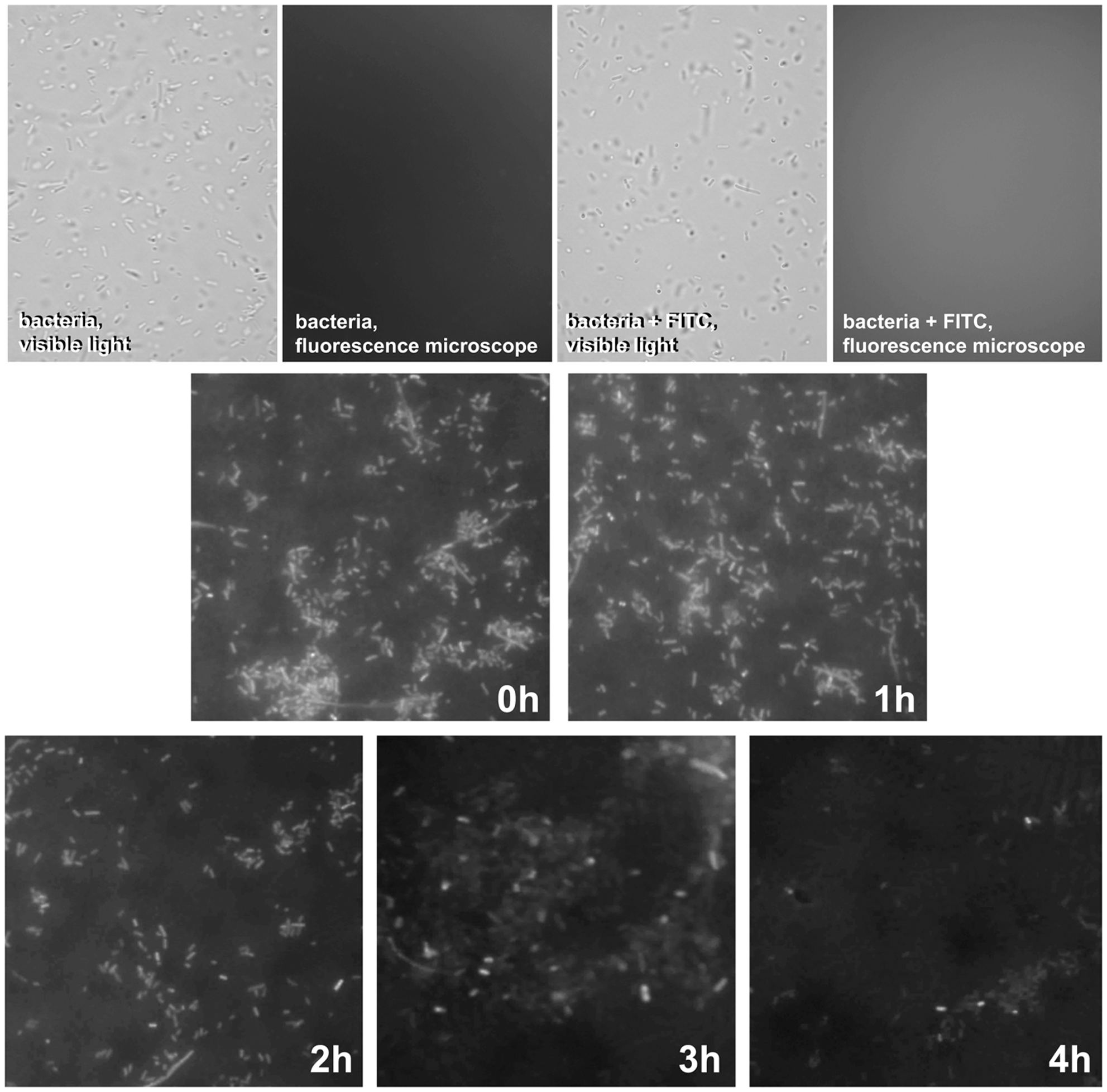
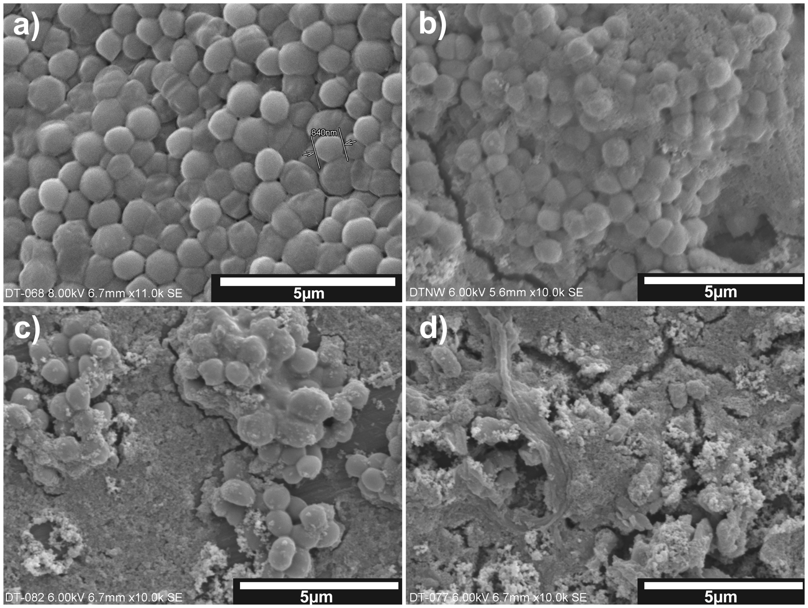
Further investigations aiming on the visualization of the bacteria – ZnO nanoparticle interactions were carried out by SEM analyses. SEM analyses were performed to investigate morphological changes of M. luteus in the presence of ZnO particles (30–60 nm). We mixed 50 µl of the M. luteus culture with autoclaved LB broth medium and ZnO nanoparticles dispersion. Cultures were grown at 37℃ under agitation. Sample of these solutions were taken, placed on a support, and dried in a vacuum before the investigation by SEM.
Figure 6(a) shows cultures of bacteria not treated with ZnO. The cell radius is in the range of ∼840 nm and therefore much larger than the ZnO particles employed for the following samples. Comparing this micrograph with the one showing bacteria that were in contact with the nanoparticles one observes a coverage of the bacteria and a distinct lumping (compare to Figure 6(b)). By the time, a reduction of the number of bacteria can be observed as shown in Figure 6(c) and (d), supporting the results of the fluorescence microscopy.
Investigation of the effects responsible for the antibacterial action
The results presented above show that if bacteria and zinc oxide particles are available in an aqueous medium there will be coverage of the bacteria with the oxide particles; obviously a lumping occurs to a certain degree and the number of intact bacteria is drastically reduced within hours. Electrostatic interaction between the surface charge of ZnO particles and the membrane charge surface of bacteria could contribute to the antibacterial behavior of the ZnO dispersions. It has been reported that the point of zero charge for ZnO is at a pH of 9.0. 25 Since the pH of all solutions employed here is 7.0, a positive charging of the particle’s surface is expected while the surface of the bacteria is expected to be negative. The observations described are in good agreement with the observations shown above, e.g. in Figure 6(b). Certainly this does not explain the mechanisms that are responsible for the antibacterial activity of the ZnO-modified hybrid polymer coatings that were applied to the textiles. The particles immobilized on the textile surface will not be available for covering the bacteria. The halos in the tests described before prove that there must be a mechanism that does not need a direct contact between ZnO particles and the bacteria. In the following paragraph, experiments are described that were carried out to investigate possible explanations for the antibacterial properties. It should be noted that antibacterial activity of ZnO in our work has been studied in a normal laboratory environment and without ultraviolet (UV) light, which might be important since ZnO might show a photo-catalytic activity.
To exclude the possibility that zinc ions are released into the aqueous ZnO suspension were responsible for the antibacterial activity, control experiments were carried out by dissolving a zinc salt (zinc acetate, 0.045 M) in water. Tests were carried out for antibacterial activity against E. coli and it was found that under the conditions used in this study, zinc (II) ions are not responsible for the strong antibacterial activity observed for the ZnO nanoparticle systems.
Amount of H2O2 generated from ZnO nanoparticles (30–60 nm)
Effect of catalase for the antibacterial activity of ZnO dispersions

(+): distinct antibacterial activity is observed
(−): no antibacterial activity observed
To support that H2O2 is in fact involved in the antibacterial activity, antibacterial activity of ZnO dispersion in presence of catalase was tested. Catalase is an enzyme that decomposes hydrogen peroxide to water and oxygen. We found that the antibacterial activity decreased when catalase is present in the solution (cp Table 3) which is a strong hint supporting the explanation that hydrogen peroxide is at least jointly responsible for the antibacterial properties of ZnO.
Another suggested mechanism responsible for the antibacterial activity of ZnO nanoparticles is the generation of oxy- or hydroxyl radicals. If available, these radicals will be able to be involved in the decomposition of the cell membrane, for example. For proving the existence of radicals 2,2-diphenyl-1-pikrylhydrazyl (DPPH Relative absorption of aqueous ZnO dispersions containing a given mass of ZnO with varying particle sizes. Initial dye concentration was 20 mg DPPH per 100 ml dispersion. Changes occurring in ambient light have been observed for up to 90 min by measuring absorbance. Absorbance was measured at the maximum of aqueous DPPH solutions, which is at a wavelength of 514 nm.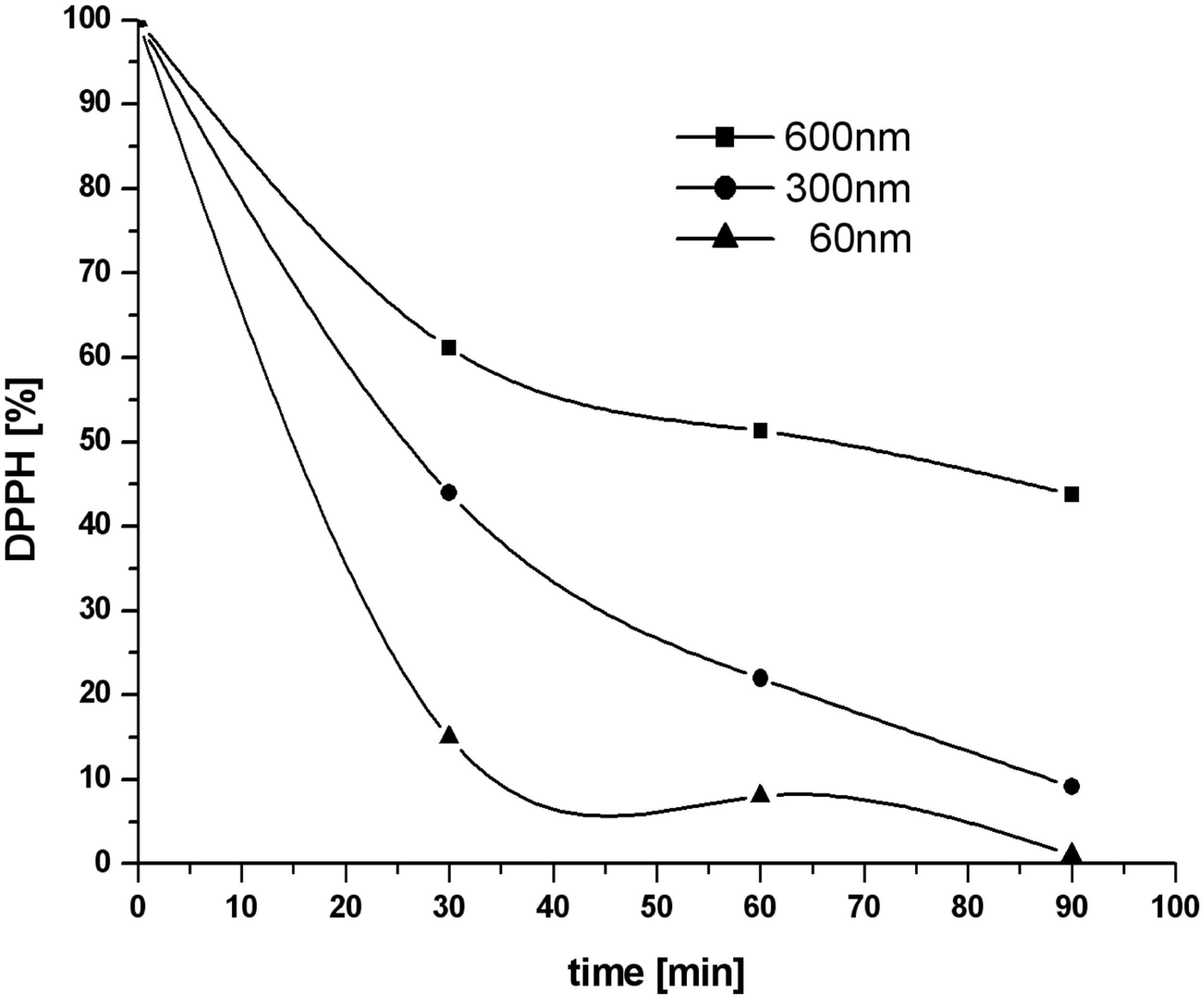
Conclusion
Nano sized zinc oxide particles were successfully synthesized, applied and added to inorganic–organic hybrid polymer sols. The resulting hybrid materials can be applied to textile materials by a simple pad–dry–cure method – here shown as exemplarily for cotton (100%) and polyester/cotton (65/35%) fabrics.
The resulting textile materials exhibit antibacterial activity. The antibacterial performance of fabrics coated with different amounts of active ZnO particles or different particle sizes was investigated respectively. The antibacterial activity of textiles treated with ZnO nanoparticles increases when the amount of nanoparticles deposited is increased and it increases with decreasing particle size. The enhanced bioactivity of smaller particles is attributed to the higher surface area to volume ratio; up to 98.8% of E. coli and 97.3% of M. luteus were killed within 5 h by the best samples prepared for this report.
Further experiments were carried out to explain the mechanism responsible for the antibacterial action. It was proven that the activity is not due to a release of zinc ions. Results showed that hydrogen peroxide and/or radical species are developed by the ZnO-modified hybrid polymer and may contribute to the antibacterial activity. SEM analyses show that direct interaction between ZnO particles and the membrane surface of bacteria may lead to extensive coverage of the bacteria followed by a lumping which obviously also contributes to the observed decomposition of the bacteria.
The results are very promising since the antibacterial activity of the textile material is comparably high; ongoing work aims at a combination with other antibacterial additives (e.g. biopolymers) to further improve the performance.
Footnotes
Acknowledgment
We thank Prof Dr Nickisch-Hartfiel, HS-Niederrhein, for her kind support in carrying out the antibacterial tests.
Funding
The authors wish to convey a very special thanks to the Egyptian Missions Department for giving Asmaa Farouk the opportunity and the financial support to pursue a PhD degree in Germany. Further financial support by the Ministerium für Innovation, Wissenschaft, Forschung und Technologie des Landes Nordrhein-Westfalen (Department of Innovation, Science, Research and Technology of the state of Nordrhein-Westfalen) is also gratefully acknowledged.