
Abstract
Viscose non-woven was treated by NH3 plasma. Different exposure times were used in order to find the optimum conditions for simultaneously improved hydrophilicity and antimicrobial activity, as desired effects by wound dressings. Both chemical and morphological modifications were studied by X-ray photoelectron spectroscopy and atomic force microscopy, respectively, revealing functionalization with nitrogen groups as well as formation of rich morphology at sub-micrometer scale. The wetting rise curves increased from 0.04 g2 s–1 for non-treated material to 1 g2 s−1 after prolonged treatment. The water contact angle decreased almost linearly with treatment time from 90° for non-treated samples to about 40° for samples treated for 140 s and remained rather constant thereafter. The AATCC 100–1999 standard test revealed reduction on all used bacteria, more pronounced for Gram-negative, that is, E. coli and P. aeruginosa, then for Gram-positive, that is, a significant for S. aureus and a marginal for E. faecalis.
The use of textiles in wound care has a long tradition. Viscose, in the form of woven or non-woven textiles, is mostly used. Although the sorption capacity, as well as the wettability rate, of viscose is much better than for many other similar materials (e.g. lyocell, modal) these properties are still not optimal and definitely below the capacities of alginate. Various techniques,1–3 including chemical processes,4–7 are usually used to improve cellulose absorption properties. However, such chemical processes often degrade the mechanical–technological properties of treated materials.
The use of low-temperature gaseous plasma is an environmentally friendly alternative. Such a medium is found in the thermodynamically non-equilibrium state of gas.8–11 Charged particles are accelerated in electrical fields and gain certain kinetic energy. Plasma is created by different electrical discharges but electrode-less high-frequency discharges are very popular since they allow for production of huge numbers of neutral excited reactive particles, such as radicals, neutral atoms and molecules excited to both electronically and vibrational excited states. 12 Light particles (electrons) can follow changes in the local electrical field in such discharges so they gain kinetic energy well above the energy of random movement at room temperature, but heavy particles (neutral as well as positively and negatively charged atoms and molecules) cannot, providing the electric field frequency is more than about 1 MHz. The electrons can transfer only a negligible amount of their kinetic energy to heavy particles kinetic energy so they remain essentially at room kinetic temperature. Neutral atoms are often stable in the gas phase at reasonably low pressure (below a few mbar) and with the absence of metallic electrodes they are not lost by heterogeneous surface recombination so the dissociation fraction of gaseous molecules at room kinetic temperature may reach several percent, if not more.8–10 The reason for good stability of atoms at low pressure is a low probability for three body collisions that can only lead to recombination of atoms to a parent molecule in the gas phase. The particles created in plasma are chemically extremely reactive so such a medium is suitable for modification of surfaces, including functionalization of organic materials and formation of nanostructured materials13–16 without influencing the bulk properties of solid materials.
In the past, the traditional theory has always been that wound should be kept dry so that a scab may form over the wound: the wound should be exposed to the air and sunlight as much as possible. The clear disadvantages of these principles are that the scab, which is made up of the dehydrated exudate and drying dermis, is a physical barrier to healing which is then delayed, because the epidermal cells cannot move through the scab formed. Exposure to air reduces the surface temperature of the wound, causing peripheral vasoconstriction affecting the flow of blood to the wound, which further delays healing. In 1962, Winter 17 was able to show that the wounds healing under moist conditions healed faster than the wounds under dry conditions, open to air.
The removal of excessive exudate from the wound without allowing the wound to dry out, thereby maintaining a moist environment alone, is an insufficient benefit in modern wound dressings. Additional properties promoting wound healing are required, for example the ability to prevent infection. Controlling bacterial or fungal growth on fabric can be achieved using biologically active polymers,18–24 by binding drugs onto the polymers25,26 or by introducing various functional groups.18,19,27–29 It has been found that the quality and quantity of protonated amino groups mainly influence the antimicrobial properties of functionalized fiber-oriented polymers.30,31 Many metal ions have antimicrobial activity,32–34 but are either very toxic or not safe for patients35–38and the environment.
Based on the above considerations, the wound infection control in the wound environment would be a bonus, and wound dressings possessing both properties would be of benefit. Most available procedures combine different functionalization steps one after another, that is, for improving sorption1–7 and antimicrobial properties.18–29 In addition, more than 400 individual advanced hydrophilic wound dressings, including 25 alginates, 55 foams, 50 hydrocolloids, 51 hydrogels, 24 transparent films 39 and dressings that contain and release antimicrobial agents at the wound surface, exist in the marketplace. 40 Still, little literature has been published addressing a one-step procedure resulting at the same time in an excellent adsorption and bacterial load management, which leads to longer wear time of dressings and faster patient’s wound healing.
The present study aims at examining effects, often not simultaneously present, of environmentally friendly treatment on hydrophilicity and antimicrobial activity, trying to define the practical balance between high-hydrophilicity and antimicrobial activity. It is known that surface composition41,42 and morphology after applied plasma could be altered depending on used gases and treatment duration. 43 Baring this in mind, viscose non-woven fabric was exposed to ammonia gas at different time intervals. The surface properties were analyzed by X-ray photoelectron spectroscopy (XPS), while morphological changes were determined by atomic force microscopy (AFM). The effect of plasma modification on hydrophilicity was evaluated by water rise monitoring and contact angle, while the antimicrobial activities were determined by AATCC 100-1999 standard test. 44
Experimental details
Materials
A cellulose material, as regenerated cellulose fibers (CV) in its non-woven form was used, as kindly supplied by KEMEX, The Netherlands, a leading European and world supplier and manufacturer of medical non-wovens and laminates. The fibers used in the non-woven form were of medical grade. The non-woven fabric was produced by needle-punching technology, that is, the web of fibers is bonded by means of needles. The surface mass of the non-woven fabric was 175 g/m2 (SIST ISO 3801) and the thickness under ambient conditions was about 1.7 mm (SIST EN ISO 5084). The non-woven material was used as it was received; no additional pre-treatment cleaning was performed.
Treatment procedure
Ammonia plasma treatment
A vacuum dried (20℃, 100 mbar, 24 h) reference viscose sample (CV) was cut to pieces of 3 cm × 12 cm and treated by non-equilibrium plasma created in NH3 gas in a reactor made from borosilicate glass. The reactor was a 60-cm long cylindrical tube that was connected to the gas inlet system on one side, while on the other side it was pumped continuously with a two-stage rotary vane vacuum pump with the ultimate pressure well below 1 Pa and a nominal pumping speed of 0.022 m3s−1 constant in a range of pressures from 1 to 104 Pa. A rather powerful pump allowed for fast removal of gaseous molecules desorbed from the surface of the samples. NH3 gas was leaked into the system from a high-pressure container through a manually controlled leak valve. Continuous leakage on one side, as well as pumping on the other one, allowed for establishment of the pressure of about 150 Pa inside the plasma reactor. The pressure gradient along the reactor was almost negligible due to a large ratio between the conductance of the glass tube at this pressure and for this particular gas, and the nominal pumping speed. The inner diameter of the plasma reactor was 3.6 cm and the estimated gas drift velocity along the tube was calculated from known parameters as
Plasma parameters determination
Plasma parameters were determined using an electrical probe, mass spectrometry and optical emission spectroscopy.
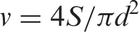
A simple double electrical probe was immersed into the plasma reactor in order to estimate the density of charged particles. The probe was kept at a floating potential, since the biasing of electrodes was realized using a battery instead of a grounded direct current (DC) power supply. Floating conditions are necessary in order to avoid high-frequency interferences that would otherwise make measurements impossible. The diameter of the probe electrodes was 1.5 mm in order to meet conditions where the Debye length is at least an order of magnitude smaller than the probe diameter. Such a configuration allowed for rather reliable measuring of the density of charged particles. At the pressure of 150 Pa and the power of 100 W the density of charged particles as determined using the electrical probe was about 5 × 10¹⁵ m−3. The electron temperature has been estimated from the I = I(UB) characteristics of the double probe as well and was about 35,000 K. Here, I (A) represents the measured electrical current through the double probe circuit and UB (V) is the biasing voltage – the voltage applied between the electrodes. A simple electrical circuit was used to bias electrodes to different voltages up to of the voltage supplied by the battery. Such a density and energy of electrons allow for extensive dissociation of NH3 molecules. The optical spectrum of such plasma after 30 s of plasma treatment is presented in Figure 1 and it reveals a high concentration of excited hydrogen atoms. The fact that atomic nitrogen lines are absent does not mean the atoms in the ground state do not abound, but is rather due to high excitation energy of nitrogen atom radiation states. The accuracy of the low-resolution spectrometer Avantes AvaSpec 3648 is not great at about 0.5 nm, but since only few spectral features appear in the spectrum it is sufficient for this sort of experiment. Furthermore, the technique is qualitative so no attempt was made to calibrate the instrument for spectral response prior to measurements. The integration time was set to 5 s. In order to quantify the dissociation of NH3 molecules in the plasma, mass spectrometry was applied in addition to optical emission spectroscopy.
A typical optical spectrum of plasma created in NH3 gas at the pressure of 150 Pa and radio frequency power of 100 W.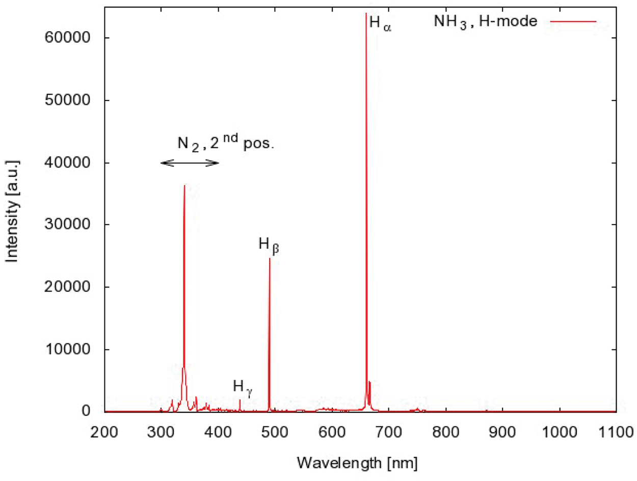
A differentially pumped mass spectrometer was attached to the plasma reactor. We used a commercial residual gas analyzer Pfeiffer PrismaPlus QMG 220. A rather powerful turbomolecular pump with the nominal pumping speed of 0.5 m3 s−1 was used to achieve suitable vacuum in the mass spectrometer. A narrow glass tube was mounted between the reactor and the spectrometer so the gas pressure in the spectrometer chamber was about 3 × 10⁻³ Pa when the pressure in the discharge chamber was 150 Pa. Mass spectra were acquired in the range up to 35 amu. Typical spectra for gas in thermal equilibrium and gas transformed into the plasma state are presented in Figure 2.
Two mass spectra of NH3 gas in equilibrium state (a) and in plasma state (b).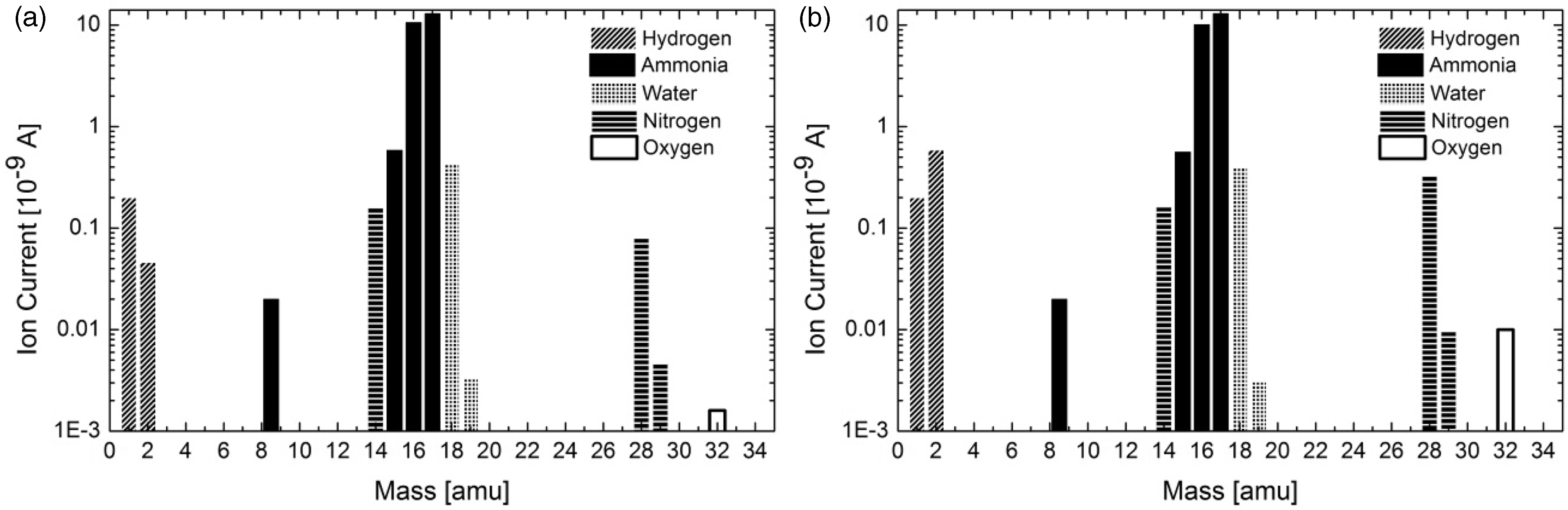
As expected, the masses at Z = 17 and 16 prevail for NH3 gas in the equilibrium state, and the next highest mass at Z = 18 is attributed to water vapor, which represents the residual atmosphere in the differentially pumped chamber where the mass spectrometer is mounted. Hydrogen and nitrogen peaks observed in the spectrum of equilibrium gas are artifacts of the mass spectrometer. Namely, the electrons used for gas ionization in the spectrometer also cause partial dissociation of NH3 molecules and creation of hydrogen and nitrogen molecules upon surface recombination. The spectrum for non-equilibrium gas (when discharge is on) is similar to the equilibrium one, except that the peaks corresponding to hydrogen and nitrogen molecules at Z = 2 and 28 are higher. The origin of the small peak between Z = 8 and 9 is probably a double ionized molecule at Z = 17. The difference between the two spectra is explained by dissociation of NH3 in the plasma region to N and H radicals and subsequent recombination to hydrogen and nitrogen molecules on the way from the discharge to the mass spectrometer. The height of the most representative peaks versus the power of the RF generator is presented in Figure 3.
The height of typical mass peaks versus the power of the radio frequency generator.
The peaks at the mass Z = 17 and 16, which are typical for NH3 gas, decrease slightly with increasing RF power and the opposite behavior is observed for Z = 2 and 28. The ratio between the height of the H2 or N2 peaks and the NH3 peak allows for determination of the lower limit of the dissociation fraction of NH3 molecules. For this particular system used in this study, the dissociation fraction was at least 4%. This is the most conservative estimation. In fact, H and N atoms are capable of association back to NH3 molecules, too. Unfortunately, the association mechanisms are not known, so any discussion of the actual value of the dissociation rate is beyond the scope of this paper. From the mass spectrometry measurements it is only possible to conclude that the dissociation fraction was above 4%, which is favorable since N and H atoms are known to be chemically very reactive and interact with viscose materials.
Methods
Determination of surface chemical composition by XPS
Surface analysis by XPS was performed using a TFA XPS Physical Electronics spectrophotometer. The measurements were done using monochromatized Al Kα1,2 X-ray source (hν = 1486.6 eV). The samples were illuminated with X-rays over an area of 400 × 400 µm2. The take-off angle was 45° measured with respect to the surface of the sample. Survey (wide scan) spectra were obtained with pass energy of 187 eV using an energy step of 0.4 eV. The measured spectra were analyzed using MultiPak v7.3.1 software from Physical Electronics, which was supplied with the spectrometer. High-resolution C1s peaks are not representative for this particular experiment, since the major sub-peak corresponding to the C-O functional group overlaps with the C-N bond.
Determination of surface morphology by AFM
AFM was used to study the surface morphology. The three-dimensional topographical images of the sample surface were obtained using the microscope Solver PRO, NT-MDT, Russia, in the tapping mode in air at room temperature. The surfaces were analyzed with a standard Si cantilever with a force constant of 10 N/m and at resonance frequency of 170 kHz. The scanning rate was around 1 Hz.
Hydrophilicity determination by the capillary rise method
Experimental methods based on capillary rise are widely used for characterization of porous materials, in particular for determination of the contact angle of a liquid on a solid surface, and surface energy evaluation. Among them, measurement of mass gained by wetting liquids versus time is one of most common. The simplest way to analyze the results obtained by this technique is the Washburn equation:
46

The Washburn equation presents linear dependence of the square of height penetration of penetrating liquid in the tube versus time. In the case of porous materials, assuming the model as a bundle of cylindrical capillaries, a modified Washburn equation applies as:
Equation (3) is used to analyze resulting data of the capillary rise method for our materials. By replacing the height of the wetting front h (mm) by the increase in weight m (g) due to the penetration of the liquid through a bundle of n capillaries, the Washburn equation becomes, more simply:


The “c” is a diffusion coefficient related to the measured sample and the physicochemical properties of the test liquid. Therefore, a surface treatment that modifies r (this term in fiber networks means an equivalent radius of the capillary porous structure) and the contact angle causes some variations in this coefficient. There are two unknown parameters in Equation (4), namely c and θ. The common way to solve this problem, that is, to define c, is to use a liquid that completely wets the solid (θ = 0). For our particular materials we used n-heptane as such a liquid. The fabric samples were cut into rectangular pieces (2 cm × 5 cm) and hung on the sample holder in the Tensiometer Krüss K12 apparatus. The n-heptane was used as a test liquid to determine the constant c. The obtained values amounted between 1.07 e−3 cm5 (for the non-treated sample) and 6.25 e−5 cm5 (for treated samples exposed for the longest time to ammonia gas).
Once the constant c was known, systematic measurements of sorption properties for water were performed. Mili-Q water was used to analyze the hydrophilic properties by measuring the weight increase during liquid penetration and calculating the water contact angle. At least 10 measurements were performed for each sample in order to obtain statistically significant results.
Antimicrobial properties determination
The antimicrobial finishes of textile materials – AATCC 100-1999 standard test
44
was used as a quantitative procedure for evaluating the degree of antimicrobial activity. The tested and control samples were inoculated with challenge bacteria and, after a period of incubation, the bacteria were eluted from the swatches with known volumes of extraction solution. Then the numbers of visible bacteria present in the extraction solution were determined as a percentage of reduction:
Four challenged bacteria species, two Gram-positive, that is, Staphylococcus aureus (ATCC No 25923™) and Enterococcus faecalis (ATCC No 19433 Vitroids™) and two Gram-negative, that is, Escherichia coli (ATCC® 700926™) and Pseudomonas aeruginosa (ATCC® CRM9027™) were used throughout the experiment, since they are recognized as one of the most common causes of wound infections. The bacteria were incubated in nutrium broth at 37 ± 2℃ for 1–3 days. The bacteria cultures were diluted with trypticase soy agar slants (TSA, BBL® No. 11768 trypticase soy broth, and 2.0% agar) that provided 2 × 10⁸ CFU/mL. Diluted broth cultures were serially diluted to a final bacterial density of 1–2 × 10⁵ CFU/mL. 1 mL of inoculum was dispersed over the samples jar. After all the samples were inoculated the jars were incubated at 37 ± 2℃ for 24 h before being analyzed for bacterial density. The extract of bacteria from samples was diluted with 100 mL of sterilized water and put onto the shaker for 1 min. Then the aliquots were removed and plated to dishes. No antibiotics were used and plates were incubated at 37 ± 2℃ for 24 h before the plates were counted.
Results
Surface chemical composition
The success of producing nitrogen-functionalities onto sample surface was investigated using XPS. The obtained XPS spectra of non-treated and ammonia plasma-treated viscose samples are presented in Figure 4. The XPS survey spectrum in Figure 4(b) presents a typical spectrum for a sample exposed to NH3 plasma for 20 s. Similar spectra were obtained for samples treated with plasma at other exposure times (not presented here).
X-ray photoelectron spectroscopy spectra of non-treated (a) and NH3 plasma-treated viscose sample for 20 s (b).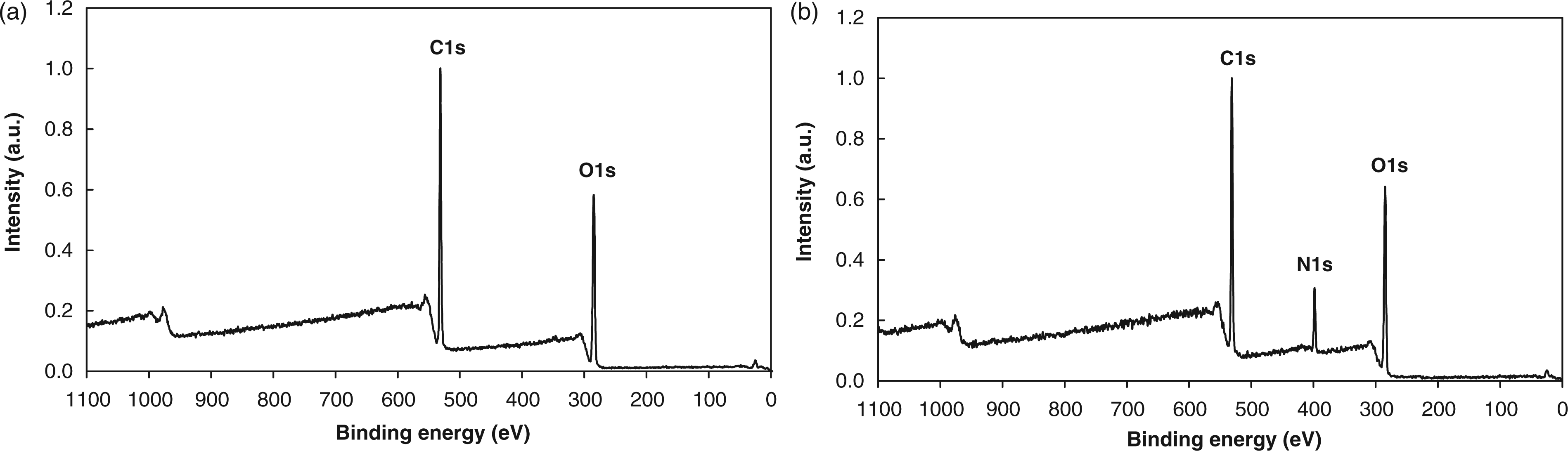
The elemental survey spectrum of the ammonia plasma-treated viscose sample shows an N peak with a binding energy (BE) of 399.2 eV, what is a good evidence for the presence of nitrogen atoms on surfaces.
Elemental surface composition of non-treated and plasma-treated viscose substrates in ammonia gas for 20 s as determined from X-ray photoelectron spectroscopy
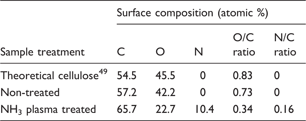
For such a short plasma exposure time, Table 1 shows higher atomic percent carbon and lower atomic percent oxygen, while atomic percent nitrogen reaches a value of 10%. This value does not reflect the real content of nitrogen on the surface of the sample, since the escape depth of photoelectrons should be taken into account. The real concentration of N atoms on the surface is probably higher. The longer treatment time and lower discharge power favor high N content, as also evinced by other authors. 50
It is, however, essential that a high-resolution scan signal could also be acquired and properly charge compensated to establish the peak BE. It is worth mentioning that in our particular case this technique is not useful due to the rich morphology of our particular samples. Namely, as known from literature, best results using high-resolution XPS spectra are obtained for almost flat and homogeneous surfaces, and rich morphology leads to broadening of peaks so characterization of materials using high-resolution XPS on the C1s peak fails in cases of heterogeneous materials with complex morphology. 51
Surface morphology
The surface morphology changes after applied NH3 plasma treatment were investigated by AFM. Figure 5 shows the three-dimensional AFM images of non-treated and plasma-treated viscose samples by ammonia gas for 100 s.
Atomic force microscopy images of (a) non-treated and (b) ammonia plasma-treated viscose sample for 100 s.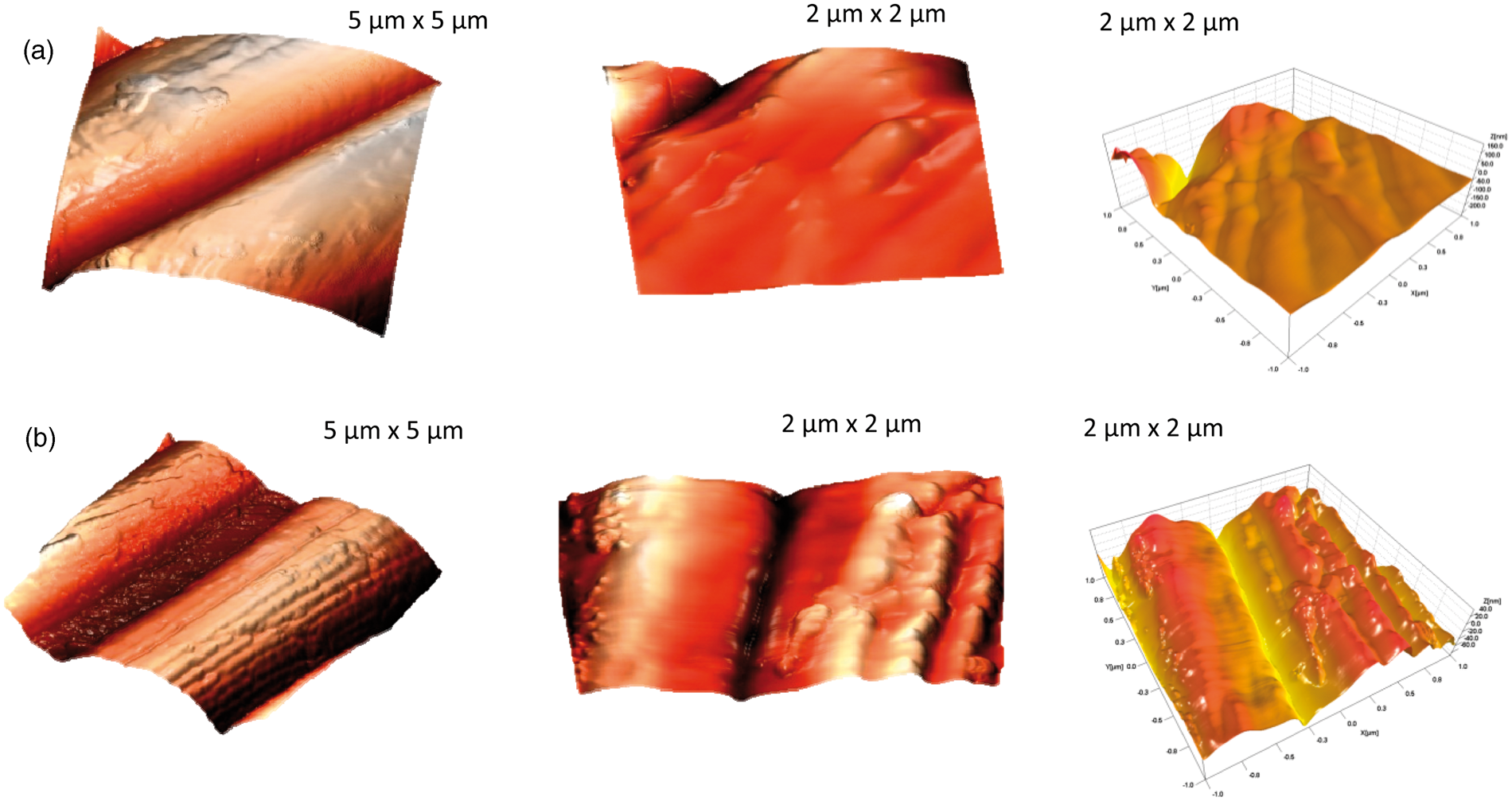
A substantial difference between non-treated and plasma-treated samples is observed: the morphology of the plasma-treated sample is much richer. Such behavior has been observed for many polymers, 52 but the phenomenon is not yet fully understood. One possible explanation is that the small inhomogeneity of the original material reflects in selective etching, since the etching rate depends on the degree of crystallinity of polymers, as demonstrated by Junkar et al. 53 More complex mechanisms have been reported recently by Kontziampasis et al. 54 Whatever the mechanism is, such nanorough and functionalized surfaces obviously do not allow for accommodation of bacteria.
Hydrophilic properties
The results of water rise in non-treated and plasma-treated viscose samples depending on exposure time to ammonia gas are reported in Figure 6 in terms of gained mass versus time. Absorbency capacities presented as the amount of liquid uptake in equilibrium are also presented in Figure 6. Similar results have also been obtained for other exposure times and are not presented here.
Water rise on non-treated and plasma-treated viscose samples at different exposure times to ammonia gas, that is, 20, 60, 80, 140, 200, 260 and 300 s.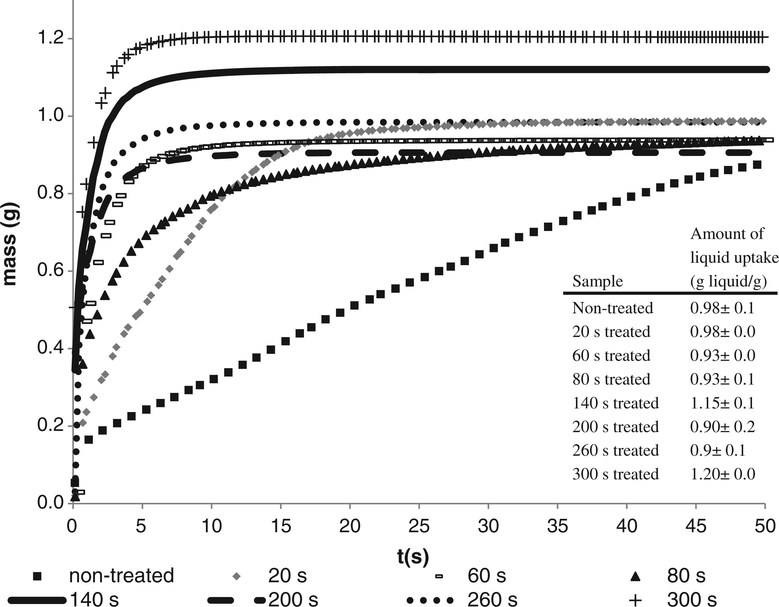
The slowest water rise was evidenced by the non-treated sample, since the complete wetting was observed only after about 100 s. The amount of water uptake in equilibrium amounted to about 0.98 g. The ammonia plasma-treated samples were able to reach the equilibrium value faster. Even a brief treatment for 20 s allowed for reaching the equilibrium uptake in a much shorter time, say about 20 s. The water uptake curves presented in Figure 6 indicate a monotonous decrease of the time needed for reaching the equilibrium with increasing plasma treatment time. For prolonged plasma treatment this time is only few seconds and the equilibrium amount of water uptake is above 1 g.
On the other hand, in the initial stage of water uptake, the mass
2
data plotted versus time, as presented in Figure 7, are fitted by straight lines, according to the modified Washburn equation. The rise of curves at the beginning of water uptake, that is, for the first 3 s, indicates the lowest imbibition of water by a non-treated sample. For all NH3-treated samples the rate of water uptake was faster and depended on plasma treatment time. The differences between samples are most pronounced for short treatment times but at the longer treatment times they are not so important.
Square mass data of water rise on non-treated and plasma-treated viscose samples at different exposure time to ammonia gas, that is, 20, 40, 80, 120, 140, 200, 220, 260 and 300 s.
The absorbency rate is, beside the absorbency capacity, also an important performance parameter to be considered for absorbent application of non-woven materials. The absorbency rate, presented as mass
2
versus time, is presented in Figure 8.
Absorbency rate of non-treated and plasma-treated samples depending on exposure time to ammonia gas, that is, 20, 40, 80, 120, 140, 200, 220, 260 and 300 s.
The absorbency rate is governed by the balance between the forces exerted by the capillaries and the frictional drag offered by the fiber surfaces. The results shown in Figure 8 demonstrate a dramatic increase of the absorbency rate by plasma-treated material as compared to the non-treated sample. The absorbency rate by plasma-treated samples depends on the treatment time, that is, the longer the exposure time the higher is the rate. Obtained values of mass
2
versus time were used in the modified Washburn equation. Calculated values of water contact angle are presented as average values in Figure 9.
Water contact angles of non-treated and plasma-treated viscose samples depending on exposure time to ammonia gas, that is, 20, 40, 60, 80, 100, 120, 140, 160, 180, 200, 220, 240, 260 and 300 s. To make the dependency hydrophilicity versus exposure time clearer, the dashed line is added as an eye guide.
The hydrophilicity is enhanced by plasma treatment. The non-treated sample shows a water contact angle of about 89°. The contact angles constantly decreased with increasing plasma exposure time up to about 140 s. Thereafter, the contact angle remains fairly constant at the value of about 40°. Obviously, some kind of saturation of surface wettability appears at prolonged treatment times.
Antimicrobial activity
Reduction R (%) of the bacteria, mostly present in the infected wound, for non-treated and treated viscose sample exposed to ammonia gas for 60 and 300 s
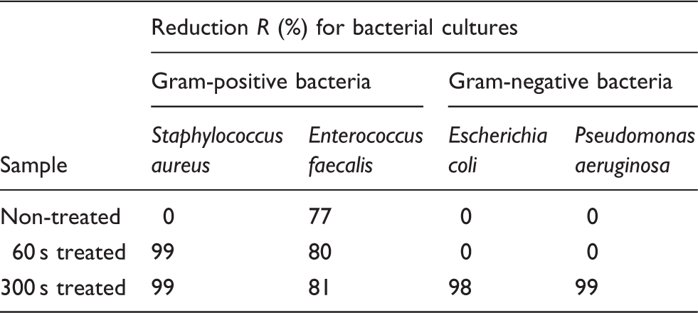
The AATCC test method 100-1999 reveals the percent of reduction as result R = 0 or R < 60 in the situation where the sample does not possess bacteriostatic properties. The mentioned was evident by non-treated sample, except the 77% reduction with E. faecalis was a surprise. Plasma-treated samples, regardless of used treatment time, indicated antimicrobial properties by Gram-positive bacteria, which were more pronounced for S. aureus. The exposure plasma time had significant influence by Gram-negative bacteria, since the reduction was evident only for samples imparted by ammonia gas for 300 s.
Discussion
Influence of plasma parameters
As mentioned earlier, the plasma is rich in both charged and neutral reactive particles. Also, plasma created in hydrogen-containing gases is a rich source of ultraviolet (UV) radiation. Although such radiation is not really revealed from the optical spectrum presented in Figure 1, it exists due to the following reasons. Firstly, very intensive hydrogen atomic lines are observed in the spectrum of Figure 1. The lines originate from transitions of H atoms from the higher to the first excited states (Balmer series). As usual, the highest line, marked Hα, corresponds to the transition from the second to the first excited state of hydrogen atoms. If these transitions are intensive, even more intensive will be the transitions to the ground state (Lyman series). Namely, H atoms do not have metastable excited states, so the excitation of any state is due to the direct transfer of kinetic energy of electrons (electron impact excitation). Furthermore, strong radiation is expected also from the relaxation of excited molecules formed by heterogeneous surface recombination of H atoms. Such recombination definitely appears in the experimental system and is proved by the mass spectrum presented in Figure 2. The proof for such intensive UV radiation has been reported recently by Kregar et al. 55 The reason such emission is not observed in Figure 1, is the fact that the sensitivity of our optical spectrometer in the UV range is much lower than in the visible range, and the fact that transmission of borosilicate glass for light quanta decreases rapidly with decreasing wavelength in the UV range. Energetic photons therefore abound in the reactor and cause destruction of molecular bonds in our materials, resulting in formation of free radicals on surfaces. 56 The penetration depth of photons is not great but large enough to cause modification of viscose material not only on the surface but also in the bulk of our material.
Plasma always contains positively charged molecules and atoms. The ions are accelerated across the sheath next to the sample and cause modification of surface functionalities on the surface of the samples. If the surface was flat and the probability for interaction between an impinging ion and a carbon atom on the surface was 1, the surface would saturate with ion-induced functionalities in a short time, which can be estimated taking into account the flux of ions onto the surface and the surface density of atoms in solid materials:

Here, Ns is the surface density of atoms in solid materials (the order of magnitude is 1019 m−2 for all materials), n+ (m−3) is the density of charged particles in the unperturbed plasma and v+ is the Bohm velocity if ions, that is, v+ = √ (kTe/m+) ≈ 4 × 10⁴ m s−1. The time needed for saturation in the upper approximation is therefore solely τ = 0.05 s. In practice, the surface is not at all flat but very rough due to fibril structure of the sample, so the real surface area is orders of magnitude larger than the geometrical time. Still, if ions were responsible for modification of the surface properties the saturation would appear in a much shorter treatment time than about 140 s. Obviously ions are not the only plasma particles responsible for surface modifications.
Apart from charged particles whose density in plasma was measured with the double electrical probe, neutral reactive particles also abound in our plasma. Figure 3 reveals that a substantial number of NH3 molecules dissociate in plasma at reasonably high RF power. At the pressure of 150 Pa the density of molecules is about 4 × 10²² m−3. The density of reactive nitrogen atoms is therefore at least about 1 × 10²¹, thus five orders larger than the density of ions, and they definitely play a role in functionalization of our materials.
Plasma surface chemical modifications
Ammonia plasma induces N-containing functional groups, whose distribution and density can be tailored with plasma parameters. In fact, saturation of the materials with nitrogen is observed for most materials even after a brief exposure to N-atom rich atmosphere. 57 Our XPS results just confirm this theory, since the surface of our samples contains a substantial amount of nitrogen as revealed from Figure 4. Any attempt to acquire a high-resolution C1s peak failed due to the nature of the samples, as well attenuated total reflectance (ATR) Fourier transform infrared (FT-IR) spectroscopy cannot be used to identify the chemical groups if the thickness of a plasma polymer material is in the micrometer range.58,59 Therefore, it is not possible to extract the exact functional groups appearing on the viscose samples upon ammonia plasma treatment. According to relevant literature, a variety of functional groups may appear on the surfaces and the effect of post-plasma oxidation could convert amines into amides. 60 It is interesting, however, that the concentration of oxygen on the surface as revealed from our XPS results dropped by almost a factor of 2 for prolonged plasma treatment. Such a dramatic decrease in oxygen concentration could be explained by formation of oxygen-free nitrogen-functional groups, especially when taking into account the chemical structure of non-treated viscose and the final depth resolution of XPS devices.
Surface topographical changes
Plasma particles are capable of etching polymer materials and the etching is often highly selective.52,53 The surface morphology (Figure 5) of the plasma-treated sample shows a dramatic increase of the surface roughness compared to non-treated material. While the fibers are rather flat prior to plasma treatment they obtain rich morphology after the treatment. The change of the morphology definitely influences both adsorption and absorption of water and other fluids. The results summarized in Figures 6 and 9 are therefore explained by combined effects of surface functionalities and roughness. Namely, the contact angle of a liquid on a solid surface depends on these two parameters. Increased roughness allows for excellent hydrophilicity of materials with high surface energy. A great advantage of weakly ionized plasma created by electrode-less high-frequency discharges is that the neutral reactive particles abound and their density easily reaches values over 1021 m−3. 11 While excellent functionalization of the sample surface is easily obtained even by gaseous ions from plasma, the neutral atoms play an important role in etching. The synergistic effects of ions and neutral nitrogen atoms allows for etching of polymer materials even at room temperature. Such etching is limited to the fibers on the very surface of the samples, since the ions cannot penetrate through interfibril spaces. Neutral atoms, on the other hand, easily penetrate deep into pores due to poor probability for heterogeneous surface recombination. Although not as reactive as ions, they also cause slow etching of polymer materials and are thus capable of modifying surface morphology of fibers even within the bulk of materials. As shown recently, neutral atoms are capable of etching polymers at room temperature in the absence of any ions. 61 The etching therefore allows for increased roughness of fibers, which is a definite benefit.
Plasma-induced hydrophilicity
Hydrophilic properties do not depend only on the functionalization of the sample on the surface, but also on the functionalization of the bulk of the material. The used non-woven samples were a few millimeters thick and composed from randomly oriented viscose fibers. In order to obtain optimal hydrophilicity it is therefore essential to modify the bulk of the sample. In Figure 7, a huge difference between non-treated and ammonia plasma-treated samples is observed. With non-treated material the water rise (defined as the squared mass gain in a unit time) is rather low. For the first 3 s it is only about 0.04 g2 s−1. The rather low rise is due to the moderately hydrophobic properties of non-treated materials. For the next sample tested (treated with plasma for 20 s) the water rise is improved significantly and is about 0.16 g2 s−1 during the first 3 s of uptake time. The water rise curves keep increasing with increasing plasma treatment time, and for treatment times exceeding, say, 140 s the material saturates with water already in a few seconds of uptake time, as revealed from Figure 6. The corresponding initial water rise curves approach values above 1 g2 s−1, which are about 30 × larger values than for non-treated material. Such a huge improve in the water rise cannot be explained solely by improved adsorption of water on the surface of samples, but bulk effects should be taken into account. Obviously, the prolonged treatment of samples with plasma allows for optimization of the hydrophilic properties of viscose material. The optimization is explained by modification of surface properties throughout the bulk of the sample.
The high-energetic photons penetrate deep into our materials and cause bond scission in solid material. This interaction leads either to reorganization of the atoms in cellulose material or to chemical interaction with hydrogen and nitrogen atoms that abound in our plasma reactor. Furthermore, neutral atoms in the ground state are capable of forming functional groups on the surface of polymers even in the absence of UV radiation. Namely, experiments performed in late-flowing afterglows, where only the most stable particles created in plasma (predominantly neutral atoms in the ground state apart from metastable molecules with long radiation relaxation times) are present, showed similar results in terms of polymer surface functionalization as the experiments in plasmas themselves. 61 The functionalization in the bulk is therefore governed by diffusion of neutral reactive plasma particles and that is the reason for rather slow improvement of the water rise versus plasma treatment time (Figures 6 and 7). Namely, Equation (7) clearly indicates that the sorption properties should improve in a second of plasma treatment if functionalization of the surface only is taken into account.
Here it is worth mentioning that there is not a strict parallelism between the water uptake amount and the exposure time of samples with ammonia gas. The deviation is roughly within the limits of statistical errors that are presented in Figure 6. Taking into account the systematic error, which is also estimated to about 10%, the curves lay well within the total error for this sort of measurements. The poor parallelism between the water uptake amount and the plasma exposure time could be, therefore, explained with the accuracy of this specific experimental procedure.
The contact angle by non-treated material, as presented in Figure 9, is rather large at about 90°. Even a short exposure to plasma for 20 s causes a drop of the contact angle for several degrees and the contact angle keeps decreasing monotonously (almost linearly) with increasing treatment time until it reaches a value of about 40° after roughly 140 s of plasma treatment. Thereafter, the water contact angle remains fairly unchanged. Obviously, some kind of saturation appears after 140 s of plasma treatment. Taking into account upper considerations, it is possible to conclude that this treatment time allows for throughout functionalization of the material and further exposure does not affect the wettability of our samples.
The penetration of the fragmented gas particles into the voluminous textile is also influenced by the textile geometry, that is, the distance between fibers in the textile. The non-woven sample consists of a great number of fibers disorderly gathered together, which also forms spacing between fibers. At low pressure, the mean free path (the distance traveled by a molecule or atom between successive collisions) in the gas phase is higher than the textile distances, and the collision of gas molecules with the fiber surface is enhanced as compared to gas–gas collision, thus favoring a good penetration of plasma species into the textile. This means that the obtained hydrophilicity of the plasma-treated sample has improved significantly due to the looser structured non-woven material, as also evidenced by Hossain et al. 62
Plasma-enhanced antimicrobial properties
Within the limits of the experimental error, the reduction rate for non-treated material is zero, while surprisingly enough a 77% reduction for E. faecalis was evident. Plasma treatment for shorter time indicates a sufficient antimicrobial effect as long as S. aureus is the merit. Longer exposure time to ammonia gas revealed sufficient antimicrobial effect for Gram-negative bacteria and does not seem to affect the growing of E. faecalis. It is not straightforward to explain the observed effect, since the interaction between the bacterial cell wall and plasma-treated materials is generally not known. The nitrogen-rich functional groups on the surface of viscose materials may cause a stress to bacteria and thus inability to grow and multiply in such an environment.30,31 The stress could be attributed to specific interactions between nitrogen-functional groups and the bacterial receptors, but any discussion on exact mechanisms is beyond the scope of the paper. Since the type and concentration of different nitrogen-containing functional groups cannot be revealed from XPS results, any further discussion would not be fruitful.
E. coli and both Gram-positive cultures are non-motile, while P. aeruginosa is a motile bacteria. Therefore, the reason for “discrepancy” in reduction, regardless of used treatment, could be in bacteria cell size and shape. 45 S. aureus is cocci with a diameter of 1 µm and mostly arranged like irregular clusters. 63 E. faecalis is a spherical bacterium with a cell size of 0.5–1 µm and observed as singly or in pairs with short chains. Both Gram-negative bacteria are straight or slightly curved rods, 1.5–2 µm long and stacked up next to each other.
Compared to larger arrangement settled cultures, the smaller individual bacteria such as E. faecalis could easily penetrate into the porous sample, while others appearing as clusters or pairs could not.
Factors influencing bacteria adhesion
Bacterial adhesion is an extremely complicated process that is affected by many factors including the environmental factors, such as the associated flow conditions, the presence of serum proteins or antibiotics, the bacterial properties and the material surface characteristics. 64 The factors influencing bacteria adherence to a biomaterial surface include chemical composition of the material,65–71 surface charge, 72 hydrophobicity 73 and simply surface roughness or physical configuration. 74 Surface chemistry influences bacterial adhesion and proliferation. Materials with different functional groups change bacterial adhesion in a manner depending on material hydrophobicity and charge.
If the surface chemistry is changed or modified, for example, plasma treatment,73,75–77 bacterial adhesion to these surfaces is hindered. Balazs et al. 73 and James and Jayakrishnan 78 showed that decreased bacterial adhesion occurred due to significant alteration in the hydrophilicity (i.e. decreasing of contact angle).
It has been found that the irregularities of polymeric surfaces promote bacterial adhesion and biofilm deposition, whereas the ultrasmooth surface does not favor bacterial adhesion and biofilm deposition. 79 This may happen since a rough surface has a greater surface area and the depressions in the roughened surfaces provide more favorable sites for colonization. The surface morphology as presented in Figure 5(b) showed a significant increase in surface roughness, whereas the data presented in Table 2 indicate satisfactory antimicrobial effect. The same results were also obtained by Taylor et al. 80 where a small increase in surface roughness had a significant increase in bacterial adhesion, while larger roughness increases produced had no significant effect in bacterial adhesion compared to the smooth surface. The cause of this non-linear dependence of bacterial adhesion on surface roughness is a question for further studies, such as a broader range of surface roughness, surface area measurement or analysis of surface configuration.
Generally, bacteria with hydrophobic properties prefer hydrophobic material surfaces; the ones with hydrophilic characteristics prefer hydrophilic surfaces. However, it has been shown that material surface hydrophobicity plays a more important role in bacterial adhesion than bacterial surface hydrophobicity.
Bacteria in aqueous suspension are almost always negatively charged. The surface charge of bacteria varies according to bacterial species and is influenced by the growth medium, the pH and the ionic strength of the suspending buffer, bacterial age and bacterial surface structure. However, the relative contribution of bacterial surface charge to bacterial adhesion has not been clearly understood.
However, since bacterial adhesion is a very complicated process affected by many factors, such as bacterial–material properties and environment and, furthermore, the experimental evaluation of the relative contributions of these factors is extremely difficult, more investigations are still needed to advance our understanding of the mechanisms of bacterial adhesion.
Conclusions
Ammonia plasma resulted in important morphological modifications as well in the introduction of nitrogen-functional groups. Both morphology changes and gained functionalities lead to improved hydrophilicity, since the water rise was improved up to 30 times. In addition, plasma-treated viscose non-woven material simultaneously shows also reduction on both Gram-positive bacteria, mostly responsible for wound infections. The viscose non-woven material treated for 300 s shows promise in a wound treatment management by providing fluid capability along with antimicrobial activity.
Plasma surface modification using pure NH3 gas was proven to be a successful treatment technique that could draw great interest in wound dressings. Ammonia plasma-treated wound dressing could offer a balance between cost and benefit by decreasing the infection rate and deliver cost-effective care. Still, the choice of dressing product should base on the amount of wound fluid, wound size and bed conditions, as well as on the cause of the wound. The study attempted to address the concept of treatment procedure as providing beneficial properties promoting wound healing. However, still some optimization of plasma procedure is challenging us to achieve effective 45 and broad-spectrum antimicrobial activity.
Footnotes
Funding
This work was supported by the Ministry of Education, Science, Culture and Sport of the Republic of Slovenia through the contract No. 3211-10-000057 (Centre of Excellence for Polymer Materials and Technologies).