
Abstract
H2O2/ultraviolet (UV) radiation treatment was proposed to simulate burial-induced degradation of natural dyes. The method was applied to the major pigments of Phellodendron bark, madder, and indigo plant, and their silk dyeings, and the degraded samples were analyzed using high-performance liquid chromatography-diode array detector-mass selective detector (HPLC-DAD-MS). Retention times of the HPLC chromatogram, UV-visible λmax obtained from DAD analysis, and major ion detected by the MS analysis were used to identify berberine, palmatine, alizarin, purpurin, indigotin, and indirubin included in dye solution and the extraction from silk dyeing. Alizarin, purpurin, and indirubin were more susceptible to degradation by H2O2/UV than berberine and palmatine. Indigotin was completely degraded with 5 minutes of treatment, producing isatin as the degradation product. Alizarin, purpurin, and indigotin were more resistant to degradation when they were affixed to the fibers.
Keywords
Low colorfastness of natural dyes has been one of the major problems in the conservation and preservation of museum textiles.1,2 When natural dyed textiles originate from burial excavations, colorfastness becomes an even more critical issue since most excavated textiles completely lack their original color and look tan to brown in color regardless of the original hue. 3 This is primarily due to the chemical degradation of dye within the burial environment, which results in the loss of dye and possibly staining from the microenvironment formed by soil and cadavers.3,4 In a very few excavations of the 18th century burials of Korea, it was observed that a tint of scarlet or blue color was visible as the silk textiles were excavated and exposed to open air. 5 The color immediately disappeared on site or it disappeared as the textiles were transported and were housed in the museum conservation department. 5 The visibility of some colors at the excavation site suggests that similar to the colorfastness of natural dyes in the aerobic environment, 1 different natural dyes have different rate of degradation even when the textile is buried in a coffin for a prolonged period of time.
While numerous literatures have examined the colorfastness of natural dyes, there are only a few that have dealt with the change of color related to the burial degradation of textiles. Needles et al. 6 used the American Association of Textile Colorists and Chemists (AATCC) Test Method 30-1981 in examining the burial-induced color change and strength loss of wool and silk fabrics dyed with natural dyes. Peacock 7 used the British Standard (BS) 6085 to examine the color and physical change of undyed cotton, linen, wool, and silk fabrics by burying them 10 cm below the surface of standard soil beds. Lee et al. 8 examined the color change of cotton and silk dyed with natural dyes after burial for 50 weeks. While burying the textiles may be a suitable method for simulating the burial degradation of the textiles, it is difficult to standardize the soil condition and the variables related to nature, such as seasonal weather conditions and micro-habitants are uncontrollable. 9 Therefore, a laboratory experimental design that simulates burial degradation conditions can be used as a model for actual burial.
Fungal decolorization of wastewater produced from synthetic dyes has long been the target of research in various related fields of study. 10 It has been found that there is a good correlation between the decolorization of dye by fungi and their extracellular production of peroxidases or H2O2. 11 The white-rot fungi such as Pleurotus eryngii are known to excrete aryl-alcohol oxidases that generate H2O2 in nature. 12 Pleurotus eryngii is an edible mushroom that grows in the underground roots and dead wood. 13 It is one of the largest mushroom species that grows widely in Europe, North Africa, or Asia and its production is third in volume in the world production of mushrooms. 13 Considering the widespread growth of Pleurotus eryngii, it is highly likely that there is an active generation of H2O2 in different soil microenvironments. As an example, it was found that the plantation soil samples of Oak Ridge, Tennessee, showed a dominant peroxidase generation when the soil extracellular enzyme activities were examined. 14 H2O2 is also introduced into the soil by wet and dry atmospheric deposition, with the rainwater being the most representative source. 15 Considering that H2O2 accumulates in soil by various routes, it is suggested that some part of textile degradation in the burial is due to the action of H2O2, which initially attacks the dyes. Textiles that were in the burial environment rich in H2O2 production would be directly exposed to H2O2. When such textiles are excavated, ultraviolet (UV) light in the atmosphere will react with H2O2 to produce the hydroxyl radical (·OH), initiating decomposition of the dyes. This might be why some colors observed in textiles at the point of excavation very quickly fade and disappear after they are unearthed. 5
As a way to simulate the degradation of dyes by H2O2 in the soil, we suggest the application of a laboratory oxidation process using H2O2 with UV radiation, the process that has been used to treat dye wastewater. 16 The method was previously used to examine the degradation behaviors of alizarin and curcumin using gas chromatography mass spectrometry.17,18 We found that the H2O2/UV method was a suitable tool for examining the long-term degradation of dye while thermal treatment, that is, leaving the dye in 110℃ oven, was more suitable for examining a short-term degradation effect.
The aim of this research was to investigate the degradation behaviors of major dyes of Phellodendron bark, madder, and indigo plant using the H2O2/UV process followed by analysis of degraded dyes using the HPLC-DAD-MS instrument equipped with a high-performance liquid chromatography (HPLC), a diode array detector (DAD), and a mass selective detector (MS). The HPLC-DAD-MS instrument has been used successfully in the analysis of dyes in historic textiles.3,19–22 Phellodendron bark, madder, and indigo were the most representative dyestuffs of the past to impart yellow, red, and blue color in textiles. Record of their usage as dye materials can be found in historic references, such as Ben Cao Gang Mu, 23 that originate from China in the 16th century. Traditional recipes for dyeing textiles using these plant dyes can be found in Gyuhap chongseo, 24 a 19th century Korean reference.
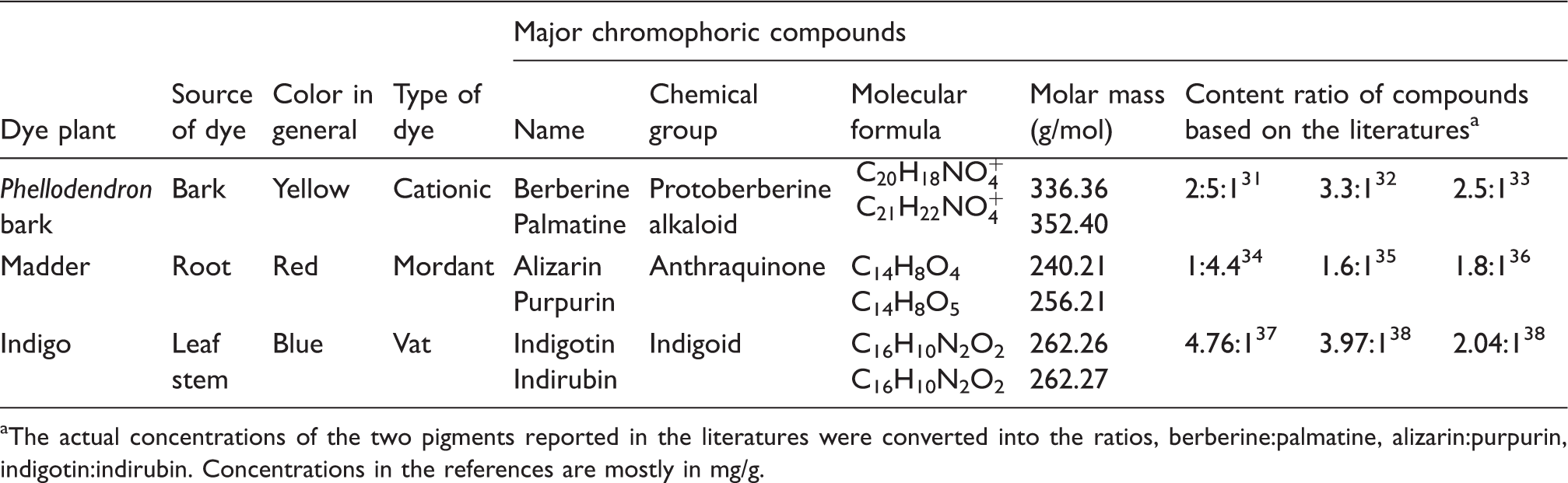
The H2O2/UV degradation was conducted on the six dyes: berberine, palmatine, alizarin, purpurin, indigotin, and indirubin (Table 1; Figure 1). Berberine and palmatine are the main chromophoric compounds present in the bark of Phellodendron tree (cork tree), Phellodendron amurense Rupr., and Phellodendron chinense Schneid. Berberine and palmatine exhibit similar biochemical activities due to their quaternary nitrogen and the polycyclic planar structure.
25
Berberine and palmatine exist in plants in their salt forms,
26
and in this research the chloride forms of the two pigment compounds were used as they were sold. Alizarin and purpurin are the main chromophoric compounds present in the roots of madder plant such as Rubia tinctorum or Rubia cordifolia.27,28 Alizarin and purpurin are anthraquinones, and they are the most representative mordant dyes that have no direct affinity toward fibers but are fixed to fibers by the aid of mordants.
28
Indigotin is the coloring compound of the indigo plant, and fiber is dyed with it by the vat dyeing process. Indirubin, a red pigment, is a minor coloring component in the indigo plant that is produced from the reaction of isatin and indoxyl in the indigo leaf
29
or occurs as the degradation product of indigotin.20,30
Characteristics of the six dyes under investigation The actual concentrations of the two pigments reported in the literatures were converted into the ratios, berberine:palmatine, alizarin:purpurin, indigotin:indirubin. Concentrations in the references are mostly in mg/g.
Degradation behaviors of six dyes were first examined by degrading the liquid dye solution. Investigation was conducted on the individual dye solutions and in a mixed dye solution containing all six compounds. Next, the degradation behaviors of six dyes were examined by degrading the silk sample that had been dyed consecutively with berberine, palmatine, alizarin, purpurin, and indigotin after mordanting with alum type mordant. The results obtained from the dyes in solution were used as reference for interpreting the HPLC-DAD-MS results of the dyes in degraded silk samples. Silk was chosen as the dyeing medium since the majority of the textiles excavated from the Korean burial sites were found to be silk owing to the fact that the preserved textiles mostly came from the burials of the high social status individuals.3,5 Aluminum potassium sulfate was used as the modern method of alum mordanting. 3 Investigation on degradation of individual and mixed dye solutions, and the silk dyed with the composite dyeing of five of the six dyes provides information on degradation behaviors of the chromophoric compounds that are relative to long-term burial. The results of this study could provide crucial information for designing the excavation procedures of textiles from burials, and the results would also assist museum curators in protecting the valuable textile assets from possible degradation during conservation and preservation.
Experimental details
Materials
Berberine chloride form (berberine in the following) (97%), palmatine chloride hydrate (palmatine in the following), alizarin, indigotin (95%, synthetic), and indirubin were purchased from Sigma-Aldrich. Purpurin (85%) was purchased from Tokyo Chemical Industry. Hydrogen peroxide (30%, ACS grade) and sodium hydrosulfite, HPLC grades of dimethyl sulfoxide (DMSO), acetonitrile, and HPLC water were purchased from Fisher Scientific. Formic acid (88%, ACS grade) was purchased from Macron Chemicals. Aluminum potassium sulfate [AlK(SO4)2·12H2O] was purchased from Shinyo Pure Chemicals (Osaka, Japan). Each HPLC sample was filtered using a glass fiber-enhanced 0.45 µm syringe filter by Alltech Associates. Silk used for dyeing was the Standard Adjacent Fabrics for Colorfastness (KS K0905) purchased from KATRI (Seoul, Korea). Water used for dyeing was purified using Milli-Q Integral System by EMD Millipore.
Methods
Preparation of individual and mixed pigment solutions
For individual dye solutions, 0.016 g of berberine, palmatine, alizarin, and purpurin, 0.008 g of indigotin, and 0.004 g of indirubin were each dissolved in 80 mL of DMSO. For the mixed dye solution, 0.01 g each of berberine, palmatine, alizarin, and purpurin, and 0.005 g of indigotin and indirubin were dissolved in 300 mL of DMSO.
Degradation treatment of individual and mixed pigment solutions
For the degradation of individual dye solutions, 2 mL of each dye solution were added to a glass vial (i.d. 25 mm, height 95 mm) for each degradation time interval. In each vial, 0.5 mL of 30 wt% hydrogen peroxide was added, and this corresponded to the 4:1 v/v ratio of dye solution and H2O2. Sample vials were placed under a 365 nm UV lamp (UVL-18, Upland, CA) so that the distance between the surface of the liquid and the lower edge of the light source was 9.3 cm, the UV radiation was applied vertically from above the mouth of each vial. We used the UV lamp, which was fixed to 365 nm wavelength since 365 nm is part of the UVA ray that penetrates the ozone layer and reaches the surface of the Earth. All vials were tightly covered with Saran Wrap® wrapping film to minimize evaporation of the solvent, and other light source was blocked by covering the degradation set-up with aluminum foil. Each sample was treated for the designated degradation time and filtered before it was analyzed using HPLC-DAD-MS. For the degradation of mixed dye solution, 120 mL of the mixed dye solution and 30 mL of H2O2 were added to a 500 mL Pyrex beaker. This reaction beaker was placed under a 365 nm UV light for the designated degradation time and the UV radiation was applied vertically from above the mouth of the beaker. The beaker was covered with Saran Wrap® and other light sources were blocked by aluminum foil. For each degradation time, 2 mL were pipetted from the reaction beaker and then filtered for the HPLC-DAD-MS analysis. Two samples were prepared for each degradation time for the degradation of individual dye solutions and the mixed dye solution.
Dyeing of silk
A composite dyeing was carried out on the silk using berberine, palmatine, alizarin, purpurin, and indigotin. Indirubin was not used in the dyeing process. A 370-mL dyebath was prepared in purified water for each of berberine, palmatine, alizarin, and purpurin so that each bath contained 0.025 g of corresponding pigment. One piece of approximately 40 cm × 20 cm sized (2.45 g) silk fabric was used for dyeing. This corresponded to 1% o.w.f. (on weight of fiber) dye concentration in 1:30 liquor ratio for each pigment bath. The silk was dyed using the four dye baths consecutively using the pre-mordanting procedure, that is, mordanting–berberine dyeing–mordanting–palmatine dyeing, etc. Mordanting was done with 0.49 g (20% o.w.f.) of aluminum potassium sulfate for 1 hour at 60℃ and a fresh mordant bath was used for each mordanting step. After each mordanting, the silk was thoroughly rinsed with running tap water then placed between the layers of Kimwipes® wiping paper for experiments to remove excess water. Dyeing was conducted with berberine, palmatine, alizarin, purpurin baths for 1 hour at 60℃. After each dyeing, the silk was thoroughly rinsed with running tap water, soaked in the purified water for about 10 minutes, and then dried between the Kimwipes®. The dyed silk was cut into approximately 1.7-g specimens for the indigotin dyeing. A 255-mL indigotin dyebath was made with 0.017 g dye and 0.05 g sodium hydrosulfite. The dyebath was adjusted to pH 11 using NaOH. Indigotin dyeing was conducted for 30 minutes at room temperature. After the dyeing was completed, the silk was then thoroughly rinsed with running cold water and air dried so that the leuco form of indigotin would freely oxidize to the insoluble indigotin within the silk.
Degradation of dyed silk
The dyed silk was cut into 2.5 cm × 2.5 cm sized samples and a 500 mL stock of 9:1 v/v H2O:H2O2 solution was prepared. A 20 mL of the stock solution and a piece of the silk sample were put into individual clear glass vials (i.d. 25 mm, height 95 mm) for each degradation time. With the vials tightly covered with Saran Wrap®, the vials were placed under the UV lamp and at the end of the degradation time, the silk sample was taken out, rinsed, and dried between the layers of Kimwipes®.
Extraction of dye from silk
Approximately 0.5 cm × 0.5 cm sample of each degraded silk specimen was place in a small beaker with 0.4 mL of mixed solution of HCl/methanol/water (2:1:1 v/v/v). The beaker was placed in a 110℃ oven for 10 minutes, cooled, and then placed above NaOH pellets inside a vacuum desiccator until the liquid was completely removed. An aliquot of 1.5 mL of DMSO was added to the completely dried specimen in the beaker for the final extraction process. The DMSO extract was filtered and analyzed using the HPLC-DAD-MS.
HPLC-DAD-MS analysis
Chromatographic conditions
An Agilent 1200 series binary HPLC-DAD-MS system (Foster City, CA) equipped with a DAD and a MS consisting of a single quadrupole mass analyzer was used for sample analysis. The mass detector was operated in the SIM mode using the atmospheric pressure chemical ionization (APCI) source in the positive mode to detect the chromophoric compounds. LC separation was achieved by Agilent ZORBAX SB-C18 column (length 50 mm × I.D. 2.1 mm, particle size 1.8 µm). The gradient elution applied in the analysis using solvent A (acetonitrile) and solvent B (1% formic acid in water) was: 0–5.7 min, 90–20% B; 5.7–10 min, 20–61% B, 10–15 min, 61% B. The flow rate was 1.0 mL/min, and the injection volume was 20 µL. Detection wavelength for the DAD was set for 265, 255, 288, 606, 542 nm. The column temperature of the MSD was 25℃ and the ionization source was operated with drying gas flow 12.0–13.0 L/min, drying gas (N2) temperature 150–350℃, vaporizer temperature 250℃, nebulizer pressure 60 psi, capillary voltage of 5 kV in positive ion mode, charge voltage 1.3 kV, fragmentor voltage 95 V, and mass range m/z 200–400. The SIM program was run with group 1 0–8 min m/z 336, 338, 352, 354; group 2 8–9:50 min m/z 241, group 3 9:50–10.70 257, m/z 263, and group 4 10.70 m/z 263.
Analysis of relative concentration of dyes
An ion chromatogram was generated for each ion m/z 338, m/z 354, m/z 241, m/z 257, m/z 263 (indigotin), and m/z 263 (indirubin) in the mass spectra obtained from the APCI positive ion SIM mode. Based on the R t range of each pigment compound, the abundance of the major peak was obtained using the integration results of the Agilent ChemStation (Agilent Technologies, 2001–2006). This abundance was used as the relative concentration of the corresponding pigment compound detected. Fitting equation y = y o + ae −bx (R2: 0.90–0.99) was used to fit the data for Figures 3, 7, 8, and 10 using Sigma Plot.
Experimental results
HPLC-DAD-MS analysis of the untreated dyes
Results of high-performance liquid chromatography-diode array detector-mass selective detector (HPLC-DAD-MS) analyses of individual pigments before treatment

APCI: atmospheric pressure chemical ionization.
Figure 2 shows the HPLC-DAD chromatogram of the mixed dye solution before degradation treatment. The UV-visible absorption spectra and the mass spectra generated from each pigment compound in the mixed dye solution are presented elsewhere.
39
The retention times of the six dyes, DAD UV-absorption maxima, and their major molecular ions were all identical to the results obtained from the HPLC-DAD-MS analyses of the individual dye solutions.
39
Overlapping of the peaks of indigotin and purpurin was expected since their retention times fell within the same R
t
range. UV-visible absorption spectra of the two dyes in the visible wavelength also show that the λmax of indigotin (606 nm) and the λmax of purpurin (481 nm) were concomitantly detected in both dyes. However, the identification of the two compounds was possible based on the major molecular ion detected in the HPLC-MS analysis, ions m/z 263 and m/z 257 for indigotin and purpurin, respectively.
39
Berberine and palmatine, the two other pigments whose peaks were not well separated, could also be distinguished from each other by their major molecular ions [M + H2]+, which were m/z 338 and m/z 354, respectively.
39
High-performance liquid chromatography-diode array detector chromatogram of the mixed pigment solution before the degradation treatment. The peaks labeled are (1) berberine, (2) palmatine, (3) alizarin, (4) purpurin, (5) indigotin, and (6) indirubin. Change in the relative concentration of pigment compounds in the individual dye solutions by H2O2/ultraviolet treatment.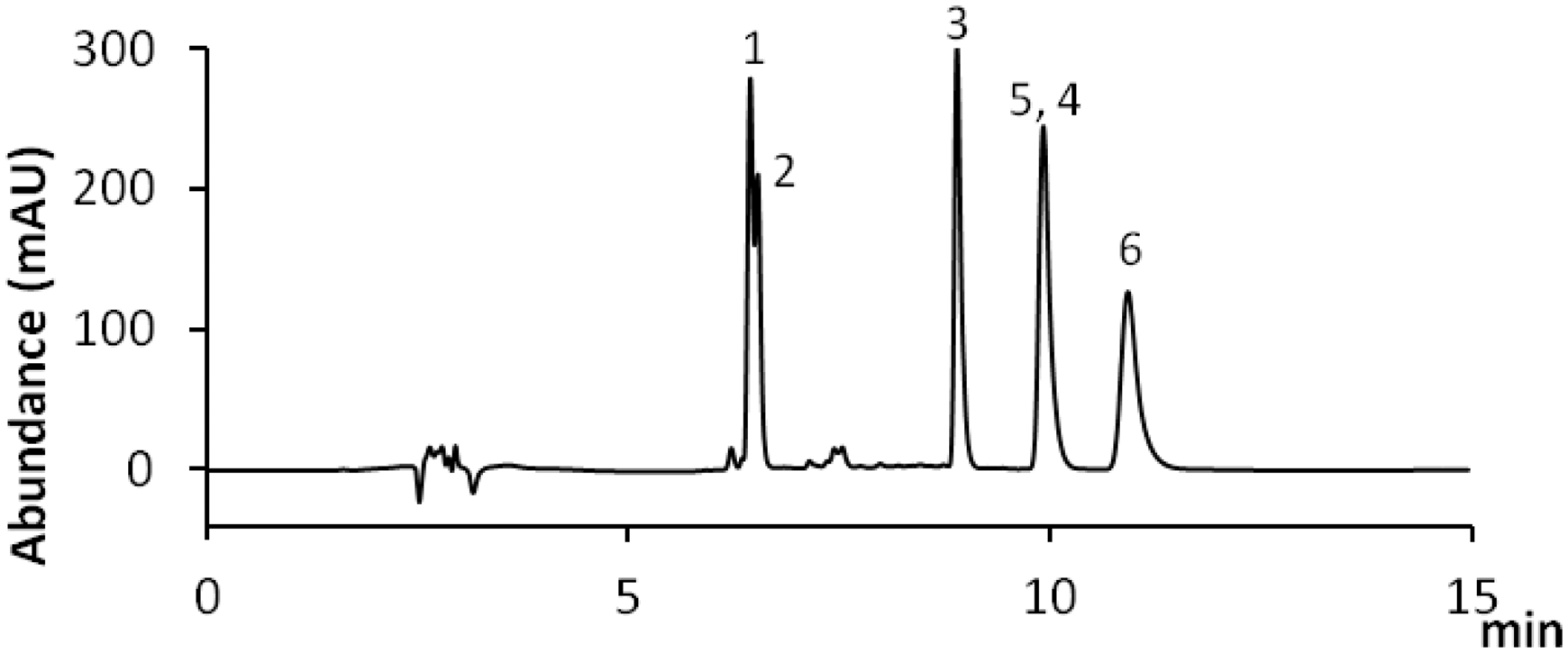

Degradation of individual pigment solutions
Based on HPLC-DAD-MS results of individual dye solutions, the six chromophoric compounds were identified in the samples treated under H2O2/UV condition. Figure 3 shows the change in relative concentration of each chromophoric compound in the individual dye solutions for 10 hours of H2O2/UV degradation. The degradation pattern of berberine and palmatine were almost identical, showing that they were fairly resistant to degradation in the H2O2/UV environment. Their concentration remained unchanged after a 20% decrease in concentration that occurred during the first hour. Alizarin, purpurin, and indirubin were much more susceptible to degradation than berberine and palmatine, with purpurin exhibiting the highest amount of degradation. Overall, the greatest amount of degradation of alizarin, purpurin, and indirubin is within the first two hours with little degradation after six hours. When the treatment was terminated, the concentrations of berberine and palmatine in the individual solutions were above 70%, and the concentration of alizarin was close to 50%, and those of indirubin and purpurin were less than 40% of their respective untreated samples.
During the experimental process, it was found that the indigotin solution became nearly colorless soon after exposure to UV light. The HPLC-DAD-MS trials of this sample showed that the abundance of m/z 263 at the R
t
range 9.9–10.5 min was zero. To investigate this further, indigotin was subjected to a 4-hour H2O2/UV degradation treatment and examined every 5 minutes during the first half hour. It was found that over 95.5% of indigotin was lost after 5 minutes of degradation treatment (Figure 4) and the indigotin peak completely disappeared in the HPLC-DAD chromatogram (Figure 5). In a separate trial, indigotin was treated only with H2O2 without the UV radiation. In this case a visible change in color occurred after about 2 hours and the solution became colorless in 3 hours. Color change was also visible when indigotin was treated with UV radiation without the addition of H2O2. In this case, however, the time consumed for the total loss of color was not measured. In the HPLC-DAD chromatograms of the degraded indigotin solutions, a new small peak appeared at 7.54 minutes in the 5-minute degradation sample (Figure 5), and it continued to be observed in the samples for the duration of the experiment. The DAD UV-visible spectra of the particular peak indicated λmax of 300 and 414 nm (Figure 6). In addition, the most representative λmax of indigotin, 607 nm in the visible wavelength region, was not present in the new peak (Figure 6), reflecting that the product does not have blue color of indigotin. When indigotin in dimethylformamide (DMF) solvent was examined with light excitation in the presence and absence of molecular oxygen, isatin was identified as the degradation product.
42
Based on the literature, the peak of 7.54 minutes appears to be caused by isatin that was eluted as a degradation product of indigotin. The λmax of isatin in the literature was 418 nm.
42
The difference between the λmax of isatin reported in the literature and our findings (414 nm) is due to the difference in the polarity of the solvent,
43
where λmax appeared at the shorter wavelength when the solvent was DMSO.
Change in the relative concentration of indigotin by H2O2/ultraviolet treatment. Change in the high-performance liquid chromatography-diode array detector chromatogram of indigotin solution by H2O2/ultraviolet treatment. Ultraviolet-visible spectra of indigotin and the peak at 7.54 minutes eluted in the high-performance liquid chromatography-diode array detector chromatogram.


Degradation of mixed pigment solution
Mixed dye solution was subjected to long-term degradation involving a 264-hour H2O2/UV treatment. The degraded samples were analyzed at 24-hour intervals. Significant amounts of dye were lost in the first 24 hours, and there was little to no change in concentration after 48 hours (Figure 7). It is possible that such a result was obtained because most H2O2 in the reaction medium was consumed by the dyes during this period.
Indigotin was completely lost, showing a zero abundance of ion m/z 263, and all other dyes notably decreased during the first 24 hours of treatment with little change over the next 240 hours. By the first 24 hours, the concentrations of dye remaining in the mixed solutions were close to or much less than 50% of the untreated solution. In the case of the two pigment compounds in indigo plant, indigotin was completely lost and only 15% of indirubin remained in the mixed solution. When the treatment was terminated, the concentrations of each chromophoric compound in the solution were slightly over 30% for berberine, palmatine, and purpurin, and 44% for alizarin. In addition, there was only 0.3% of indirubin and no indigotin remaining in the mixed solution after 264 hours of H2O2/UV treatment.
Figure 8 shows the initial stage of degradation (0–3 h) in the mixed solution. Except for alizarin, which showed a continuous decrease in concentration up to 2 hours, all other dyes showed the most significant decrease in concentration during the first 30 minutes. Indigotin was completely lost and among the other dyes the highest decrease was observed in purpurin. This result was in good agreement with the results obtained for the degradation of individual solutions (Figure 3).
Analysis of degraded silk dyeings
Figure 9 represent the HPLC-DAD chromatogram of the dye extracted from the dyed silk. The R
t
ranges of the five pigments, DAD UV-absorption maxima, and major molecular ions matched the results obtained from the HPLC-DAD-MS analyses of the individual pigment solution and the mixed dye solution shown in Table 2.
Change in the relative concentration of pigment compounds in the mixed solution in the long-term H2O2/ultraviolet treatment. Change in the relative concentration of pigment compounds in the mixed solution at the initial stage of H2O2/ultraviolet treatment. High-performance liquid chromatography-diode array detector chromatogram of the dyed silk showing the five pigment peaks. The peaks labeled are (1) berberine, (2) palmatine, (3) alizarin, (4) purpurin, and (5) indigotin. Change in the relative concentration of pigment compounds in the dyed silk by H2O2/ultraviolet treatment.


The silk dyed with five pigments was subjected to 72-hour H2O2/UV degradation treatment (Figure 10). The most noticeable feature in the results of the degradation of dyed silk was the survival of indigotin. This was different from the results obtained from the dye solutions, where indigotin was almost completely degraded in 5 minutes. The concentration of indigotin in the dyed silk after 72 hours of H2O2/UV treatment was similar to those of berberine and palmatine. Alizarin and purpurin showed the least decrease in concentration compared to the other dyes. The results indicate that the dyes affixed to the fiber react quite differently to the H2O2/UV treatment than those dissolved in the solution.
Discussion of results
Discussion on the degradation of dyes in solution
Major pigments of Phellodendron bark, madder, and indigo plant were degraded by the H2O2/UV treatment and analyzed using HPLC-DAD-MS. The theoretical background for applying H2O2/UV treatment was that the oxidative degradation will occur in pigments by the free hydroxyl radicals (·OH), which are produced from the UV photolysis of H2O2. 16 Berberine and palmatine showed relatively high resistances to H2O2/UV treatment over a period of 10 hours when they were examined in the individual dye solutions (Figure 3). As protoberberine alkaloids, both berberine and palmatine have high radical scavenging activity, thus functioning as antioxidants.31–33 Alizarin in the dye solution tended to be more susceptible to degradation by H2O2/UV during the initial stage of treatment, whereas with longer treatment the amount of degradation was similar to berberine and palmatine. This was true also for purpurin in the dye solution, although the amount of degradation of purpurin was higher than that of alizarin. Alizarin and purpurin are known as strong antioxidants, and the radical scavenging activity of purpurin has been found to be higher than that of alizarin. 27 Hydroxyl radicals oxidize alizarin red S by attacking the junction between the quinone and the 1,2-dihyrobenzene ring of the dye, producing phthalic acid as the main degradation product. 44 This same mechanism of oxidative cleavage should occur for alizarin in solution. The higher amount of degradation of purpurin can be explained by the higher radical scavenging ability of purpurin to alizarin. 27
The most notable result we observed in the degradation of pigment solution was the total disappearance of indigotin with the H2O2/UV treatment. Apart from some experimental results on the excellent lightfastness of indigotin dye on wool fiber,1,2 it has been found that indigotin degrades extensively in different solvents or in solid state.42,45 As an example, indigotin dissolved in the DMF solution degraded and produced isatin as its major degradation product when the pigment was irradiated with 335-nm light in the presence of O2.
42
It was proposed that the degradation was caused by the intermediates, such as hydroperoxy groups or hydrogen peroxide, which was formed in the DMF solution by the UV irradiation.
42
It was suggested that the reaction between the indigotin molecule and the hydroxyl radical occurs mainly through the ·OH addition to the central double bond (−C = C−), and secondly through hydrogen abstraction from the -NH site.
46
It was found that such molecular cleavage resulted in the production of lower molecular weight species, mainly isatin.42,46 In the present investigation, the H2O2/UV degradation of indigotin in DMSO solution progressed rapidly, and consistent with previous findings in the literature, isatin was produced as the major degradation product. It is suggested that isatin was produced as ·OH addition occurred to the central double bond of indigotin upon H2O2/UV degradation (Figure 11).
A scheme for the possible degradation pathway of indigotin transforming into isatin by OH addition to the central double bond.
Discussion on the degradation of pigments in silk dyeing
Silk dyed with five natural dyes was degraded using the H2O2/UV treatment, and the extract was examined by HPLC-DAD-MS (Figure 10). Results indicated that the dye affixed to the fiber reacted differently to the H2O2/UV treatment than those dissolved in solution (Figures 3, 7, and 8). Alizarin and purpurin were more resistant to degradation when they were affixed to the fibers. The resistance of alizarin and purpurin in the dyed silk to H2O2/UV degradation must be due to the fact that the silk was premordanted with aluminum salt before dyeing, which formed a stable chelated complex between the silk protein and alizarin or purpurin (Figure 12(a)).
47
This ‘fiber-metal-dye’ complex not only provided a good fixation of dye to the fibers, but it also can potentially enable the dye molecules to form larger aggregates inside the fiber (Figure 12(b)). Dye molecules in larger aggregates allow less surface area to be exposed to degradation medium than the dye molecules with smaller particle size.1,48

Among the five pigments, indigotin showed the largest difference between the degradation of pigment solutions and the dyed silk. In order to dye a fabric, indigotin must be reduced to soluble leuco-indigotin, which is then absorbed in the fiber. By oxidation in air, leuco-indigotin changes back to insoluble indigotin within the fiber, thus giving it the excellent colorfastness among the natural dyes. 49 As a result of this dyeing mechanism, indigotin in the fiber was much more resistant to degradation than the indigotin in solution. However, indigotin is not fixed to the fiber by chemical bonding, such as the covalent bond, but is trapped in the amorphous spaces within the fiber. The dye is held inside the fiber since indigotin forms intermolecular hydrogen bonding between the adjacent indigotin molecules, 49 thus forming larger aggregates. Therefore, this probably results in the higher amount of degradation of indigotin in the dyed silk than that observed for alizarin and purpurin, which are affixed to the fiber molecules by chelation. As natural cationic dyes, berberine and palmatine have high affinity for protein fibers due to the electrostatic attractive forces between the cationic nitrogen in the dye and the anionic carboxyl groups in the fiber. In silk protein, berberine and palmatine form an ionic bond with amino acids, such as glutamic acid and aspartic acid. Hydrogen bonding with serine and threonine is also possible. However, it is expected that the particle size of berberine and palmatine dye inside the fiber would be relatively smaller than alizarin, purpurin, or indigotin since berberine as well as palmatine would not form intermolecular hydrogen bonding between the dye molecules, limiting formation of larger aggregates.
Conclusion
The H2O2/UV radiation process was applied on the DMSO solutions and silk dyeings of the major dyes of Phellodendron bark, madder, and indigo plant to simulate burial-induced degradation, and the degraded samples were analyzed using the HPLC-DAD-MS. Results clearly indicate that a considerable amount of dye would be lost in the textiles recovered from the archaeological burial. The relative amount of decomposition of each dye and the survival of different dyes in a single burial will be dependent largely upon the dye–fiber bonding mechanism and the physical state of the dye molecules in the fiber. Based on our investigation of the three plant dyes, alizarin and purpurin, which are the major coloring compounds of madder, would have slightly higher probability of survival than Phellodendron bark or indigo due to the stable fiber-metal-dye chelated structure. Although different dyes exhibited different amounts of degradation when exposed to H2O2/UV treatment, it is clear that a considerable amount of dye loss is expected in the natural dyed textiles during the long-term archaeological burial. Textiles may be buried in H2O2 rich soils and when excavated, UV radiation in the atmosphere will react with H2O2 to produce the hydroxyl radical (·OH), initiating the decomposition of the dyes. Such a possible scenario suggests that textiles should be blocked as much as possible from sunlight radiation and atmospheric UV at the point of unearthing the artifacts. Excavation of textiles should be conducted in the presence of textile experts and museum curators so that complete documentation of the textiles should be made while the textiles are still intact and the surviving color is remaining. Cleaning of the textiles is also important, since if H2O2 remains in the textiles, it will continue causing degradation of the dyes. Cleaning should be carried out soon after the textiles are transported to the museum, and the museum storage environment should be free of UV exposure. It is hoped that the results of the present investigation could assist archaeologists and museum curators in designing excavation and conservation procedures that can prolong the life of textile-based heritage objects.
Funding
This research was supported by Basic Science Research Program through the National Research Foundation of Korea (NRF) funded by the Ministry of Science, ICT & Future Planning [2013028568] and by the College of Human Ecology at Cornell University.
Footnotes
Acknowledgment
The authors greatly acknowledge Professor and Chair Jintu Fan, PhD, and the administrative staffs of the Department of Fiber Science & Apparel Design of Cornell University for support for this research.