
Abstract
To conduct research into the long-term preservation of fragile wool textile relics unearthed at archeological sites, it is essential to simulate the aging process and evaluate the factors affecting the degradation of wool fabrics. An accelerated aging method is therefore proposed to simulate the degradation process using either CaCl2 or NaCl in conjunction with hydrothermal treatment. The accelerated aging of wool fabrics was investigated by color measurement, scanning electron microscopy, cross-sectional observation, attenuated total reflection Fourier transform infrared spectroscopy, and amino acid analysis. Wool fabrics subjected to NaCl-hydrothermal or CaCl2-hydrothermal treatment exhibit an apparent yellowing with increasing treatment time. Fluctuations of cysteic acid and cystine dioxide content are shown to be the most prominent, and a distinct conversion of α-helices into β-sheets is observed with increasing treatment time. These results indicate that the effect of Ca2+ was greater than Na+ in promoting the degradation of disulfide groups in the hydrothermal degradation process.
Cultural relics are of great value in science, art, and history. However, among the wide range of objects of cultural heritage, textiles, including paper and other wood products, are among the most fragile and difficult natural materials to preserve. In recent years, many precious cultural textile relics made of wool have been unearthed in China. However, after undergoing an extended time of burial, most wool fabrics are fragile, stiff, or seriously degraded, to the extent where some even disintegrate with a gentle touch. For the purpose of long-term preservation, appropriate protection and restoration methods are desperately needed. Therefore, it is of primary importance for conducting research into the long-term preservation of fragile wool textile relics to simulate the aging process and evaluate the factors affecting the degradation of wool fabrics.
Wool is a natural composite material, comprising cuticle, cortex, and medulla, with keratin as its basic component. Keratin is the major structural fibrous protein providing for the outer covering of wool fibers, and has an extremely complex morphological and chemical structure. At the molecular level, cysteine residues in the keratin are oxidized to provide inter- and intra-molecular disulfide bonds, which cross-link the protein chains and are thus responsible for the higher stability and lower solubility of keratin compared with most proteins.1–5 Many factors contribute to the deterioration of wool, including acid, alkali, water, light, heat, mineral salts, and microorganisms. Thus, burial environments are generally not conducive to the survival of wool fabrics.
In previous studies, methods of simulating the aging process, such as alkaline hydrolysis,1,5 ultraviolet irradiation,6–10 ozone,11,12 thermal aging, 10 biodeterioration,13–16 and high humidity,16–20 have been investigated. Keratin undergoes various photochemical reactions when wool fibers are exposed to ultraviolet radiation, causing yellowing2,21 and main-chain scission of proteins. 10 Moreover, damage to the inter-structure is mainly due to the destruction of disulfide bonds, which is caused by the oxidation of cystine.6,10,22 Like silk fibroin, keratin contains significant amounts of photoactive protein residues, including tryptophan, tyrosine, and phenylalanine. These amino acids produce chromophores upon exposure to ultraviolet radiation. 2 It was found that ozone could act as an oxidizing agent, resulting in damage to the disulfide bonds within the fiber structure. 12 Ozonation was found to decrease the pilling propensity of wool fabrics and improve the degree of whiteness and dyeability of Angora rabbit fiber. Thermal treatments of untreated and pretreated wool fabrics with alkaline peroxide, sodium dithionite, and sodium hydroxide at 115℃ over six days produced no change in the concentration of oxidized sulfur or dehydroalanine residues. 10 However, continued heating resulted in the degradation of wool fiber. The heating of wool fibers in the presence of water vapor induces changes in the physical properties of the fibers; this degradation becomes more severe with increasing water content. 23 Microorganisms can affect all stages of textile processing and storage, with fungi being the microorganisms most active in textile biodeterioration processes. 13 However, it was found that fungi did not affect the peptide bonds to a large extent, but still caused breaks and decomposition of the cuticle layer. 14 Wool textiles unearthed from wet archeological sites, which were water-saturated to the extent that all pore spaces were filled with water, were degraded by microorganisms that consumed the material and weakened the physical structure to some degree. 16 The chemical changes associated with the degradation of keratin by water and heat have been systematically studied.18,19 It was demonstrated that the hydrothermal treatment of wool with deionized water at 50–100℃ or steam at higher temperatures results in the fission of some peptide bonds. The rate of hydrolysis of peptide bonds increases with increasing treatment temperature and time. Furthermore, it was confirmed that hydrothermal treatment results in the conversion of cystyl residues to thionyl residues and alanyl residues. An investigation of the steam explosion of wool showed a strong decrease in the concentration of disulfide bonds associated with the amino acid cystine. 24 Another method commonly used to degrade wool fabric is alkali aging, which is a common method of hydrolyzing keratin by the cleavage of peptide bonds, primary amide bonds, and cystine disulfide bonds.1,5
Wool fabrics recently unearthed from Xiaohe Cemetery in Xinjiang Province dating back to the Bronze Age (1985–1485 BC) have been found to be in various states of degradation. Some of the relics remain in good condition, with good color, luster, and strength, whereas some are in a very fragile state, and may even disintegrate at a gentle touch. After close inspection, white contaminants were observed to be adhered on the surface of the most fragile wool textiles. Therefore, it was supposed that this white contaminant might be correlated with the damage sustained by the wool fabrics. The Xiaohe Cemetery is located along the former shoreline of Lop Nur, 60 km from the south valley of the lower Peacock River. Lop Nur was once the second largest saltwater lake in China. Paleo-environmental studies suggest that, over the past 3000 years, this area has experienced increased droughts, shriveled rivers and lakes, and a decrease in biodiversity. 25 With strong sun and obvious temperature changes between day and night, these wool textile relics were mostly in a dry condition when unearthed. The white contaminants were determined to be soluble salts, such as NaCl and CaCl2.
Therefore, in this study, we have attempted to determine the effect of NaCl and CaCl2 on the degradation of wool textile relics. Both CaCl2-hydrothermal and NaCl-hydrothermal artificial aging were conducted, and the physico-chemical properties of wool fabrics during the aging process were investigated.
Experimental
Materials
Modern wool fabrics were supplied by Hangzhou Fusi Industrial and Trading Co., Ltd. All wool fabrics were beige-white and cut into single strips (20 cm × 5 cm) unless otherwise stated. All the water used in the experiments was distilled water. Samples of archeological wool relics unearthed from the Xiaohe Cemetery were kindly supplied by the Xinjiang Institute of Relics and Archeology. The samples were tested without further treatment.
Preparation of NaCl-hydrothermal and CaCl2-hydrothermal aging samples
Wool fabric strips of the same color were immersed in 2 mol/l NaCl solution or 2 mol/l CaCl2 solution with a bath ratio of 50:1 at 100℃ for, at most, 32 days. During this period, distilled water was added every 12 h to retain the original bath ratio, and samples were removed every eight days. The NaCl-hydrothermal and CaCl2-hydrothermal aged samples were then air-dried at room temperature. The NaCl-hydrothermal aged samples treated for 8, 16, 24, and 32 days are denoted Na-8d, Na-16d, Na-24d, and Na-32d, respectively, whereas CaCl2-hydrothermal aged samples treated for 8, 16, 24, and 32 days are denoted Ca-8d, Ca-16d, Ca-24d, and Ca-32d, respectively.
Characterization of wool fabric relics and artificially aged wool fabrics
Surface morphological observation and energy dispersive X-ray spectroscopy
The surface morphological structure of wool fabrics before and after aging was observed by means of scanning electron microscopy using a JEOL JSM-5610 microscope. Samples were sputtered with gold for 60 s at 15 mA, and then measured at a typical accelerating voltage of 10 kV. The elemental compositions of the wool fabric relics were determined by energy dispersive X-ray spectroscopy, the spectrometer for which was attached to the scanning electron microscope.
Cross-sectional observation
The wool fibers after aging were embedded with viscose fibers. Fiber sections of 20 µm were subsequently obtained using a Type Y172 fiber slice cutter. The cross-sectional shapes and aging conditions of the sections were examined using a universal measuring microscope (VANOX AHB-K1).
Color measurement
Color differences were measured using a Konica Minolta spectrophotometer (CM-700D, Japan), with the color of the untreated sample employed as the standard color. During the aging treatment, the NaCl-hydrothermal and CaCl2-hydrothermal aged samples were removed every two days, and their color was measured using the color space CIELAB system. This provides a psychometric index of lightness L*, a chromatic measure taking values ranging from 0 (black) to 100 (white), and two color coordinates a*, a green–red continuum (positive values for reddish colors and negative values for greenish ones), and b*, a blue–yellow continuum (positive values for yellowish colors and negative for the bluish ones). The possible ranges of the a* and b* coordinates have no specific limits, depending on the color space that one is converting from. Color differences between the untreated sample and the aged samples were determined as
Results are given in terms of the arithmetic mean of at least five parallel experiments per sample.
Fourier transform infrared spectral analysis
The infrared absorption spectra of aged samples were measured by attenuated total reflection Fourier transform infrared spectroscopy (ATR-FTIR; Nicolet 5700, US) in the wavenumber range of 700–4000 cm−1. The fabric samples were analyzed directly using the spectrometer.
Amino acid analysis
The amino acid contents and concentrations of the samples were analyzed (Waters 2695, America). To break down the proteins into individual α-amino acids, the dried samples were hydrolyzed with a 6 M hydrochloric acid solution. After hydrolysis for 24 h at 110℃, the hydrated solution was dried by nitrogen. The hydrolyzates, together with an internal standard substance, were then dissolved in a derivation liquid and evaluated.
Results and discussion
Energy dispersive X-ray spectroscopy of wool fabric relics
Figure 1 shows that the fibers from the wool fabric relics are brown, and that accumulations of white contaminants are adhered to the surface of the wool fibers. Energy dispersive X-ray spectra provide semi-quantitative information regarding the elemental composition of the surface of a sample. As indicated in Figure 2(a) and Table 1, the primary elemental constituents of the fiber sample are C and O, which are the main elements in keratin. The Na, Cl, and Ca contents are also high, which, owing to the nature of the site, might be the result of high concentrations of inorganic salts (NaCl and CaCl2) in the burial environment. Also observed are various elements such as Si, Mg, Al, S, K, and Fe, presumably owing to traces of soil binding on the surface of the wool relic samples. As shown in Figure 2(b) and Table 1, the primary elemental constituents of a representative white contaminant are Na and Cl, with much smaller relative concentrations of O, Al, K, Ca, and Si. It is therefore supposed that the soluble salts in the contaminants of the wool fiber surfaces are predominantly NaCl. It can be postulated that, under conditions of strong sunlight along with the upward migration of groundwater from below, varying amounts of soluble salts were deposited in the different strata of Xiaohe Cemetery, resulting in varying deterioration levels of unearthed wool fabrics. In fact, the wool fabric relics unearthed from different depths of the soil layer in Xiaohe Cemetery are in various deterioration conditions. In addition, the wool fabric relics unearthed from shady slopes and sunny slopes are in various deterioration conditions.
Micrograph of fibers from the wool fabric relics. Energy dispersive X-ray spectra of fibers from the wool fabric relics for (a) a wider scope and (b) contaminants covered on the fiber surface. Relative elemental composition analysis of the wool textile relic sample surface using energy dispersive X-ray spectroscopy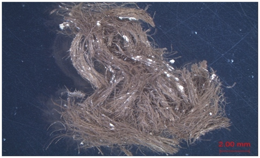

Cross-sectional observation
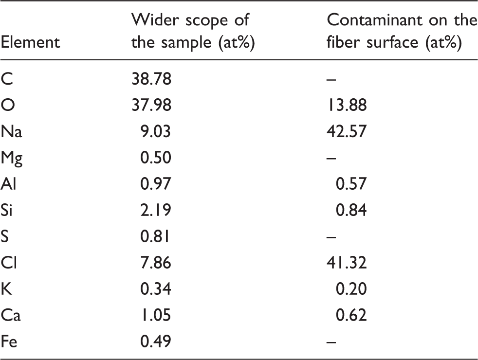
Cross-sectional scanning electron micrographs of relic wool fabric fibers and artificially aged samples are shown in Figure 3. Compared with the untreated sample (Figure 3(a)), the cross-section of the relic wool fiber sample (Figure 3(b)) is characterized by marked deterioration, as indicated by the blurred cross-sectional edges. Moreover, numerous micro-holes and some cracks are observed in the medulla of the relic wool fibers. The medulla and the cortex, which determine the physical and chemical properties of wool fibers, have suffered serious degradation, which would lead to a substantial decline in the sample’s mechanical properties. As shown in Figure 3(c) and (d), wool fibers of Na-8d are arranged loosely, while the fiber diameters of Na-24d are reduced significantly and the fibers are more compactly arranged. As can be seen from Figure 3(e) and (f), the wool fibers of Ca-8d are arranged loosely with relatively clear cross-sectional edges. However, the cross-sectional edges of Ca-24d have practically disappeared, and the cross-sections of the wool fibers appear somewhat as irregular polygons with a large number of micro-holes and few cracks.
Images of the cross-sections of wool fibers of (a) untreated, (b) relic, (c) Na-8d, (d) Na-24d, (e) Ca-8d, and (f) Ca-24d artificial aging (1: micro-holes; 2: cracks).
Cross-sectional analysis indicates that NaCl or CaCl2 and the hydrothermal condition caused severe changes to the cross-sectional structure with increasing treatment time. Wool fibers were arranged loosely in the early aging period, but were arranged more compactly following prolonged aging treatment. The most likely cause of this phenomenon would seem to be the hydration of salt in the initial stages of aging. A large amount of water absorbed in the interior of the fiber resulted in fiber expansion, leading to the destruction of secondary bonds inside the wool fiber and a decrease in intermolecular forces. As the aging treatment progressed, salts entered the fibers and assisted keratin degradation, leading to substantial loss of mass and a decrease in fiber diameter. Wool fabrics subjected to the NaCl- or CaCl2-hydrothermal aging treatment exhibit a surface morphology that is closer to that of the relic wool fibers.
Surface morphological observation
Artificially aged samples were subjected to scanning electron microscopy to evaluate morphological changes that might have occurred during prolonged treatment, leading to changes in physical and mechanical properties. As shown in Figure 4(a) and (c), no significant changes in the morphology of Na-8d were observed, and cuticle scales were clearly distinguishable. However, as shown in Figure 4(b) and (d), a number of fibers of Na-32d were broken, and a few small particles and some slight damage were observed on the scale surface, appearing as a slight shrinkage. Scanning electron micrographs of wool fibers treated with CaCl2 for different times are shown in Figure 5. As shown in Figure 5(a) and (c), the surface of the wool fibers of Ca-8d exhibited some slight morphological alterations, as demonstrated by damaged scales and some attached particles. As can be seen from Figure 5(b), some wool fibers of Ca-32d were broken. Moreover, treatment with CaCl2 resulted in a marked fiber shrinkage phenomenon, and a large number of crystallized salt particles were observed on the scale surface, as shown in Figure 5(d).
Scanning electron micrographs of (a, c) Na-8d and (b, d) Na-32d. Scanning electron micrographs of (a, c) Ca-8d and (b, d) Ca-32d.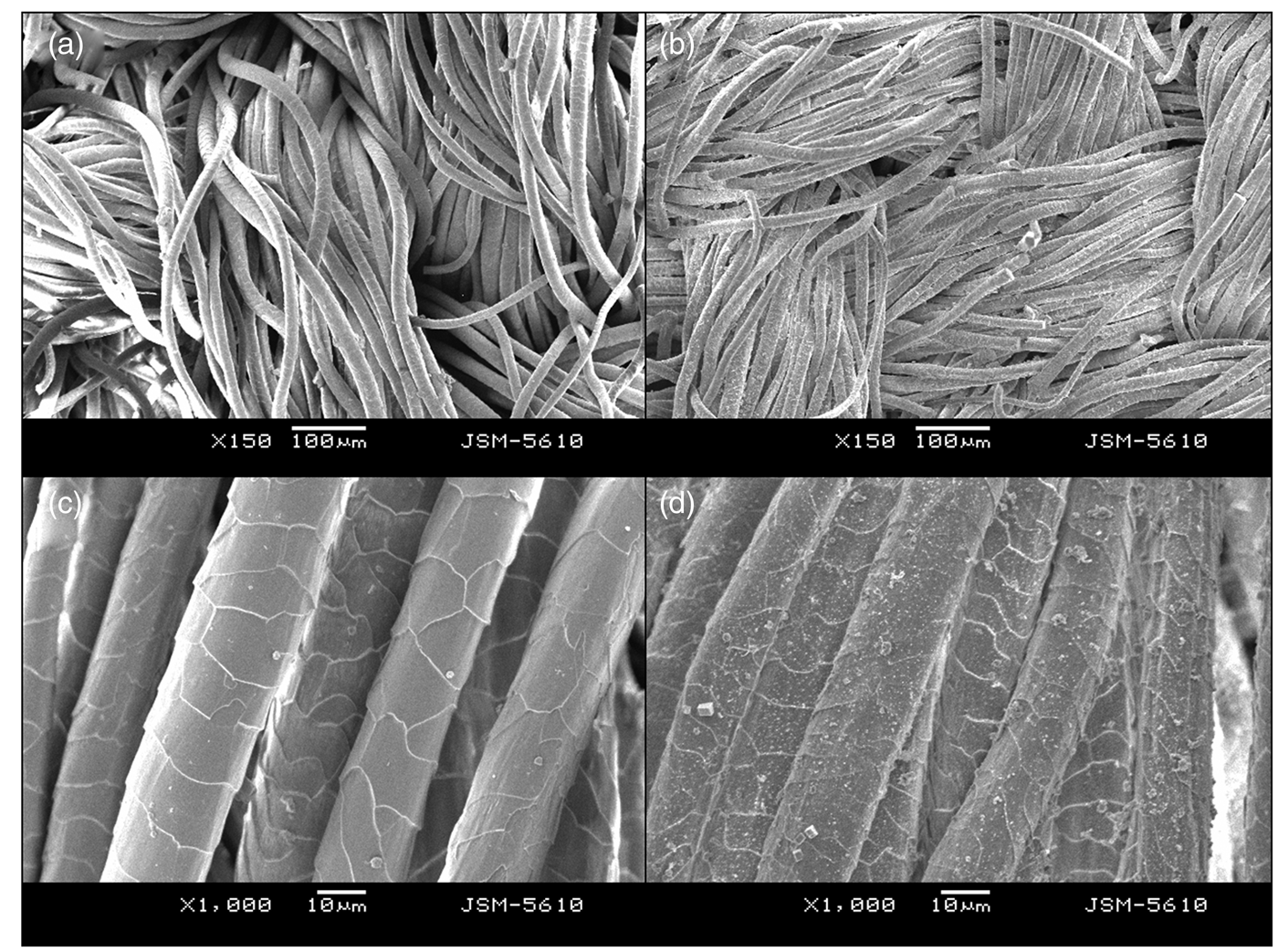

The presence of surface damage was more apparent for CaCl2-hydrothermal aged samples than for NaCl-hydrothermal aged samples. The fission of fibers of the CaCl2-hydrothermal aged samples was also more marked, and the edges of the scales were almost completely exfoliated. In addition, some crystallized salt particles were observed on the wool fiber surfaces. These results suggest that the presence of Na+ and Ca2+ causes aggravated denaturation of keratin during hydrothermal treatment. It has been reported previously that most proteins are more easily hydrolyzed when in a denatured form, relative to the case in their native form. 26 In a denatured form, the peptide bonds of keratin might be exposed, making them available for hydrolysis to small peptides during hydrothermal treatment. These results also suggest that Na+ and Ca2+ promote the degradation of disulfide groups in wool by hydrolysis during heating.
Color difference analysis
As shown in Figure 6, wool fabrics yellowed at a significantly increased rate during prolonged treatment. In the initial six days of aging treatment, the color difference of wool fabrics increased dramatically, while the rate of color change reduced during subsequent treatment time. It was also observed that the color change was accompanied by fiber embrittlement. Keratin contains specific amino acids, such as tryptophan, tyrosine, and phenylalanine. These aromatic amino acid residues within the keratin structure are the major chromophores in wool. Therefore, the most likely cause of the observed color change in the wool fabrics would appear to be associated with the oxidative breakdown of keratin following NaCl-hydrothermal or CaCl2-hydrothermal aging treatment. Under these hydrothermal conditions, some yellow substances were formed, presumably as a result of the oxidative degradation of tryptophyl, tyrosyl, and cystyl residues, perhaps coupled with the hydrolysis of certain peptide bonds. More seriously, the results indicate that the peptide chains of keratin were broken, producing more free amino acid residues. The decrease in the rate of color change is expected to be directly related to the decreased content of exposed tryptophyl, tyrosyl, and cystyl residues.
Color difference curves of NaCl-hydrothermal aging samples and CaCl2-hydrothermal aging samples.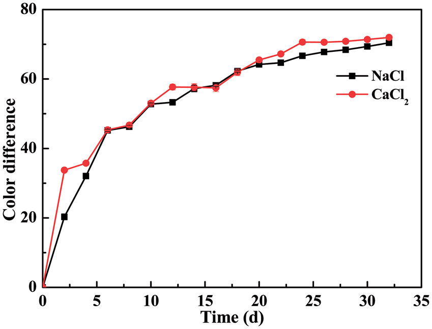
Fourier transform infrared spectral analysis
ATR-FTIR spectroscopy was used to investigate the protein molecular structure of samples subjected to NaCl-hydrothermal or CaCl2-hydrothermal aging treatment for different times, as shown in Figure 7. Characteristic absorption bands assigned to peptide bonds (-CONH-) were denoted Amide I (1700–1600 cm−1), Amide II (1540–1520 cm−1), and Amide III (1420–1400 cm−1 and 1300–1230 cm−1).1,27 In this study, changes in the structure of wool fibers owing to NaCl-hydrothermal or CaCl2-hydrothermal treatment were analyzed using the Amide I band shape, which is known to be especially sensitive to the secondary structure of the proteins.
28
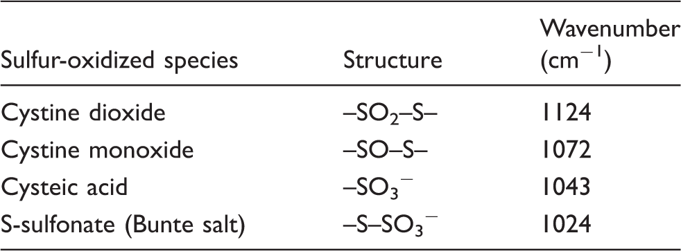
The absorption at 1657–1651 cm−1 suggests the presence of the α-helix structure, whereas the bands related to the β-sheet structure fall in the 1638–1610 cm−1 range. Peaks corresponding to a random coil structure can be detected in the 1697–1670 cm−1 range.29–31 The Amide I absorption bands were therefore fit to individual Gaussian bands according to these various spectra. In addition, cystine residues play an important role in stabilizing the structure of wool fiber and maintaining its strength. However, the disulfide bond (S–S) is the most reactive component of keratin, and, after being initiated in air, gives rise to several sulfur-oxidized species, such as S-sulfonate, cysteic acid, cystine monoxide, and cystine dioxide.
32
Table 2 lists the characteristic infrared absorbance frequencies assigned to the sulfur-oxidized species.10,32,33
ATR-FTIR spectra of (a) NaCl-hydrothermal aged samples and (b) CaCl2-hydrothermal aged samples. Characteristic wavenumbers of sulfur-oxidized species for the untreated wool fabric
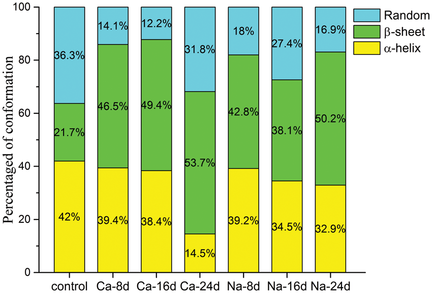
The results with respect to treatment time of the ATR-FTIR analysis represent a relative variation of the individual sulfur-oxidized species when compared with the control sample, as shown in Figure 8. Following NaCl-hydrothermal treatment, the amounts of cystine monoxide and cystine dioxide decreased below the initial level and S-sulfonate disappeared over the first eight days, while the amount of cysteic acid reached a maximum that was 3.8 times higher than that of the control sample. However, the amount of cysteic acid was reduced to a minimal level and that of cystine dioxide reached a maximum level after treatment for 24 days. Over the same period, amounts of cystine monoxide and S-sulfonate initially increased and then slightly decreased. With the exception of cystine dioxide, the content of the other monitored products did not exceed initial levels. For CaCl2-hydrothermal aged samples, the content of cystine monoxide, cystine dioxide, and cysteic acid initially reached a maximum level after the first eight days while that of S-sulfonate decreased below the initial level. Following prolonged treatment, amounts of cysteic acid and cystine monoxide decreased while those of cystine dioxide and S-sulfonate initially decreased and then increased. Cysteic acid and cystine dioxide were the dominant S-oxidized species in the wool throughout the CaCl2-hydrothermal aging period. Figure 9 shows the quantitative characteristics of the changes observed in the ATR-FTIR spectra for Amide I. During both NaCl-hydrothermal and CaCl2-hydrothermal aging treatments, two processes can be observed: a percentage decrease of disordered forms and a distinct conversion of α-helices into β-sheets.
Relative variations of oxidized sulfur species in aged wool fabrics following different treatment times. CA, cysteic acid; CDO, cystine dioxide; CMO, cystine monoxide; S-sulf, S-sulfonate. Percentage area of the secondary structure elements of control and aged samples calculated from the fitting of Gaussians in the Amide I band.
Amino acid analysis
Amino acid compositions of the cultural relic, untreated, and NaCl-hydrothermal and CaCl2-hydrothermal aging samples (mol%)
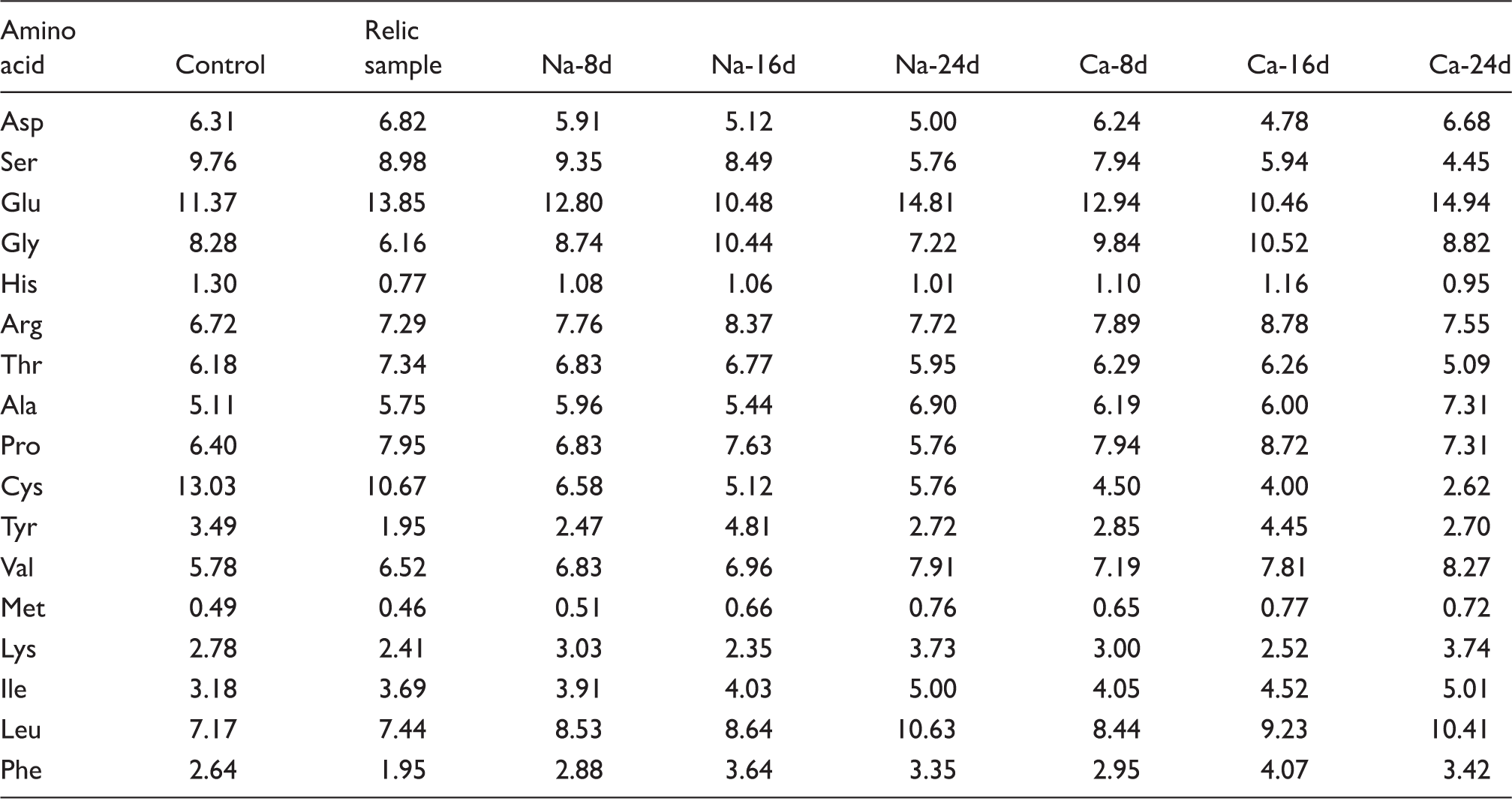
Asp, Ser, Glu, Gly, His, Arg, Thr, Ala, Pro, Cys, Tyr, Val, Met, Lys, Ile, Leu and Phe are the abbreviations of aspartic acid, serine, glutamic acid, glycine, histidine, arginine, threonine, alanine, proline, cystine, tyrosine, valine, methionine, lysine, isoleucine, leucine and phenylalanine, respectively.
These results demonstrate that NaCl- and CaCl2-hydrothermal treatment causes a decrease in the cystine content of the wool with increasing aging time. Clearly, the oxidative damage of disulfide groups led to a sharp decline in the content of cystine. The cuticle proteins are rich in cystine, and the decline of the cystine content eventually results in the breakage of the cuticle layer. Compared with NaCl-16d, the cystine content of NaCl-24d increased slightly. This increase may be caused by experimental error. Furthermore, the cystine content of the CaCl2-hydrothermal aged samples was less than that of the NaCl-hydrothermal aged samples for equivalent treatment times. This result also demonstrates that the effect of Ca2+ was greater than Na+ in promoting the degradation of disulfide groups in wool by hydrolysis during heating.
Conclusion
In this study, the effects of NaCl- and CaCl2-hydrothermal aging treatment on wool fabrics were investigated, with the aim of simulating wool fiber degradation for conducting research into the long-term preservation of fragile wool textile relics unearthed at archeological sites. Energy dispersive X-ray spectra of the contaminants adhered to wool fiber surfaces of relics obtained in the desert soil near the Xiaohe Cemetery indicated that these were soluble salts, predominantly NaCl. According to surface morphological observations, the influence of NaCl- and CaCl2-hydrothermal treatment on wool fibers was different. In CaCl2-hydrothermal aged samples, broken scales were more obvious than for NaCl-hydrothermal aged samples for equivalent treatment times. The obtained results from FTIR analysis indicated that NaCl- and CaCl2-hydrothermal treatment of wool fabrics led to a transformation of the secondary structure from that of α-helices to β-sheets. Moreover, the fluctuation of cysteic acid and cystine dioxide contents was observed to be the largest. In addition, cross-sectional observation, color measurement, FTIR, and amino acid analysis also revealed that Ca2+ played a greater role in promoting the degradation of disulfide groups during aging treatment than did Na+. It is therefore supposed that a greater CaCl2 content in the soil would lead to a greater level of damage for wool fabrics buried therein.
Footnotes
Declaration of conflicting interests
The authors declared no potential conflicts of interest with respect to the research, authorship, and/or publication of this article.
Funding
The authors disclosed receipt of the following financial support for the research, authorship, and/or publication of this article: This research was financially supported by National Key Technology R&D Program (grant number 2013BAH58F01-02), Special Funds from the Administration of Cultural Heritage of Zhejiang Province (grant number 2013211) and The Young Researchers Foundation of Key Laboratory of Advanced Textile Materials and Manufacturing Technology, Ministry of Education, Zhejiang Sci-Tech University (grant number 2013QN03).