
Abstract
Wet-laid blend nonwovens of calcium carboxymethyl cellulose fibers and chitosan powder were prepared as a new efficacious hemostatic agent, and their water absorption behavior, rupture strength in the dry and wet states, blood clotting properties, calcium ion release behavior, and cytotoxicity were investigated. The calcium carboxymethyl cellulose and chitosan nonwovens lost their structural integrity in the wet state due to water absorption and subsequent gelation, whereas the calcium carboxymethyl cellulose/chitosan blend nonwovens maintained their structural integrity. The formation of polyelectrolyte complexes between calcium carboxymethyl cellulose and chitosan after the absorption of water is believed to have contributed to the structural integrity in the wet state. In the in vitro blood clotting tests, the calcium carboxymethyl cellulose/chitosan blend nonwovens immediately absorbed blood and exhibited efficacious blood clotting characteristics. The calcium carboxymethyl cellulose/chitosan blend nonwovens were not cytotoxic. Considering both the structural stability in the wet state and blood clotting performance, calcium carboxymethyl cellulose/chitosan (7/3 w/w) blend nonwoven was found to be the most effective hemostatic agent of those assessed in this study, and has good potential for commercial application.
Keywords
Hemorrhage commonly occurs during surgical operations. 1 In general, 20% of total blood volume or more is lost during surgery. 2 Severe hemorrhage can also occur for other reasons, including trauma. Uncontrolled hemorrhage is a significant, major cause of traumatic death.3,4 Therefore, various hemostatic agents have been developed for surgical procedures and emergency use to control hemorrhage rapidly and effectively. For such products to be efficacious, they need to make effective contact with the bleeding surface, exhibit fast blood absorption, and be easily handled and prepared. 5 While hemostatic agents exist in a variety of compositions and dosage forms, they can incorporate collagen, chitosan, oxidized cellulose, kaolin, or thrombin.6–9 Moreover, they can be formulated as a powder, hydrogel, foam, knit, or nonwoven sheet. Powder- and hydrogel-type hemostatic agents can cover irregular surfaces. They are limited to relatively severe bleeding and have a higher difficulty of use compared to other forms. Foam-type hemostatic agents can absorb larger amounts of blood than other forms and can be removed easily from the wound. Nonwoven-type hemostatic agents are easy to handle and can control severe bleeding over wider wounds. They can also be used with pressure application. The most appropriate hemostatic agent is determined according to the situation and the degree of hemorrhage. 6
Polyelectrolyte complexes (PECs) are formed by electrostatic interaction between the polycations and polyanions, which are generally water insoluble and form hydrogels. Some PECs have been studied for wound dressing applications.10–12 Representative natural polycations and polyanions are chitosan (CS) and carboxymethyl cellulose (CMC), respectively. CS has been studied widely for biomedical applications owing to its biocompatibility, biodegradability, hemostasis, and antimicrobial activity.13–17 Regarding its hemostatic behavior, CS bonds with platelets and red blood cells to form a gel-like clot that seals a bleeding vessel.18,19 CMC has been widely used in pharmaceutical, cosmetic, and food applications owing to its biocompatibility, biodegradability, and non-toxicity.20–22 CMC is generally used in the form of its sodium salt (Na-CMC), but it is also used as a calcium salt (Ca-CMC) for the disintegration and dispersion of tablets. Na-CMC is soluble in water, whereas Ca-CMC is not completely soluble but swells to several times its original volume upon contact with water and turns into a gel. 23 Because calcium ions can accelerate coagulation through platelet aggregation, a dressing containing calcium ions can be effective for hemostasis by releasing them upon contact with a wound.24–27
In the present study, blended nonwoven-type hemostatic agents of Ca-CMC and CS were prepared using a wet-laid nonwoven process for rapid hemostasis. Most studies on hemostatic agents focused on a single material.8,28,29 Hemostatic agents composed of multiple materials have not been studied satisfactorily. The mechanical properties, blood clotting effect, calcium ion release behavior, and cytotoxicity of the Ca-CMC/CS blend nonwovens were investigated systematically according to the Ca-CMC/CS blend ratio. In addition, their blood clotting effects were compared with those of commercial nonwoven-type hemostatic agents.
Materials and methods
Materials
Viscose rayon fibers (2.1 dtex, 6 mm) were purchased from Kelheim fibers GmbH (Germany). Chitosan powder (HFP®, degree of deacetylation: 85%, molecular weight (MW) ∼200 kDa) were acquired from Smart Safe Zone Co. Ltd. (Korea). Monochloroacetic acid (MCA, 99%), acetic acid (99.7%), and calcium chloride (CaCl2) were obtained from Daejung Co. (Korea). Sodium hydroxide solution (25 wt%), tert-butanol (TBA, 99.5%) and methanol (99.5%) were purchased from Samchun Co. (Korea). QuikClot® and Celox™ Rapid, which are commercialized nonwoven-type hemostatic dressings, were purchased from Z-Medica Corporation (USA) and Medtrade Products Ltd. (UK), respectively.
Preparation of Ca-CMC fibers
Ca-CMC fibers were prepared by treating Na-CMC fibers with CaCl2. Na-CMC fibers were first prepared by a two-step carboxymethylation process of viscose rayon fibers, i.e. alkalization and etherification. A 100 g sample of viscose rayon fibers was treated with 1.5 L of a 25 wt% sodium hydroxide solution at room temperature for 20 min to produce alkali cellulose fibers. The alkali cellulose fibers were then added to 2.7 L of TBA and stirred using an overhead stirrer at 1000 rpm and heated to 70℃. A 300 mL volume of TBA, mixed with MCA, was added to the fiber/TBA mixture for the etherification reaction, and the mixture was additionally stirred at 1000 rpm for 20 minutes. The reaction temperature was maintained at 80℃ during the etherification reaction. After etherification, the mixture was washed with an excess of methanol/water (9/1, v/v) and neutralized by acetic acid. Finally, the fibers were rewashed with methanol. The washed Na-CMC fibers were dried in a vacuum oven at 50℃ for six hours. The degree of substitution (DS) of Na-CMC was determined according to modified ASTM D1439-03, as described elsewhere. 30 The MW of Na-CMC was measured by gel filtration chromatography (GFC, Agilent 1100, Agilent Technologies).
The Na-CMC fibers were converted to Ca-CMC fibers by treating them with CaCl2 in an organic solvent. A 3 L volume of ethanol/water (8/2, v/v) containing CaCl2 (0.05 mol/L) was prepared. A 30 g sample of the Na-CMC fibers was weighed, immersed in the prepared CaCl2 solution, and stirred at room temperature for 40 minutes. Subsequently, the fibers were washed with ethanol/water (8/2, v/v) and rewashed with methanol. The washed fibers were dried in a vacuum oven at 50℃ for six hours. The sodium and calcium contents in the fibers were analyzed by inductively coupled plasma optical emission spectrometry (ICP-OES, ULTIMA II, Jobin-Yvon).
Preparation of blend nonwovens
Blend ratio and dispersion medium of wet-laid nonwovens
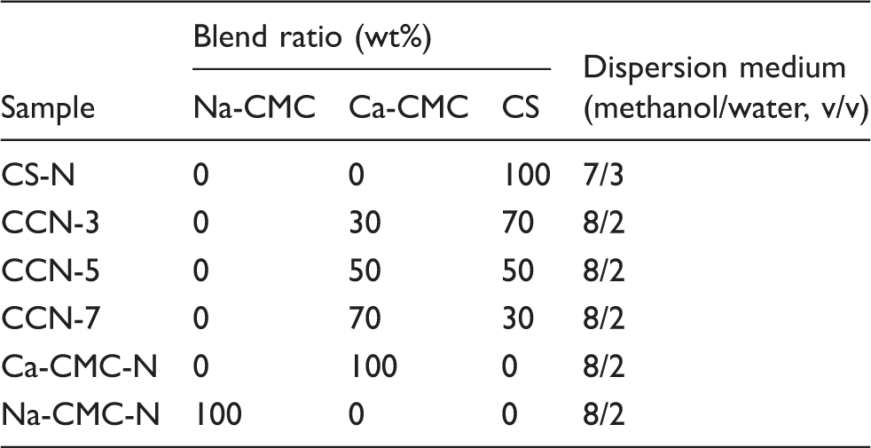
Na-CMC: sodium carboxymethyl cellulose; Ca-CMC: calcium carboxymethyl cellulose; CS: chitosan; N: nonwovens.
Characterization of blend nonwovens
The Fourier transform infrared (FT-IR) spectra of the samples were obtained using the attenuated total reflectance (ATR) technique to examine the changes in the molecular structure during the carboxymethylation and ion exchange processes. The surface morphologies of the samples were observed by field emission scanning electron microscopy (FE-SEM; Hitachi SU-8010, Hitachi High Technologies Co. Japan). The samples were sputter-coated with gold for 200 seconds using a 15 mA current prior to imaging. The thickness of the samples was measured five times by a thickness tester (ID-C112, Mitutoyo Co., Japan). The water absorption time and contact angle were measured by placing a drop of water on the blend nonwovens using a drop shape analyzer (DSA100, KRÜSS, Germany).28,31 The water absorption time was defined as the time required for the drop to become flat on the nonwoven surface. The volume of water droplet was 3 µL. The contact angle and water absorption time were measured five times. A penetration test was performed to investigate the mechanical properties in both the dry and wet states. Dry nonwovens were cut into 2 × 2 cm 2 sized pieces. The rupture strengths, defined as maximum forces during penetration, were measured using a TA-XT plus Texture Analyzer (Stable Micro Systems, Godalming, Surrey, UK) equipped with a 5 mm diameter cylinder probe (P/5). 32 The measuring probe was lowered at a speed of 1 mm/s. For the penetration tests in the wet state, the dry samples were immersed in distilled water at room temperature for five minutes. The specimens were then removed from the water and placed gently on a filter paper for 30 seconds to remove the excess water. Penetration tests in the wet state followed the same procedure as that used in the dry state. All penetration tests were repeated three times and the results were averaged.
Blood collection
Human blood collection was approved by the Institutional Review Board (IRB# P01-201604-33-001). Human blood was collected from heathy volunteers, and nine parts of whole blood were mixed with one part of 3.8% sodium citrate as an anticoagulant.
In vitro blood clotting test
The in vitro blood clotting tests were conducted using research methods reported elsewhere.28,31,33 Nonwoven samples were cut into 1 × 1 cm 2 pieces and placed into 50 mL tubes. A 100 µL sample of blood was dropped slowly onto each sample, followed by the addition of 10 µL of 0.2 M CaCl2 solution to trigger blood coagulation. The tubes were incubated at 37℃ for 0.5, 1, 2, 3, and 5 min. Subsequently, 12.5 mL distilled water were added to each tube and blended carefully without disturbing the clot for hemolyzing the untrapped red blood cells in the clot. A 1 mL sample of the solution was then collected from the tube and placed in a 1.5 mL tube. Every 200 µL of the solution was added to a 96-well plate and the level of hemoglobin in the unclotted blood was determined by measuring the absorbance at 540 nm using a microplate reader (iMark™, Bio-Rad Laboratories, Inc.). The blood clotting test was repeated three times.
Morphological changes of the blend nonwovens during blood coagulation were also investigated. Nonwoven samples were cut into 1 × 1 cm 2 pieces and placed on dishes. A 100 µL sample of blood was slowly dropped onto each sample, followed by the addition of 10 µL of a 0.2 M CaCl2 solution. The dishes including the samples were maintained at 37℃ for five minutes. After incubation, the samples were fixed in 2% paraformaldehyde/phosphate-buffered saline (PBS) at room temperature for two hours. The fixed samples were washed repeatedly in ethanol/water with a gradually increasing ethanol ratio and dried at room temperature. The morphologies of the fixed samples were observed by field emission scanning electron microscopy (FE-SEM; Hitachi SU-8010, Hitachi High Technologies Co. Japan). The samples were sputter-coated with gold for 200 seconds using a 15 mA current prior to imaging. 31
Calcium ion release
The calcium ion release tests were performed only for CCN-5. A 2 × 2 cm
2
piece of CCN-5 was immersed in 25 mL of a 0.9% saline solution at 37℃ and agitated in a shaking bath at 100 shakes per minute. A 20 mL volume of the solution was collected at 0.5, 1, 2, 3 and 5 min after immersion, and the content of calcium ions was measured by ICP-OES.
34
The release ratios of calcium ions were calculated using the following Equation (1):

In vitro cytotoxicity
Cell culture
L-929 cells derived from mouse fibroblasts were obtained from the KCLB (Korean Cell Line Bank). The cells were grown as monolayer cultures in the culture medium of RPMI media (RPMI-1640 Medium (1X) with 2.05 mM L-Glutamine, 0.1 µm sterile filtered, HyClone®) further supplemented with 10% fetal bovine serum (FBS, HyClone®) and 1% penicillin-streptomycin solution (HyClone®). The cells were cultured in an incubator in a humidified atmosphere containing 5% CO2 at 37℃.
Cytotoxicity
The cytotoxicity was evaluated from the extracts of the nonwoven samples according to ISO 10993-5. The nonwovens were sterilized using an EO Gas Sterilizer (MK-EO 30, MKT Co.,LTD, Korea) prior to the cytotoxicity tests. The sample extracts were prepared according to ISO 10993-12. The nonwoven samples were incubated with the RPMI media at an extraction ratio of 0.02 g/mL at 37℃ for 24 hours. The L-929 cells were seeded at a density of 1 × 10⁴ cells/well in a 96-well tissue culture plate with 100 µL of growth medium and incubated (5% CO2, 37℃, >90% humidity) for 24 hours. After 24 hours incubation, the culture medium was aspirated from the cells and replaced with 100 µL of the pretreated extracts in each well. The RPMI media and 1% acetic acid were used as the blank control and positive control, respectively. The 96-well plate, including the cells and extracts, was incubated (5% CO2, 37℃, >90% humidity) for 24 hours. After incubation, the extracts from the cells were removed and replaced with 100 µL/well of growth medium, and 20 µL/well of CellTiter 96® Aqueous One Solution Cell Proliferation Assay (Promega, Madison, USA) were then added.
35
After additional incubation for four hours, the optical density was measured at a wavelength of 490 nm using a microplate reader (iMark™, Bio-Rad Laboratories, Inc.). The cell viability (%) was calculated using the following Equation (2):
35

Results and discussion
Properties of the Ca-CMC/CS blend nonwovens
Ca-CMC fibers were prepared by treating Na-CMC fibers (DS = 0.36, MW ∼ 100 kDa) with a CaCl2 solution. Figure 1 shows the FT-IR spectra of the viscose rayon, Na-CMC, and Ca-CMC fibers. A strong absorption band at 1605 cm−1 assigned to the stretching vibration of the carboxylate group was observed in the Na-CMC and Ca-CMC fibers, indicating that the carboxymethylation of viscose rayon fibers had proceeded well.
36
The Na-CMC and Ca-CMC fibers were also analyzed by ICP-OES to confirm the exchange of sodium ions to calcium ions. The sodium content in the Na-CMC fibers was 3.21 ± 0.18% but the calcium content was below the limit of quantification. In contrast, the sodium content in the Ca-CMC fibers was 1.10 ± 0.06% and the calcium content was 1.92 ± 0.05%. A significant amount of sodium ions in the Na-CMC fibers had been replaced with calcium ions.
FT-IR spectra of viscose rayon, sodium carboxymethyl cellulose (Na-CMC), and calcium carboxymethyl cellulose (Ca-CMC) fibers.
Figure 2 presents SEM images of various nonwoven specimens. The CS powder had a flake form. Na-CMC and Ca-CMC fibers seemed to have no significant morphological difference. CS-N could be fabricated due to the partial gelation of CS powder by water in the dispersion medium. More Ca-CMC fibers were observed than CS powders on the surface of blend nonwovens with increasing blend ratio of Ca-CMC fibers, which means that the blend nonwovens were well fabricated (Figure 2(b)–(d)).
SEM images of various wet-laid nonwovens: (a) CS-N, (b) CCN-3, (c) CCN-5, (d) CCN-7, (e) Ca-CMC-N, and (f) Na-CMC-N. Na-CMC: sodium carboxymethyl cellulose; Ca-CMC: calcium carboxymethyl cellulose; CS: chitosan; N: nonwovens.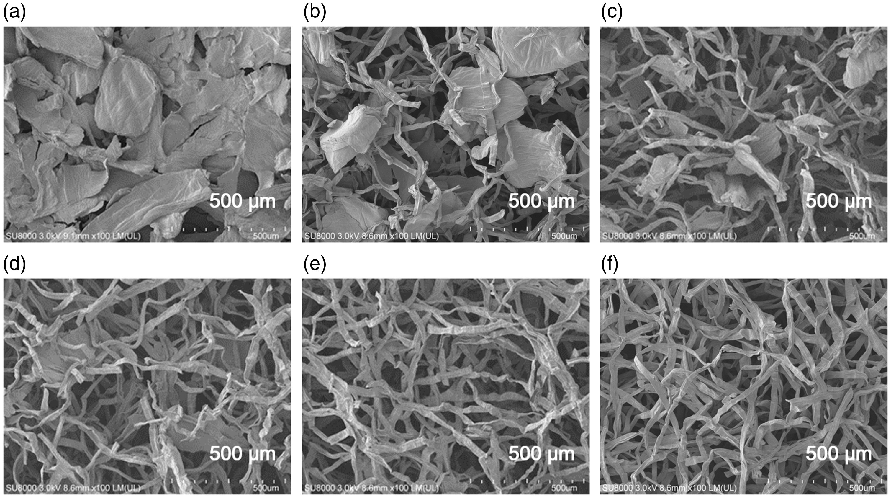
Thickness, initial contact angle, and absorption time of water droplet on wet-laid nonwovens
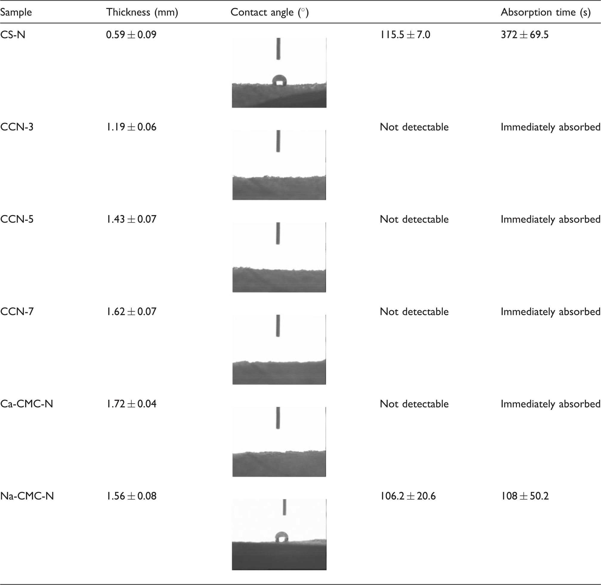
Na-CMC: sodium carboxymethyl cellulose; Ca-CMC: calcium carboxymethyl cellulose; CS: chitosan; N: nonwovens.
Mechanical properties
The mechanical properties of hemostatic dressings are very important in terms of handling and blood coagulation. In particular, their mechanical properties in the wet state are crucial because they become wet with blood from the bleeding site.
39
Figure 3 shows the rupture strengths of the various nonwovens measured using a texture analyzer in the dry and wet states. Ca-CMC-N had the highest rupture strength in the dry state. The rupture strengths of CS-N and Ca-CMC-N in the wet state could not be measured because they lost their structural integrity in the wet state due to water absorption and subsequent gelation. In the cases of CCN-3, CCN-5, and CCN-7 in the dry state, their rupture strengths increased with increasing Ca-CMC content. The inter-fibrillar friction by Ca-CMC fibers is believed to have enhanced the rupture strength. Interestingly, CCN-3, CCN-5, and CCN-7 maintained their shapes without disintegration in the wet state, and their rupture strengths could be measured, even in the wet state. The PECs of Ca-CMC and CS are believed to have formed and contributed to the structural stability in the wet state. Figure 4 shows pictures of CCN-3, CCN-5, and CCN-7 after soaking in distilled water for five minutes. CCN-7 showed the best structural integrity, as evidenced by its rupture strength. As a result, Ca-CMC/CS nonwovens containing more than 50% Ca-CMC are expected to be desirable in terms of wet stability.
Rupture strengths of various nonwovens in the (a) dry and (b) wet states. Ca-CMC: calcium carboxymethyl cellulose; CS: chitosan; N: nonwovens. Photographs of CCN-3, CCN-5, and CCN-7 after immersion in distilled water for five minutes.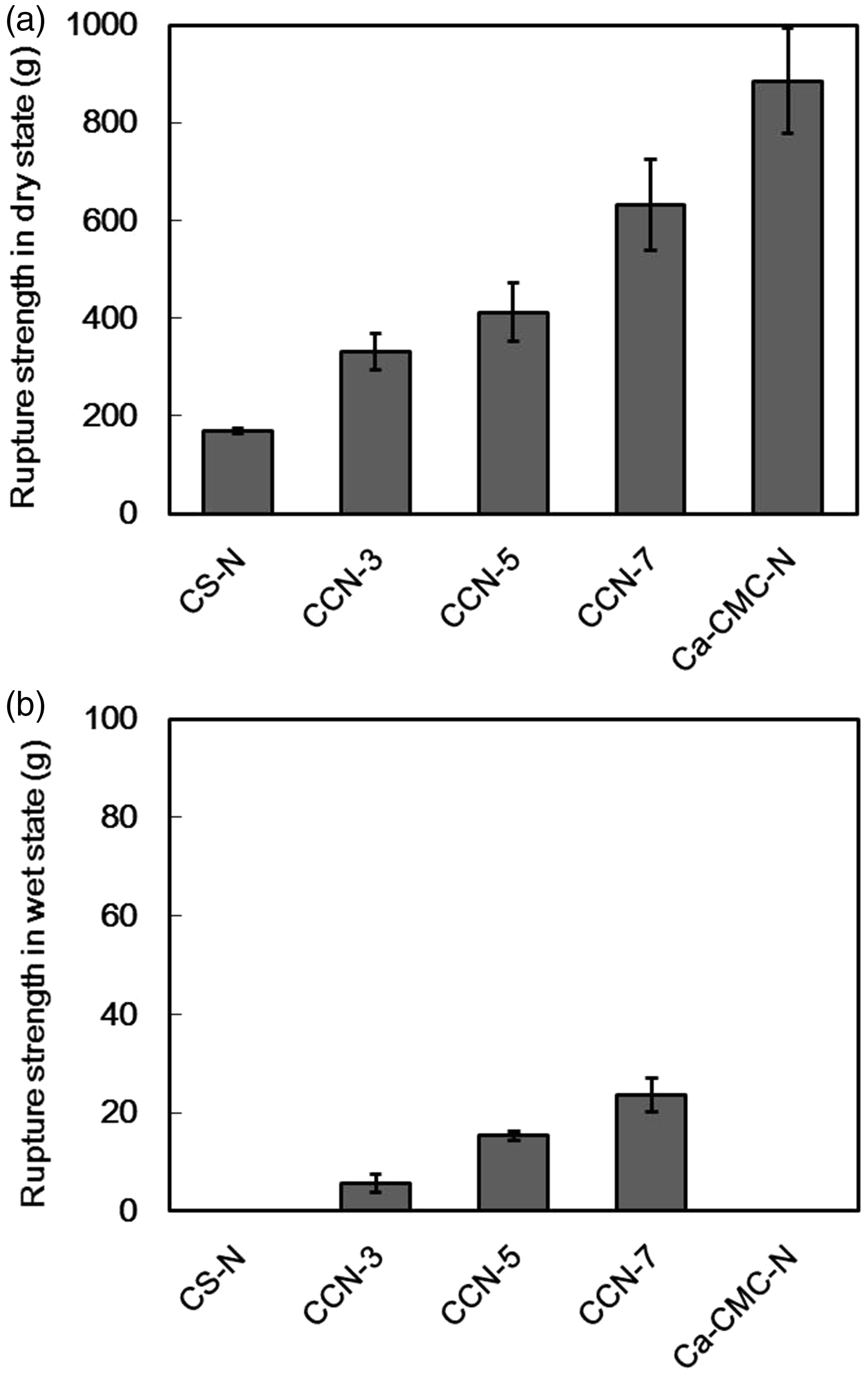

In vitro blood clotting tests
Blood clotting tests were conducted to examine the hemostatic effects of Ca-CMC/CS blend nonwovens. The level of hemoglobin in the unclotted blood was determined by measuring the absorbance at 540 nm using a microplate reader. A lower absorbance at 540 nm indicates fewer blood cells in the test solution, meaning more effective clotting. The whole blood not applied to a nonwoven was used as the control. Figure 5 shows the in vitro blood clotting of various nonwovens and commercial hemostatic dressings with the incubation time. The initial absorption of blood is very important for achieving hemostasis in a very short time.
28
In practice, the water absorption time listed in Table 2 is closely related to the blood clotting effects. Ca-CMC-N had a lower absorbance than Na-CMC-N. Ca-CMC-N absorbed blood quickly, as shown in Table 2, and released calcium ions that could facilitate rapid clot formation through platelet aggregation. On the other hand, CS-N absorbed blood slowly (Table 2), which delayed blood clotting. CCN-3, CCN-5, and CCN-7 absorbed blood immediately, which led to significantly lower absorbance, even at 30 seconds. The Ca-CMC/CS blend nonwovens showed much better blood clotting rates than QuikClot® and Celox™ Rapid.
In vitro blood clotting tests of various nonwovens and commercial hemostatic dressings over the incubation time. Na-CMC: sodium carboxymethyl cellulose; Ca-CMC: calcium carboxymethyl cellulose; CS: chitosan; N: nonwovens.
Figure 6 shows the surface morphology of various nonwovens after in vitro blood clotting tests with five minute incubation. Figure 6(a) shows the smooth surface of CS-N with several blood cells entrapped within CS-N due to the dissolution of CS. The Ca-CMC/CS blend nonwovens showed a better blood clotting effect than the other nonwovens. In the case of CCN-3, although some complexes with blood cells could be observed, its surface was slightly smooth due to the dissolved CS (Figure 6(b)). In the cases of CCN-5 and CCN-7, complexes with blood cells and fibrins were clearly observed (Figure 6(c) and (d)). In contrast, attached blood cells and partially formed fibrin were observed on the surface of Na-CMC-N and Ca-CMC-N. Ca-CMC-N showed a slightly better result than Na-CMC-N due to the calcium ions released from Ca-CMC. Overall, CCN-5 and CCN-7 were found to be the most effective hemostatic agents in the aggregation of blood cells and fibrin formation than the other nonwovens.
Scanning electron microscopy images of various nonwovens, QuikClot® and Celox™ Rapid after in vitro blood clotting tests with five minute incubation: (a) CS-N, (b) CCN-3, (c) CCN-5, (d) CCN-7, (e) Ca-CMC-N, (f) Na-CMC-N, (g) QuikClot® and (h) Celox™ Rapid. Na-CMC: sodium carboxymethyl cellulose; Ca-CMC: calcium carboxymethyl cellulose; CS: chitosan; N: nonwovens.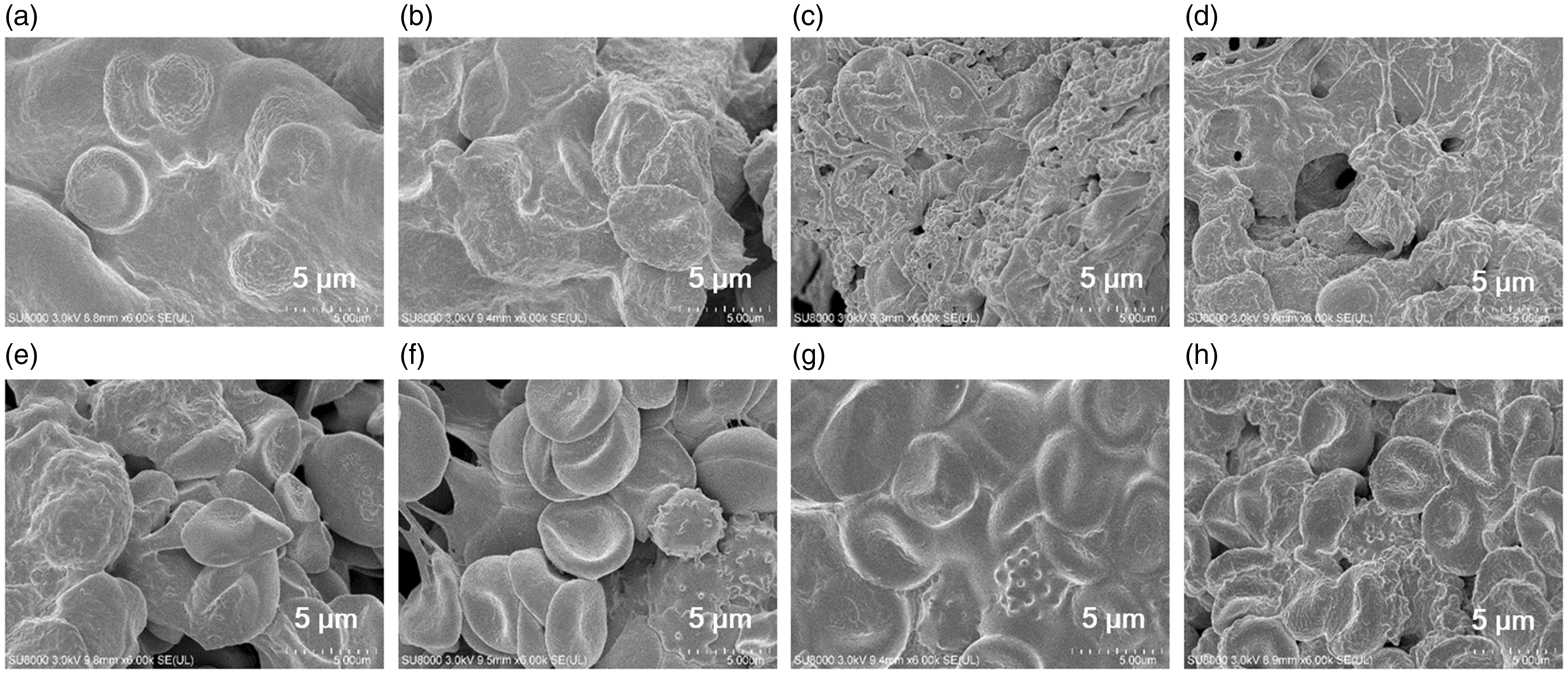
Calcium ion release into saline solution
Calcium ions accelerate coagulation during the blood clotting process. A previous study reported that calcium ions in calcium alginate for wound dressings could be released quickly because sodium ions in human body fluid could be exchanged with calcium ions.40–42 Similarly, ion exchange between sodium ions and calcium ions might also occur in hemostatic dressings containing Ca-CMC. Therefore, the concentration of calcium ions released from CCN-5 into a saline solution was measured by ICP-OES. Figure 7 shows the release profile of the calcium ions from CCN-5 over time. The release ratio of calcium ions was calculated based on the amount of calcium present in CCN-5. Most of the calcium ions in CCN-5 were released into the saline solution within 30 seconds. This suggests that the Ca-CMC/CS blend nonwovens are useful hemostatic dressings because their rapid release of calcium ions can accelerate blood coagulation by forming a fibrin clot from fibrinogen.
Release of calcium ions from CCN-5 into a saline solution over time.
Cytotoxicity
In vitro cytotoxicity tests were performed to assess the biocompatibility of the Ca-CMC/CS blend nonwovens. Figure 8 shows the cell viability of the blend nonwovens. The Ca-CMC/CS blend nonwovens were not cytotoxic, supporting their good biocompatibility. In the case of CCN-3, it was assumed that the extracted CS affected the cell viability. The cytotoxicity of CS was reported to be influenced by its MW and degree of deacetylation. A higher MW and degree of deacetylation of CS results in higher cytotoxicity and lower cell viability.
43
Cell viability of the blend nonwovens.
Conclusions
Ca-CMC fibers were prepared by the carboxymethylation of viscose rayon and subsequent ion exchange reaction. The sodium and calcium contents in the Ca-CMC fibers were 1.10 ± 0.06% and 1.92 ± 0.05%, respectively. The Ca-CMC/CS blend nonwovens were prepared using a wet-laid process. They could absorb a drop of water as soon as it was dropped, suggesting superior blood absorption characteristics. The rupture strengths of the Ca-CMC/CS blend nonwovens increased with increasing Ca-CMC fiber content and their structure integrity was maintained in the wet state. On the other hand, Ca-CMC-N lost its structural integrity in the wet state due to the dissolution and subsequent gelation of Ca-CMC fibers. The PECs of Ca-CMC and CS formed contributed to the structural integrity in the wet state. In the in vitro blood clotting tests, CCN-3, CCN-5, and CCN-7 immediately absorbed blood and exhibited efficacious blood clotting characteristics. Considering both the structural stability in the wet state and blood clotting performance, CCN-7 is the most effective hemostatic agent of the nonwovens assessed in this study.
Footnotes
Declaration of conflicting interests
The authors declared no potential conflicts of interest with respect to the research, authorship, and/or publication of this article.
Funding
The authors disclosed receipt of the following financial support for the research, authorship, and/or publication of this article: This work has been supported by Grant EO-16-0003 and EO-17-0033 from Korea Institute of Industrial Technology.