
Abstract
Bio-based polyurethane is synthesized from biodegradable polycaprolactone, methylene diphenyl diisocyanate and 1,4-butanediol. The bio-based polyurethane is blended with branched polyethyleneimine by a solution casting method and further treated with glutaraldehyde. From nuclear magnetic resonance, Fourier-transform infrared spectroscopy, leaching tests and contact angle measurements, it was found that a semi-interpenetrating polymer network structure is induced by the glutaraldehyde treatment of the bio-based polyurethane/branched polyethyleneimine blend film, which resulting from the crosslinking of branched polyethyleneimine by imine bonds formed from the amine-aldehyde reaction between branched polyethyleneimine and glutaraldehyde. In addition, the glass transition temperature, Young’s modulus and the shape retention results show that the mechanical strength of bio-based polyurethane, which is weakened by the plasticizing effect of branched polyethyleneimine, is restored by the formation of the semi-interpenetrating network structure. We found that the bio-based polyurethane/branched polyethyleneimine with a semi-interpenetrating network shows a much higher affinity for Acid Red 4 than bio-based polyurethane, and the wet fastness of dye is significantly improved by the formation of the semi-interpenetrating network.
Keywords
Since the development of Perlon U, a polyurethane (PU) fiber, by Bayer in 1937, PU has been widely used in various applications such as composites,1–5 medical materials,6,7 artificial leather, 8 insulation 9 and fibers.10,11 Many studies have been performed owing to the excellent chemical and physical properties of PU as well as its biocompatibility and wide applications to a range of different industries. In particular, interest has recently focused on bio-based PU (bio-PU) produced from vegetable12–15 and biodegradable materials.16–18 Bio-PU represents a novel material, which is environmentally friendly and safe in contact with the human body. The majority of studies into bio-PU have focused on the thermal and mechanical properties, and biodegradability of bio-PU.19–23 The dyeing properties of bio-PU are another important factor, which affect the material’s use as a fiber, but have not yet been widely studied.
PU is hydrophobic, which allows it to be dyed with disperse dyes. However, the bonding strength between PU and disperse dyes is weak and the wet fastness of dye is poor. Thus, attempts have recently been made to dye PU with acid or reactive dyes instead of disperse dyes. Wang and Tzun 24 reported hydroxyl-pendant PU with the ability to be dyed via its covalently bonded reactive dyes. Methods have also been proposed to increase the dyeability of reactive and acid dyes by spinning PU-urea elastic fibers with a hyperbranched polyester having tertiary amines as chain extenders of PU.25,26 Such approaches, however, can severely change the original physical properties of PU and are limited by the use of relatively expensive materials and the need for pre-synthesis in the early stages of PU production. Thus, it is necessary to develop new simple ways to improve the dyeability of PU without severely changing its properties.
Herein, bio-PU was polymerized using polycaprolactone diol (PCL), which is known to be biodegradable. The bio-PU was blended with branched poly(ethylene imine) (b-PEI) and further crosslinked with glutaraldehyde (GA) to endow the bio-PU with an acid dyeability and a strong wet fastness. Our results are expected to increase the application of bio-PU and will open new ways for bio-PU to be used in interior products, protective coatings and leathery claddings, as well as textiles for clothes. 27
Experimental details
Materials
PCL (Mn = 2000 g mol−1), dibutyltin dilaurate (DBTDL), Acid Red 4 and b-PEI with molecular weights of 800 and 25,000 g mol−1 were purchased from Sigma Aldrich. Methylene diisocyanate (MDI) and 1,4-butanediol (BD) were purchased from Across Organics, and GA was purchased from Daejung Chemical. The purchased reagents were used without further purification. The deionized water used in this study had a resistance above 18.0 MΩ.
Synthesis of bio-based polyurethane
PCL diol was dissolved in dimethylformamide (DMF) at 80℃ for 30 min under a nitrogen flow and reflux condensation conditions. The DBTDL catalyst was added, and then MDI was added at intervals of 30 min in six successive stages. After the final MDI addition, BD was added, stirred for 20 min, and the reaction was terminated by methanol. At this time, the molar ratio of PCL, MDI and BD was 1: 2: 1. The reaction solution was precipitated in methanol to obtain a solid component, and the resulting solid was washed with methanol, filtered and vacuum dried at room temperature.
Preparation of bio-PU/b-PEI blending film
A polymer solution of bio-PU and b-PEI (9:1, w/w) was prepared at a concentration of 5% in chloroform, stirred for 6 h, and then solution cast on a glass substrate, resulting in the bio-PU/b-PEI film. The resulting bio-PU/b-PEI film was vacuum dried at room temperature for 24 h.
Glutaraldehyde treatment
After immersing the bio-PU/b-PEI film in GA aqueous solution (10% owb.), the amine-aldehyde reaction between b-PEI and GA proceeded with an aluminum sulfate (AS) catalyst at room temperature for 10 min. The treated film was washed with water and dried at room temperature for 6 h. The bio-PU/b-PEI blended films with b-PEI molecular weights of 25,000 and 800 g mol−1 are denoted herein as MI-25000 and MI-800, respectively. The GA-treated MI-25000 and GA-treated MI-800 are denoted as MIG-25000 and MIG-800, respectively.
Characterization
1H nuclear magnetic resonance (1H NMR) and Fourier transform infrared (FT-IR) spectra were recorded on a Bruker Avance 400 spectrometer (solvent: CDCl3) and a Bruker VERTEX 70 spectrophotometer (attenuated total reflectance (ATR)), respectively, to verify the synthesis of bio-PU and MI samples. The molecular weight of the synthesized bio-PU was measured using a Waters gel permeation chromatography (GPC) system. Samples for measurement were prepared by diluting the solution to a concentration of 1% (w/v) in DMF and passing through a 0.25 µm filter. The Mn and polydispersity index (PDI) values were calculated from the resulting GPC data based on calibration with poly(ethylene glycol) standards of known molecular weights and PDI. The contact angle was evaluated with a DSA 100 (Krüss, Germany). A 4 mL portion of distilled water was dropped five times in different positions of the film, and the average value was calculated. To check whether b-PEI leached out from the MI and MIG samples, specimens were prepared with dimensions of 20 × 20 × 0.25 mm3. Then, the specimens were immersed in water with a liquor ratio of 200:1, and stirred at 100 rpm for 6 and 12 h. The degree of leaching was evaluated by gravimetric analysis. Prior to the gravimetric measurement, all samples were vacuum dried for more than 12 h.
Differential scanning calorimetry (DSC) measurements were performed under a nitrogen atmosphere over the temperature range of 90–50℃ at a heating rate of 10℃ min−1 using a Perkin-Elmer Diamond DSC (USA). The mechanical properties of the film samples were measured by the ASTM D 638-02a method with a Hounsfield (England) H10KS at a strain rate of 100 mm min−1 with a 100 N static load cell. To evaluate the shape retention characteristics, the specimens were stretched to 100% length at a strain rate of 100 mm min−1 and held for 5 min. The external force was then removed and the length after 10 min was measured. The measured results were calculated by applying Equation (1)

Evaluation of dyeing properties
To evaluate the dyeing properties, the film specimens were immersed in Acid Red 4 solution adjusted to a liquor ratio of 15:1, a concentration of 3% (owf), a dyeing pH of 3.5 (formic acid), a dyeing temperature of 25℃ and stirred at 100 rpm for 2 h. The specimens were then rinsed with water and dried at room temperature. The reflectance (R) values of the dyed film samples were then measured through X-rite Color i7 (USA), and K/S values were quantitatively derived by applying the measured reflectance values to the Kubelka–Munk formula (Equation (2))

To evaluate the migration of the dye in the samples, the dyed specimens were immersed in water with a 200:1 liquor ratio, and stirred at 100 rpm for 6 and 12 h. The degree of dye migration was estimated from the K/S value obtained by applying the reflectance value measured in Color i7 to Equation (2).
Results and discussion
The 1H NMR spectra of bio-PU, MI-800 and MI-25000 are shown in Figure 1(a). In the case of the bio-PU, proton peak i, corresponding to the urethane functional group, was clearly observed at 8.0 ppm. Peaks a, b, c and e, corresponding to the PCL main chain, were observed at 1.4, 1.6, 2.3 and 4.1 ppm, respectively. Peaks h and j, corresponding to the proton of MDI, were found at 7.1 and 7.2 ppm, respectively, and peak k, corresponding to the BD proton, was clearly observed at 1.0 ppm. The number average molecular weight (Mn) and PDI of the bio-PU observed by GPC were 15,700 and 1.06, respectively. These results revealed that the bio-PU was successfully polymerized via the formation of a urethane linkage between the PCL main chain and the chain extender BD by MDI.16,18 The 1H NMR spectra of MI-25000 and MI-800, where b-PEI was blended with the bio-PU, also showed the peaks observed in the bio-PU. MI-25000 and MI-800 also showed new peaks at 2–3 ppm that were not observed in the bio-PU, as shown in Figure 1(b). These peaks could be assigned to the 1H NMR peaks of b-PEI and indicated that b-PEI was successfully blended into the MI samples.
(a) 1H nuclear magnetic resonance (1H NMR) spectra of bio-based polyurethane (bio-PU), MI-800 and MI-25000. (b) Expanded 1H NMR spectra of bio-PU, MI-800, MI-25000 and b-PEI.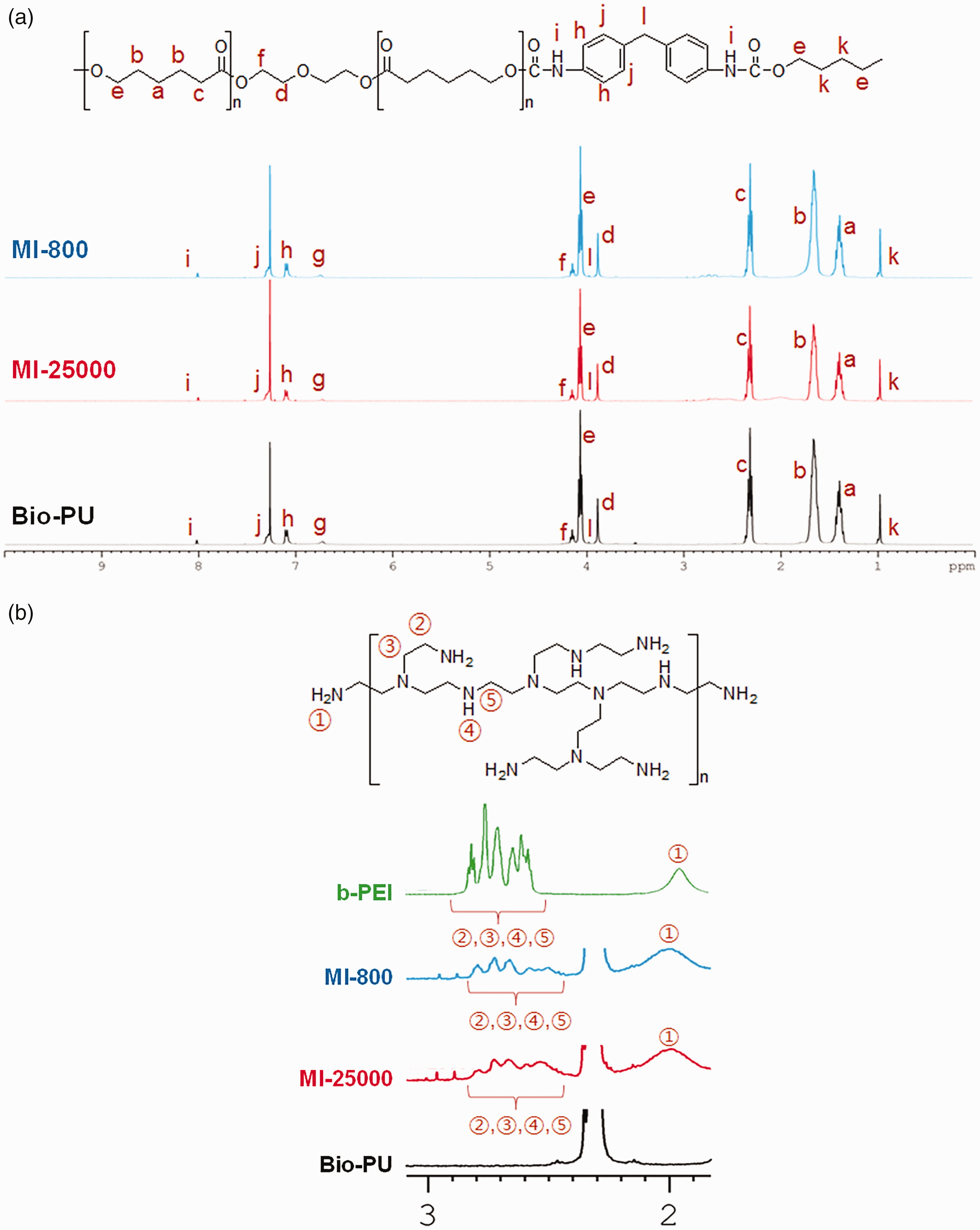
FT-IR spectroscopy measurements also showed successful preparation of the bio-PU. As shown in Figure 2(a), a free isocyanate (-NCO) peak at 2250–2270 cm−1 was not observed, indicating that the polymerization reaction proceeded and no unreacted prepolymer remained. Furthermore, the N-H stretching peak at 3340 cm−1, the carbonyl peak of the PCL at 1720 cm−1 and the C-O peak at 1050 cm−1, corresponding to urethane and ester functional groups, revealed that the targeted bio-PU was successfully formed.
18
The FT-IR spectra, shown in Figure 2(b), featured an imine peak (1650 cm−1), which was not observed in the bio-PU, MI-25000 and MI-800, but developed in the MIG-25000 and MIG-800 samples treated with GA. The presence of this peak indicated that the amine groups in the MI sample and the aldehyde of GA reacted with each other to form an imine functional group.
(a) Fourier transform infrared (FT-IR) spectra of bio-based polyurethane (bio-PU), MI-800, MIG-800, MI-25000 and MIG-25000. (b) Expanded FT-IR spectra of bio-PU, MI-800, MIG-800, MI-25000 and MIG-25000.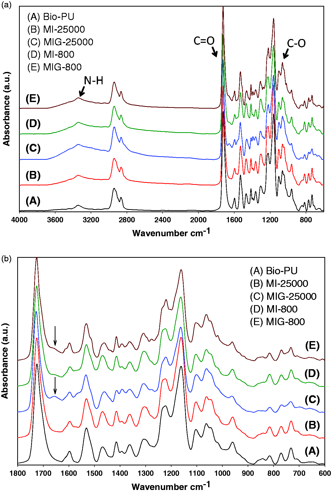
However, when the bio-PU was treated by GA in the same manner used to produce the MIG samples, peaks corresponding to imine functionality were not observed in the 1H NMR or FT-IR spectra of the bio-PU treated by GA. The 1H NMR and FT-IR spectra of the bio-PU treated by GA were no different from those of neat bio-PU, as shown in Figure 3.
Fourier transform infrared (FT-IR) spectra of the bio-based polyurethane (bio-PU) and the glutaraldehyde (GA)-treated bio-PU prepared in the same manner used for the production of the MIG (inset: FT-IR spectra of the bio-PU and the bio-PU treated by GA).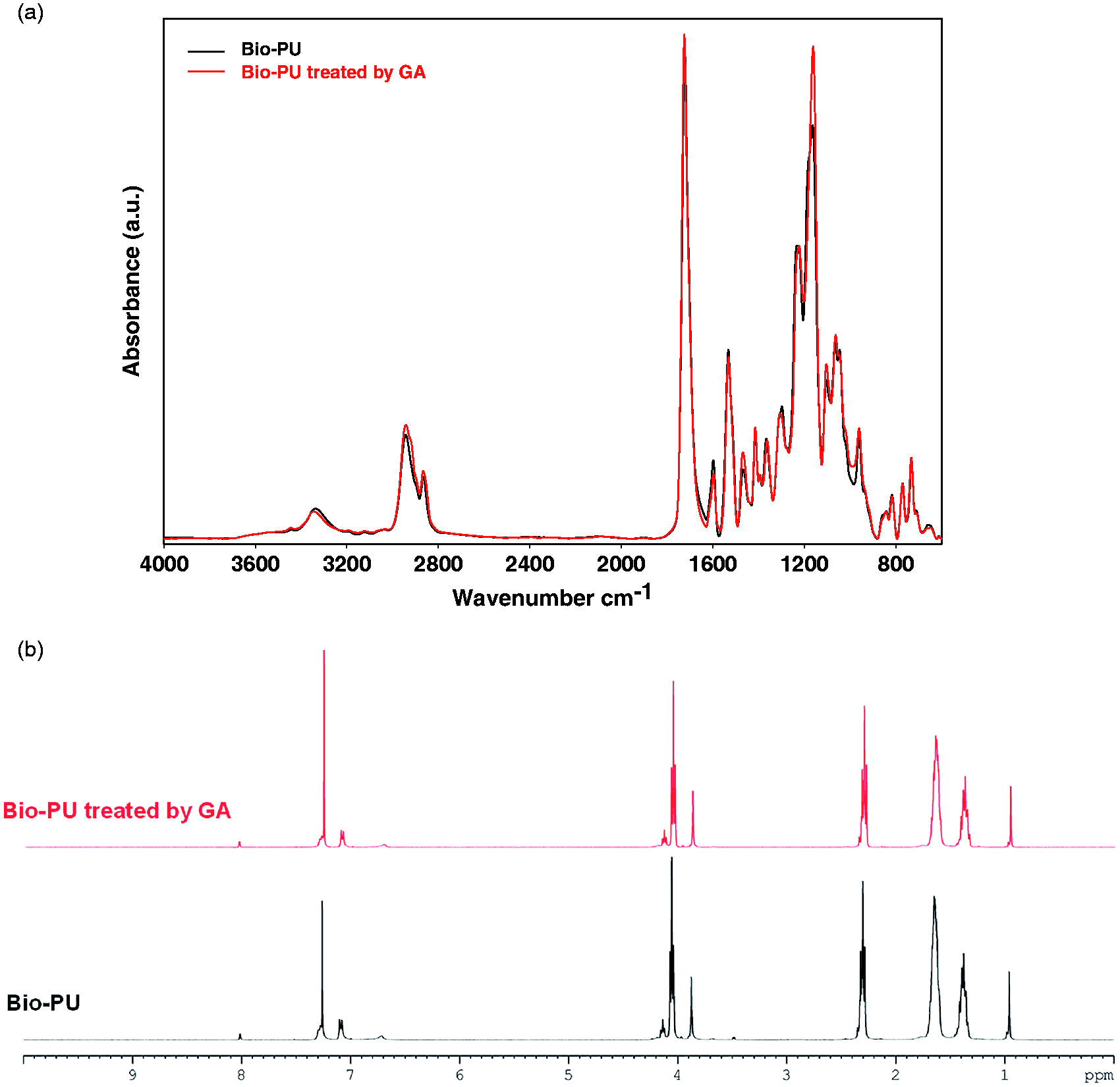
These results demonstrated that the GA molecule reacted only with the primary amine of the b-PEI in the MI sample and had no reaction with the secondary amine of the bio-PU. Thus, it was suggested that the bio-PU/b-PEI blend treated by GA, that is, the MIG sample, had a semi-interpenetrating polymer network (semi-IPN) structure resulting from imine crosslinking between the b-PEI in MI and the GA.28,29 On the basis of these results, the schematic structure of bio-PU/b-PEI semi-IPN is illustrated in Figure 4.
Schematic structure of bio-based polyurethane (bio-PU)/ branched poly(ethylene imine) (b-PEI) semi-interpenetrating polymer network formed by the glutaraldehyde (GA) treatment of the bio-PU/b-PEI blend.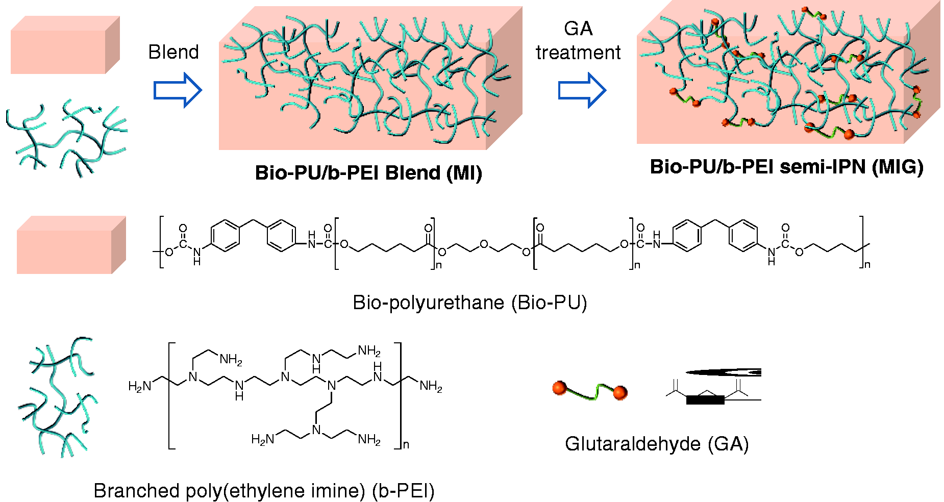
The semi-IPN structure of MIG was verified by a leaching test of b-PEI and contact angle measurements. Figure 5 shows the results of the weight change of samples after the bio-PU, MI and MIG samples were immersed in water for several hours. As shown in Figure 5, the MI-800 and MI-25000 samples showed significant weight losses at longer soaking time, while the bio-PU showed little weight loss even after 12 hours of soaking. This result can be explained by the migration of the hydrophilic b-PEI present inside the MI film toward the water phase owing to low secondary interaction forces between the hydrophilic b-PEI and the PU in the MI samples.
Weight residues of bio-based polyurethane (bio-PU), MI-25000, MIG-25000, MI-800 and MIG-800 after leaching test. First bar: before leaching; second bar: 6 h leaching; third bar: 12 h leaching.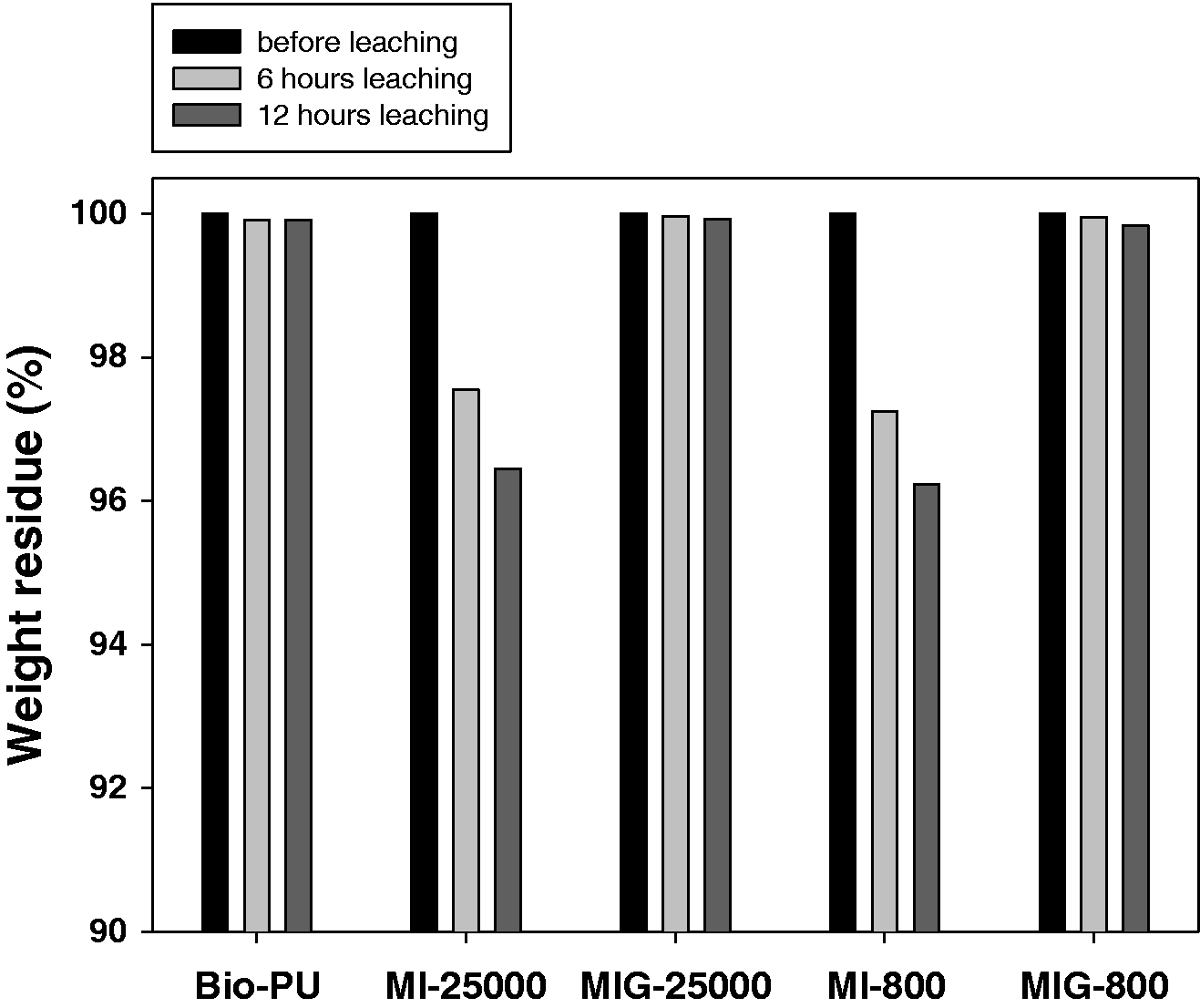
However, in the case of MIG-800 and MIG-25000 weight loss did not occur, similar to results for bio-PU. Even after immersion of the MIG samples in water for 12 h no leaching of b-PEI occurred. This result could be attributed to the formation of the semi-IPN structure in the MIG sample by imine crosslinking between the b-PEI and the GA, which suppressed b-PEI leaching into the aqueous phase. In addition, the decrease in hydrophilicity of the MIG sample caused by the decrease in the hydrophilic amine functionality of b-PEI after formation of the semi-IPN structure impeded the permeation of water into the MIG film and suppressed the leaching of b-PEI.
As shown in Figure 6, the bio-PU showed a contact angle of 90.5°, but the contact angles of MI-25000 and MI-800 were 76.2° and 74.6°, respectively, indicating an increase in hydrophilicity compared with that of bio-PU. This was thought to be caused by the presence of hydrophilic b-PEI with multiple primary amine functional groups in the MI sample.
30
The contact angles of MIG-25000 (81.5°) and MIG-800 (77.2°) increased compared with those of the corresponding MI samples, indicating a decrease in the hydrophilicity of MIG. This result was attributed to a decrease in the number of hydrophilic amine functional groups after the reaction between b-PEI and GA.
Contact angles of bio-based polyurethane (bio-PU), MI-25000, MIG-25000, MI-800 and MIG-800 films.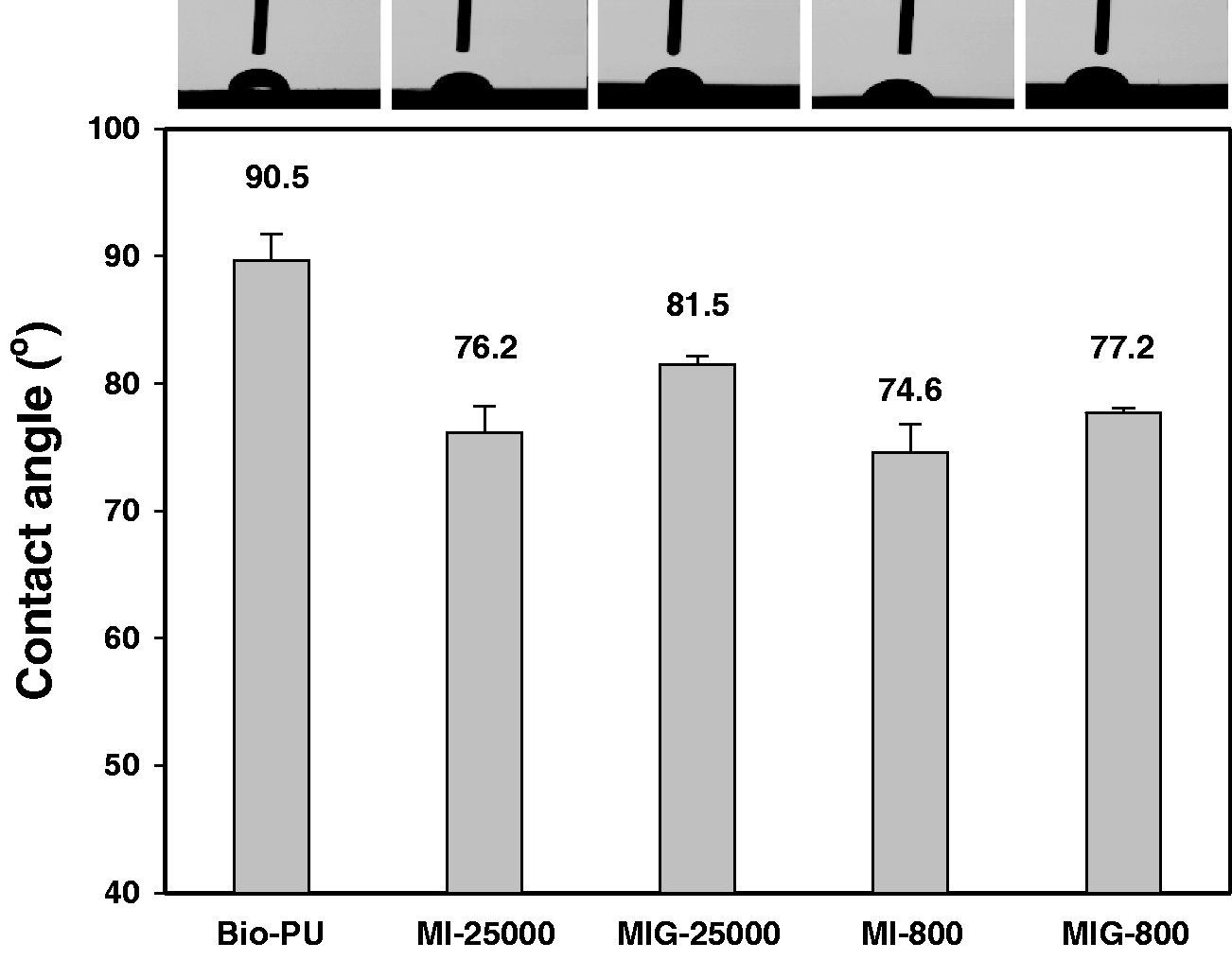
Thus, the leaching test and the contact angle results indicated that MIG formed a semi-IPN structure, which is in good agreement with the NMR and FT-IR results. Nevertheless, the contact angles of the MIG samples were lower than that of the bio-PU, which suggested that MIG is more hydrophilic than bio-PU. These properties may be expected to contribute to the dyeability and the wet fastness of the PU.
Glass transition temperature (Tg) of bio-based polyurethane (bio-PU), MI-25000, MIG-25000, MI-800 and MIG-800
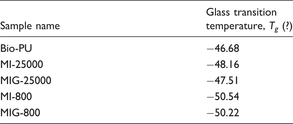
By contrast, MIG-25000 and MIG-800 treated with GA showed a slight increase of their Tg values by 0.6℃ and 0.3℃, respectively, compared with those of the MI samples before the GA treatment, MI-25000 and MI-800. This increase can be explained by a reduction in the motility of the PU chains in the MIG samples caused by the formation of a semi-IPN. However, the semi-IPN structure formed in MIG did not have a great influence on the Tg of the PU chain.
The Young’s modulus of bio-PU, MIs and MIGs was measured by tensile tests, as shown in Figure 7. The Young’s modulus values of MI-25000 and MI-800 were 0.54 and 0.4 MPa, respectively, whereas that of bio-PU was 0.66 MPa. The lower Young’s modulus of the MI samples compared with that of bio-PU was attributed to the increase in softness of the MI samples caused by the plasticizing effect of b-PEI. Furthermore, the Young’s modulus value of MI-800 decreased more than that of MI-25000, indicating considerable softened in the presence of b-PEI with a relatively lower molecular weight.
Young’s modulus of bio-based polyurethane (bio-PU), MI-25000, MIG-25000, MI-800 and MIG-800 films measured from the tensile test.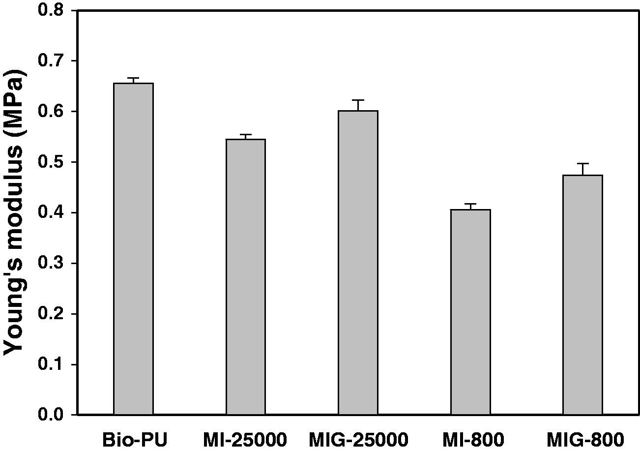
The Young’s modulus values of MIG-25000 and MIG-800 were measured to be 0.60 and 0.47 MPa, respectively, which were slightly higher values than those of the corresponding MI samples. The movement of the molecular chains was likely limited by the crosslinking of b-PEI induced by the GA treatment. These results agreed with the above-mentioned Tg results.
The shape retention characteristics of the bio-PU, MIs and MIG were evaluated by measuring the length after 100% elongated samples were allowed to recover to their original length during a certain period of time. Higher shape retention characteristics indicate greater recovery to the original sample length and a greater shape retention (%) value. As shown in Figure 8, the bio-PU had a shape retention ratio of 62.5% after three repeated shape retention characteristics tests, but MI-25000 and MI-800 showed lower retention ratios of 55% and 60%, respectively. This result may be explained by the fact that the PU chain was plasticized by b-PEI present in the MI sample. This plasticizing effect reduced the elasticity of PU and slowed the recovery after the 100% extension.
Shape retention rate (%) of bio-based polyurethane (bio-PU), MI-25000, MIG-25000, MI-800 and MIG-800 films.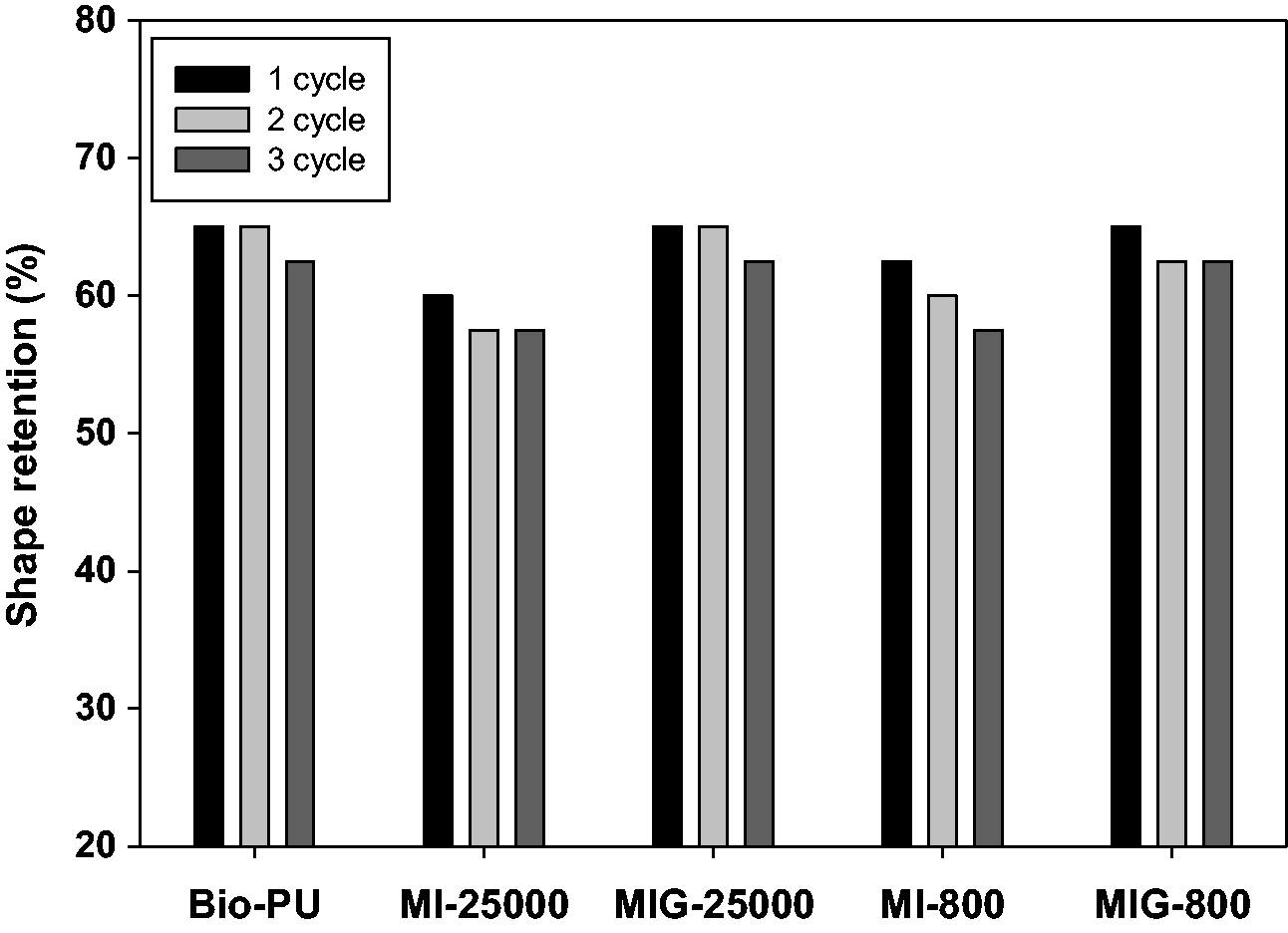
However, compared with MI-25000 and MI-800, MIG-25000 and MIG-800 treated with GA showed a larger shape retention ratio of approximately 5% based on the third experiment, and the shape retention values were found to be similar to those of bio-PU. The excellent shape retention characteristics of MIG can be attributed to the semi-IPN structure formed inside MIG, and the results were in good agreement with Young’s modulus determined from tensile tests.
To investigate the dyeing characteristics of the semi-IPN structured MIG samples, acid dyeing of bio-PU, MI and MIG films was performed using Acid Red 4 dye at room temperature. The color change of the dyed film samples, after immersion in water for 3 and 12 h, was also measured to infer their wet fastness. The color properties of the dyed samples were quantitatively evaluated by the maximum K/S value at 540 nm.
As shown in Figure 9, the bio-PU was not dyed at all. This result can be explained by the lack of sites for the acid dye to bind to in the bio-PU. Thus, acid dye adsorption could not occur during the early stages of dyeing. However, MI-25000 and MI-800, containing b-PEI, showed significantly higher K/S values compared with that of the bio-PU, which indicated that acid dyeing was achieved. The improved acid dyeability of the MI samples could be attributed to availability of dyeing sites through the conversion of primary and secondary amines of b-PEI to quaternary ammonium cations under the acidic conditions.29,31,32 Moreover, the MI samples were plasticized and made more hydrophilic by b-PEI. This effect not only increased the free volume of the MI film but also facilitated adsorption of the dyeing solution into MI film, and allowed the dye to diffuse more easily within the film. It was observed that MIG-25000 and MIG-800 also had high K/S values, indicating their improved dyeing characteristics.
K/S value at λ = 540nm of bio-based polyurethane (bio-PU), MI-25000, MIG-25000, MI-800 and MIG-800.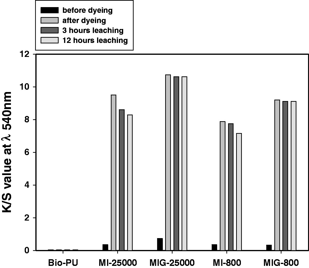
However, the K/S values of MI-25000 and MI-800 measured after the immersion treatment in water were lower than those before the immersion treatment. This tendency increased as the immersion time increased. This result may be attributed to the fact that dyes combined with b-PEI were leached into the water as the b-PEI itself leached into the aqueous phase. Furthermore, the ionic bond between the dye and b-PEI was weakened in water, permitting the dye to be easily removed from the MI. This finding indicates that dyes can leach out from the bio-PU product during typical washing or under raining conditions. These properties are unsuitable for practical use of the material, even though the bio-PU/b-PEI blend has a high dyeability. Unlike the MI samples, the MIG samples showed little change in their K/S value, even when MIG-25000 and MIG-800 were immersed in water for 12 h. This result clearly indicates that the MIG samples had much higher wet fastness than that of the MI samples. The higher wet fastness is likely because b-PEI, which provides dyeing sites for the acid dyes, is not leached into water because of the formation of the crosslinking structures of PEI inside the bio-PU matrix. Furthermore, the leaching of the dyes may also be disturbed and suppressed by the dense crosslinked structure of the MIG. These results clearly show that our approach provides bio-PU with improved dyeability and enhanced wet fastness of dye. Hence, we anticipate that these new methods will open the way for various new industrial applications of PU.
Conclusions
In this study, we have developed an acid dyeable bio-PU/b-PEI semi-IPN with enhanced wet fastness. b-PEI was blended, which features amine groups, with synthesized bio-PU and a network structure was induced by a GA treatment. From the results of NMR, FT-IR spectroscopy, leaching tests and contact angle measurements, it was found that the MIG samples, that is, the bio-PU/b-PEI blend treated by GA, had a semi-IPN structure with imine crosslinking between b-PEI and GA within the PU matrix. Furthermore, the mechanical properties of the MI samples, that is, the bio-PU/b-PEI blend, plasticized by b-PEI were restored by the semi-IPN structure, induced by the GA treatment. In particular, we showed that the bio-PU/b-PEI semi-IPN had a much higher dyeability and wet fastness than bio-PU that was not dyed at all. The differences in the properties of these materials were attributed to the increased dye interaction from the introduction of b-PEI and the formation of the b-PEI network through the GA treatment. These results are expected to increase the use of bio-PU, particularly for fiber products.
Footnotes
Declaration of conflicting interests
The authors declared no potential conflicts of interest with respect to the research, authorship and/or publication of this article.
Funding
The authors disclosed receipt of the following financial support for the research, authorship, and/or publication of this article: This work was supported by the Advanced Research Center Program (Grant No. NRF-2017R1A5A1015596) and the Basic Science Research Program (NRF-2017R1A2B4005315) of the National Research Foundation of Korea (NRF), funded by the Ministry of Science and ICT.