




Abstract
In this study, the formation of conductive nano-structured polypyrrole (PPy) on electrospun poly(ɛ-caprolactone) (PCL) nanofibers was successfully achieved using a DNA dopant (PCL/DNA-PPy) via sonication-induced layer-by-layer assembly. After PPy containing positive charges was accumulated on PCL, DNAs with negative charges deposited such that they were evenly distributed. The resulting PCL/DNA-PPy nanomembrane exhibited increased fiber diameter (PCL/DNA-PPy 5LBL: 328.11 ± 48 nm) and deformation morphology compared to pure PCL (average fiber diameter of 247.25 ± 32 nm, fibrous uniform morphology), as observed using scanning electron microscopy and atomic force microscopy. As the number of layers increased, the crystallinity of PCL/DNA–PPy nanomembranes decreased, as observed using X-ray diffraction. It was observed that the PPy-DNA deposited on the surface of PCL connected to form a nano-sheath and significantly increased the thermal stability of PCL. Moreover, the contact angle of the PCL/DNA-PPy nanomembrane (contact angle of pure PCL: 79.3 ± 1.2°) demonstrated its high hydrophilicity. The results indicate that the composites showed very good survival in a cytotoxicity test on U-118 glioma cells and excellent electrical conductivity (the highest value was 1.1 × 10⁻³ S/m). The manufactured PCL/DNA–PPy nanomembranes are considered to be promising materials for applications in the scaffold, sensor, and electronic fields.
Nowadays, novel biomaterial research is booming worldwide as the application of biomaterials in medical devices is expected to expand significantly over the next decade. Nano-structured materials and electrospun nanofibers are promising candidates for use as cell scaffolds owing to their higher surface area and porosity as compared to polymer films. Electrospinning is a remarkably simple and cost-effective technique, with a relatively high production rate of nanofibers as compared to methods such as melt fibrillation, 1 island-in-sea, 2 and gas jet 3 techniques. In the past few years, there has been significant growth in the research on electrospun nanofibrous scaffolds for tissue engineering applications. An inherent property of nanofibers is that they mimic the extracellular matrices of tissues and organs.4–6 Furthermore, the properties of polymeric nanofibers can be further optimized to satisfy different requirements, such as conductivity. Electric field stimulation could accelerate the healing of injured wounds, possibly via increased protein adsorption, 7 and manipulate cell behavior in polarized cellular growth 8 or change cell signaling pathways. 9 Several groups have attempted electric field stimulation using metallic looped wires, cuffs, or needles as electrodes to produce various stimulation patterns of tissues. 10 However, these methods contain invasive, non-biodegradable electrodes, which are not easily approved by the Food and Drug Administration (FDA) in the USA. In this work, we present the fabrication of biocompatible, biodegradable scaffolds with sufficient conductivity but without the problems associated with metallic conductors. Conjugated polymers have been one of the most broadly used materials for producing novel biomaterials owing to their ease of synthesis, excellent electrical properties, and functionality and surface characteristics.11–14 Among conducting polymers, polypyrrole (PPy), polyaniline, polythiophene, and polyphenylene contain double bonds along the backbone, resulting in their conductive nature. PPy is particularly attractive owing to the richness of its possible oxidation states, high environmental and chemical stability, and adjustable electrochemical behavior achieved by choosing the doping level, pH, and morphology. The electrical conduction in PPy originates from the electron movement occurring within delocalized orbitals and positive charged effects known as polarons. 15 These positive charges are located every three to four pyrrole units along the polymer backbone. Therefore, negatively charged DNA can form strong bonds with PPy based on the interchange of the dopant DNA molecules. 16 Although polymerized PPy materials are non-toxic, pyrrole monomer and short chain oligomers or dopants present in the starting materials can be toxic. Residues of these chemicals embedded within the final product can elicit various degrees of cytotoxicity. DNA was chosen for this study, as the generally used dopants have limited biomedical applications because they can be harmful to cells and tissues. 17 Poly(ɛ-caprolactone) (PCL) is a biodegradable aliphatic polyester with a glass transition temperature of −60℃ and melting point of 60℃ and a high level of flexibility, attractive mechanical properties such as low weight and flexibility, good dielectric properties, and the ability to be easily fabricated and blended. 18 PCL is being currently investigated extensively for biomedical applications owing to its flexibility, biocompatibility, and long-term in vivo degradation. PCL is a traditionally biodegradable aliphatic polyester polymer and has recently gained approval from the FDA for use in a load-bearing bone implant fabricated via three-dimensional (3D) printing.
In this study, we aim to develop a novel nano-structured scaffold suitable for assembling DNA and PPy in a layer-by-layer (LBL) manner on an electrospun biodegradable PCL nanofiber, which is a bioresorbable material. PCL nanofibers were fabricated using the electrospinning technique. The interfacial properties of the PCL nanofibers with PPy and DNA immobilization were investigated, and the morphological, structural, and electrochemical characterizations were performed. Further, we attempted to minimize the toxicity of the material by imparting an effective LBL morphology using PPy and DNA. Cell culture experiments were conducted on the new composite, and the results demonstrated that our composite exhibited excellent biomaterial properties and electrical conductivity, and good mechanical property, rendering it suitable for use as tissue engineering membranes. These findings may lead to the fabrication of a new conductive and biodegradable biomaterial for a broad range of tissue engineering applications.
Materials and methods
Materials
PCL with an average molecular number (Mn) of 80,000 was obtained from Sigma-Aldrich. PPy with a conductivity of approximately 10–50 S/cm was purchased from Sigma-Aldrich. DNA isolated from salmon with Mn > 100,000 was obtained from Sigma-Aldrich. Analytical grade chloroform (Aldrich, 99.8%), dimethylformamide (DMF, SAMCHUN, 99.0%), and ethylene glycol (SAMCHUN, 99.5%) were purchased. All the reagents used in this experiment were used as received without further treatment.
Fabrication of electrospun PCL nanofibers
Electrospinning was performed in a fume hood using an open cage target to collect the fibers. The relative humidity was adjusted by flushing the hood with dry air. Subsequently, 12 wt.% PCL doping solution was prepared in chloroform/DMF (9/1, W/W) at room temperature. For electrospinning, a voltage of 15 kV was applied from the metal needle (21 G) to the collector (rotational speed of 200 rpm) using a high-voltage power supply, and the distance between the needle and grounded target was 15 cm. The PCL solution was spun at a flow rate of 1.2 mL/h with a doping solution using a syringe pump.19–23
DNA and PPy immobilization on PCL nanofibers: layer-by-layer
Subsequently, 0.5 wt.% PPy solution was prepared in ethylene glycol and 0.1 wt.% DNA solution was prepared in distilled water for laminating PPy. Thereafter, each solution was sonicated for 1 h. After fixing the PCL nanomembrane using a square frame with an open center (60 mm × 60 mm), the PPy solution was initially sprinkled on both sides of the PCL nanomembrane. After allowing the sample to dry completely using a hot air dryer at 50℃, the DNA solution was dispersed on both sides and dried again at 50℃ with a hot air dryer. The preparation of a single layer is illustrated in Figure 1, and multiple layers, that is, five layers (5LBL – PCL 90.5%, DNA-PPy 9.5%), 10 layers (10LBL – PCL 81.2%, DNA-PPy 18.8%), and 20 layers (20LBL – PCL 68.8%, DNA-PPy 31.2%), were obtained by repeating this procedure.
(i) Schematic diagram for the preparation of poly(ɛ-caprolactone) (PCL)/DNA–polypyrrole (PPy) nanomembranes using layer-by-layer (LBL) processing. (ii) Digital photos and water contact images of pure PCL nanofiber and PCL/DNA–PPy 20LBL nanomembranes. NFs: nanofibers.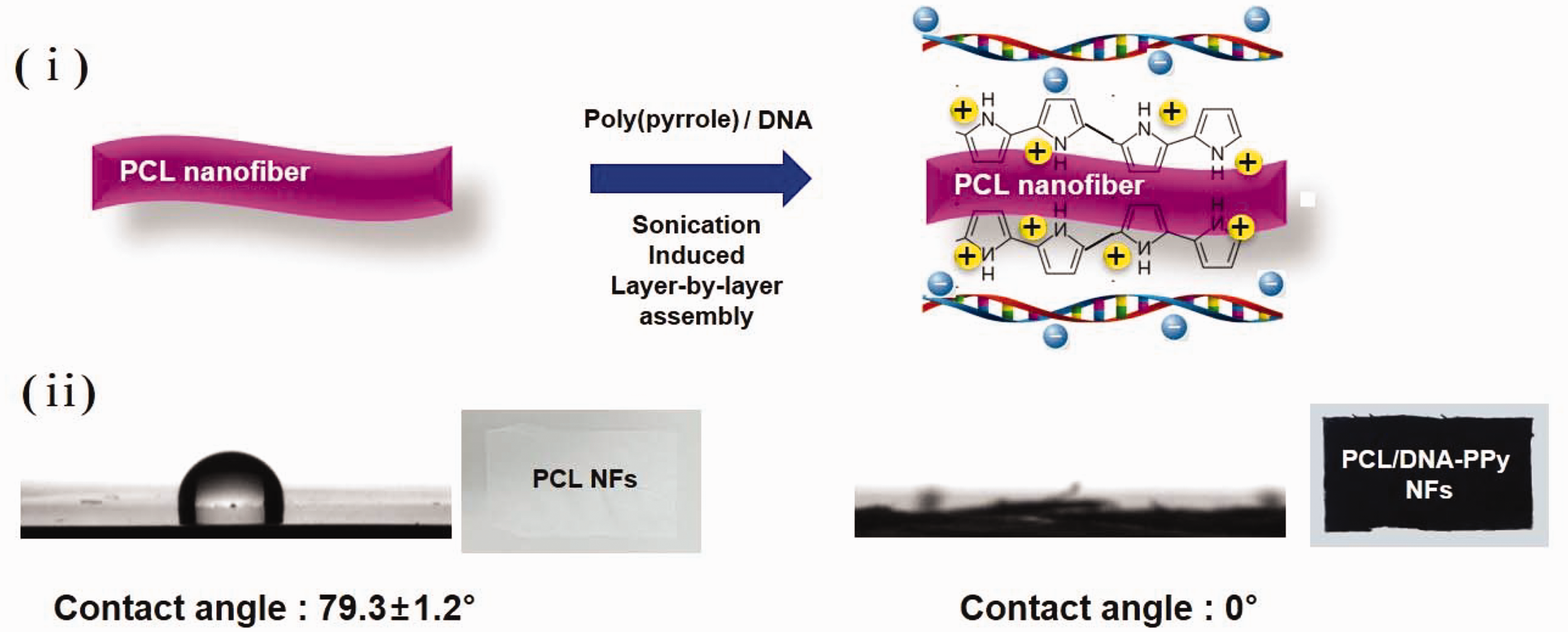
Characterization of PCL/DNA-PPy nanofiber
Morphology
The morphology of the nanofibrous membranes was characterized using field-emission scanning electron microscopy (FE-SEM; Nova 400 Nano SEM, FEI) after coating the samples with platinum and using a current of 20 mA for 120 s. The diameter of the fibers was measured from the SEM images using an image analysis software (Image J, National Institutes of Health, USA) by randomly selecting approximately 100–150 fibers in the SEM images.
Crystalline structures
Automated X-ray powder diffraction (XRD, PANalytical's Empyrean) analysis was employed to analyze the changes in crystallinity achieved by the deposition of PPy and DNA on the PCL nanofiber. The measurements were performed using monochromate Cu-Kα radiation (λ = 1.54056 Å) at 40 kV and 20 mA by collecting the spectrum at 2θ = 3−40° at a step size of 0.02.
Thermal analysis
For identifying the miscibility and thermal properties of PCL/DNA-PPy, we employed differential scanning calorimetry (DSC) and thermogravimetric analysis (TGA). The samples were subjected to a temperature rise from ambient temperature up to 600℃ at the rate of 10℃/min. TGA was performed using a Perkin Elmer thermogravimetric analyzer. Nitrogen gas was used to purge the furnace. Pyris software was used to analyze the DSC data. Further, a heat–cool cycle was applied between 25℃ and 600℃. The mass fraction of the residual moisture contained in the samples, the degradation process, changes in the glass transition temperature and melt temperature, and the degradation temperature of PCL/DNA-PPy were observed.
Atomic force microscopy
The surface roughness of different matrix samples, with an area of 10 × 10 µm2, was examined using atomic force microscopy (AFM; Model 5100, Agilent Technologies, USA). The topographic images were obtained using the intermittent contact mode employing silicon cantilevers (PPP-NCL, Nanosensors, Inc., USA). The silicon cantilevers exhibited a force constant of 40 N/m and resonating frequency of 169.52 kHz. Pico Image Basic Software (Agilent Technologies) was used to analyze the AFM images, and the surface roughness of the nanofibrous matrices was obtained in terms of the surface root mean square (RMS) roughness (Rq).
Contact angle
In order to confirm the changes in the hydrophilicity owing to the deposition of PPy and DNA on PCL, water contact angles (WCAs) were measured using a contact angle analyzer (Phoenix 300; Surface and Electro-Optics, Ansan, South Korea). Droplets of water were placed on the surfaces of pure PCL and the PCL/DNA-PPy samples. The images of the water drops on the sample surface were recorded using a camera and analyzed with a software supplied by the manufacturer. The contact angle was measured under ambient temperature and humidity of 65%, and the contact angles at five independent points on each surface were averaged and presented as the mean.
In vitro cell culture
The nanofibrous membranes were immersed in 70% (v/v) ethanol for 30 min and subsequently dried under sterile conditions. The samples were thereafter exposed to ultraviolet (UV) (UV-B 280–300 nm) radiation for 24 h and incubated with 2 mL of Dulbecco's Modified Eagle's Medium (DMEM) for 24 h before cell seeding. Human glioblastoma cell line U-118 cells (Passage 2) were seeded onto the nanofibrous membranes at a density of 1 × 10⁵ cells/well and cultured with DMEM supplemented with 10% (v/v) fetal bovine serum (FBS) at 37℃, 5% CO2, and humidity of 95%. Alternatively, nanofibrous membranes were eluted with DMEM and 10% phosphate buffered saline (PBS) in an incubator for 24 h for preparing the conditioned medium (CM, eluate solution). U-118 glioma cells were seeded on a 12-well plate (5 × 10³ cells/well). After incubating overnight, the cells were cultured by the modified CM for 24 h. Live cells stained in the culture medium were counted (Nm) using trypan blue after 24 h of incubation. For cell investigations, the cells cultured on tissue culture dishes (TCDs, high-grade polystyrene NuncTM Dishes, Thermo Fisher Scientific, Denmark) were evaluated as controls. The data collected were expressed as mean 6 SD. A P-value (p) of less than 0.007 was considered to be a significant difference.
Conductivity
After each sample was wetted with pH 7.4 PBS solution, the conductivity (SourceMeter® SMU Instruments Series 2400, KEITHLEY) of each sample was measured as in Supporting Figure 1. When measured by this method, the conductivity of pure PCL was approximately 9.4 × 10⁻⁵ S/m, and the change in conductivity was analyzed as occurring with the changes in the number of layers. The electrical conductivity of the specimen was measured by a two-terminal method by applying 21 V under the constant current of 0.1 µA. Surface resistivity was calculated using Equation (1) as

Results and discussion
We fabricated PCL/DNA–PPy electrospun fibrous webs from PCL nanofibers via the sonication-induced LBL assembly as shown in Figure 1(i). Compared with the PCL membrane, the composite membrane exhibited an apparent dark black color, which revealed the successful incorporation of DNA-PPy in the PCL membrane (Figure 1(ii)). PCL/DNA–PPy 10LBL had good flexibility and handling. However, PCL/DNA–PPy 20LBL could be easily broken into small pieces by folding because the PCL contains too many DNA-PPy layers and the two substrates are spaced apart. PCL nanofibers exhibited a thickness of 13.0 ± 1 µm, whereas the PPy-DNA-coated PCL nanofibers exhibited the thicknesses of 20.6 ± 7, 25.4 ± 4, and 34.4 ± 6 µm for 5LBL, 10LBL, and 20LBL, respectively. We confirmed a remarkable change of thickness of PCL/DNA–PPy electrospun fibrous webs after the LBL process. The crystallinity of the resultant PCL/DNA–PPy electrospun fibrous webs for use as biodegradable materials is very important because it has a decisive effect on the final physical properties (for instance, mechanical properties) and biodegradability. Figure 2 shows the wide-angle X-ray scattering (WAXD) patterns of pure PCL nanofiber and PCL/DNA–PPy nanomembranes for 5LBL and 10LBL webs. As shown in Figure 2, PCL/DNA–PPy exhibited three characteristic peaks at 2θ = 21.4° (strong), 21.9° (shoulder), and 23.6° (medium) corresponding to [110], [111], and [200] reflections, respectively, indicating the same crystalline microstructure as that of PCL. PPy is a highly rigid polymer owing to its linear structure and less flexible chain folding. However, in the presence of organic surfactants, the dopant-PPy undergoes various interactions, resulting in the organization of the polymer chains in a 3D highly ordered fashion. However, the distribution of the two crystalline reflections of PCL/DNA–PPy membranes was nearly isotropic. The increase in the number of layers of PCL/DNA–PPy can be observed by the change of PCL peak [110] and PPy peak [200]. As the number of layers increases, the peak [200] of PPy increases, resulting in a decrease of the intensity ratio I[110]/I[200] of the two main peaks, which can be observed during the uni-axial tensile deformation process of orthorhombic semi-crystalline polymers, as presented in Table 1.
24
Consequently, the crystallinity of PCL/DNA–PPy membranes was lower than that of pure PCL. Furthermore, the peak observed at approximately 2θ = 8–10° represents the enhancement in the structural order of the counterions in the polymer chains, thereby indicating a higher conductivity. The PPy main peaks observed at 22–23° could be attributed to the interplanar van der Waals arrangement of the pyrrole–pyrrole rings in the PPy chains and doped amorphous pyrrole–counterion or inter-counterion interactions.25,26 This peak is more pronounced on 10LBL membranes with an increased amount of PPy than on 5LBL membranes.
Wide-angle X-ray scattering patterns of pure poly(ɛ-caprolactone) (PCL) nanofiber and PCL/DNA–polypyrrole (PPy) nanomembranes with 5LBL and 10LBL. LBL: layer-by-layer. Intensity ratio of I[110]/I[200] for pure poly(ɛ-caprolactone) (PCL) nanofiber and PCL/DNA– polypyrrole (PPy) nanomembranes with 5LBL, 10LBL, and 20LBL based on wide-angle X-ray scattering data LBL: layer-by-layer.

Figure 3 shows the TGA and DSC curves of pure PCL nanofiber and PCL/DNA–PPy nanomembranes with 5LBL (PCL 90.5%, DNA-PPy 9.5%), 10LBL (PCL 81.2%, DNA-PPy 18.8%), and 20LBL (PCL 68.8%, DNA-PPy 31.2%). As compared to the pure PCL nanofibers (thermal decomposition temperature, Td: 342.1℃), the thermal stability of PCL/DNA–PPy electrospun fibrous webs increased up to 402.2℃ with the exception of the 20LBL membrane. PCL/DNA–PPy 10LBL exhibited the highest thermal stability. Thus, 10 layers (10LBL) were considered to be the optimum for incorporating DNA and PPy. Moreover, at the PPy-DNA loading corresponding to 10LBL, the packing of the PPy-DNA units could lead to crystallinity and a corresponding Tm transition was observed in the DSC curve. However, at 20 layers, a completely different trend was observed in the TGA curve (rapidly decreasing Td) and DSC (decreasing Tm) than that in pure PCL nanofiber. This could be attributed to the separation phenomenon of PCL fiber on PPy and DNA layers.
25
In summary, the incorporation of PPy and DNA helps the passive thermal stability of PCL. However, as the number of LBL get close to 20, PCL/DNA–PPy was physically separated from PCL membranes and caused their lower thermal stability compared with pure PCL. This result can also be explained by the lower crystallinity of PCL in the PCL/DNA–PPy 20LBL.
(a) Thermogravimetric analysis curves and (b) differential scanning calorimetry of pure poly(ɛ-caprolactone) (PCL) nanofiber and PCL/DNA–polypyrrole (PPy) nanomembranes with 5LBL, 10LBL, and 20LBL. LBL: layer-by-layer.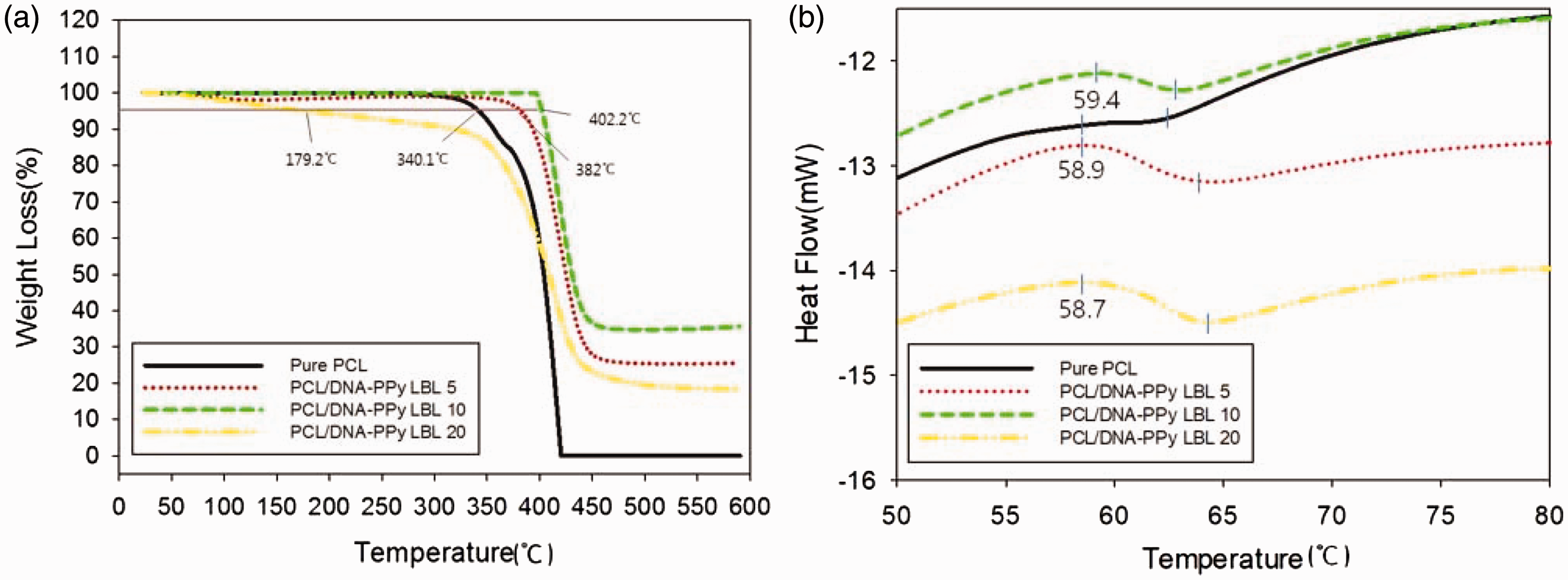
The morphological characterizations of pure PCL nanofiber and PCL/DNA–PPy nanomembranes with 5LBL, 10LBL, and 20LBL nanofibers were performed using SEM (Figure 4) and AFM (Figure 5). In PCL, the electrospun nanofibers were deposited as randomly oriented fibers
19
held together by connecting sites, such as crossing and bonding between the fibers. Furthermore, a smooth surface was observed with no bead formation. The average fiber diameter of pure PCL was determined to be 247.25 ± 32 nm. The diameters of the nanofibers increased after performing the LBL process with DNA/PPy and PCL nanofiber. The average fiber diameter of PCL/DNA-PPy 5LBL was 328.11 ± 48 nm. The PCL/DNA-PPy 10LBL and 20LBL nanofibers were hardly observed using SEM while evaluating the fiber diameters. This result indicated the presence of large amounts of DNA/PPy, as expected.
Scanning electron microscopy micrographs of (a) pure poly(ɛ-caprolactone) (PCL) nanofiber and (b) PCL/DNA–polypyrrole nanomembranes with 5LBL, (c) 10LBL, and (d) 20LBL; scale bar: 1 µm. LBL: layer-by-layer. Atomic force microscopy three-dimensional model images and root mean square roughness of (a) pure poly(ɛ-caprolactone) (PCL) nanofiber and (b) PCL/DNA–polypyrrole nanomembranes with 5LBL, (c) 10LBL, and (d) 20LBL. LBL: layer-by-layer.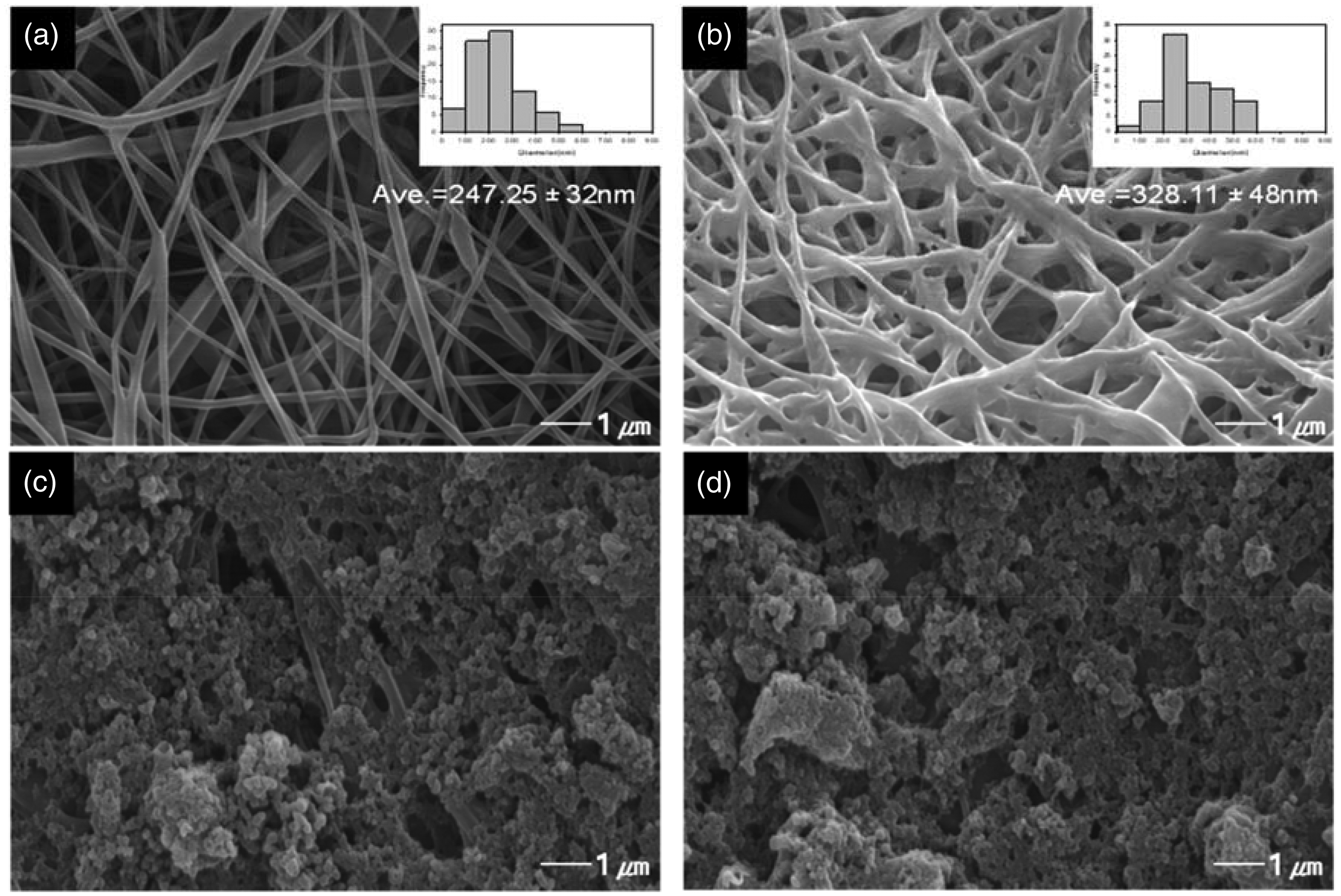

The AFM images of pure PCL nanofiber and PCL/DNA–PPy nanomembranes with 5LBL, 10LBL, and 20LBL nanofibers are illustrated in Figure 5. PCL nanofibers exhibited a RMS roughness of 296.0 ± 15 nm, whereas the PPy-DNA-coated PCL nanofibers exhibited RMS roughness of 419.6 ± 50, 457.2 ± 30, and 470 ± 45 nm for 5LBL, 10LBL, and 20LBL, respectively. This increase in the roughness achieved by coating PPy-DNA on the PCL nanofibers can be attributed to the presence of PPy and DNA on the nanofiber surface. Furthermore, the roughness of the PCL nanofiber surface increased as PPy-DNA layers increased, as the roughness benefits not only the cell adhesion, but also the hydrophilicity. The WCA measurement results of the deposition of PPy-DNA on PCL nanofibers are shown in Figure 1 and Supporting Figure 2. PCL has a high contact angle due to the presence of relatively long aliphatic chains in molecules among the biodegradable polymers. After immobilization, the contact angle of the pure PCL (approximately 79.3 ± 1.2°) changed to zero and the surface energy was changed from 64.54 to 224.13 mN/m. The fact that PCL is layered with PPy and DNA and is easily wetted in water has changed it into a material with high polarity. In short, the change in wettability correlates with the electrical conductivity of the material, so the excellent electrical conductivity of the PCL/DNA–PPy nanomembranes could be predicted. The PPy-DNA on PCL nanofiber immediately demonstrated the characteristic of absorbing water. PCL nanofibers coated with PPy-DNA have excellent hydrophilicity and are suitable for use in future biomaterials because the protein adsorption and cellular behavior are strongly dependent on the wettability of substrates. 26
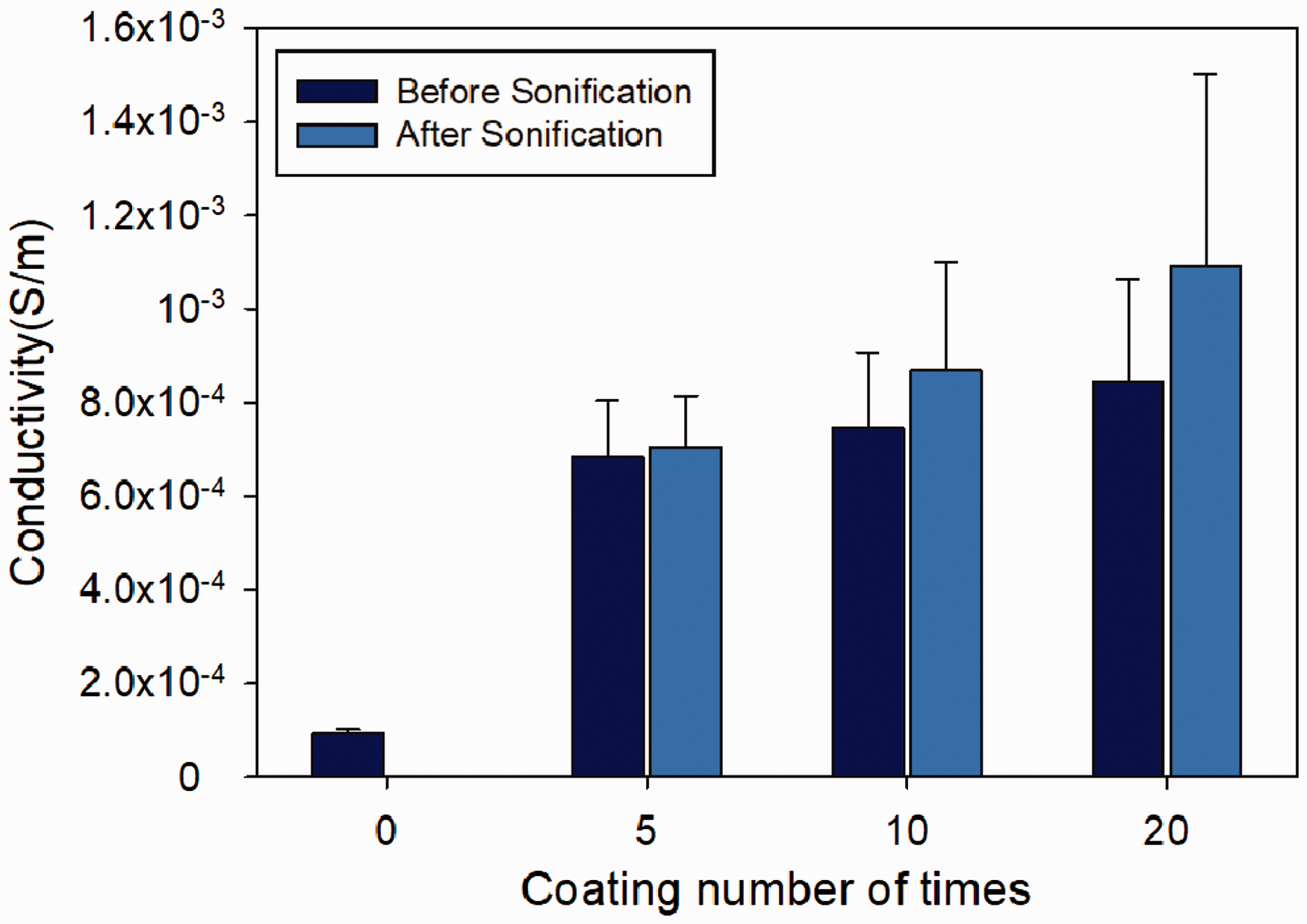
In order to assess the in vitro safety of PCL/DNA-PPy, U-118 glioma cells were incubated on the PCL/DNA-PPy nanofibers and with eluate solutions. The cells were thereafter stained to assess the cell viability, as shown in Figure 6. The cell numbers on the PCL/DNA-PPy 5LBL and 10LBL nanofibers were similar to those in the TCD, indicating good survival of cells on PCL/DNA-PPy 5–10LBL. However, the U-118 live cell numbers on PCL/DNA-PPy 20LBL were notably lower by approximately 20% than those on the TCD. This revealed that the PCL/DNA-PPy 20LBL sample contained a large dose of pyrrole monomer and PPy short chain oligomer. Although the polymerized PPy materials are non-toxic, pyrrole monomer and short chain oligomers present in the starting materials can be toxic. The residues of these chemicals might become embedded within the bulk final product and elicit various degrees of cytotoxicity. According to the results, we observed that the PCL/DNA-PPy nanomembranes possess biocompatibility, except 20LBL. Furthermore, the electroconducting properties of PCL/DNA-PPy in 0.1 M PBS solution (pH 7.4) were studied, as shown in Figure 7. Electrical stimulation was also seen to promote cell proliferation and growth in vitro. The conductivity increased with an increase in the numbers of layers. The electrical conductivity increasing is due to increasing the density and mobility of charge carriers. After applying the sonication process to PCL/DNA-PPy, the sample exhibited significantly higher conductivity as compared to the non-treated sample because sonication helps improve the overall conductivity when coating PPy particles and DNA with a high degree of dispersion. Consequently, sonication confirmed that the conductivity improves as the PPy particles are layered on the nanofibers. The sample with 20LBL exhibited a conductivity of approximately 1.1 × 10⁻³ S/m, indicating that the immobilization of PPy and DNA on the PCL nanofibers is an effective method for the fabrication of conductive nanofibrous membranes.
Viability of U-118 cells with conditioned medium (CM) eluate solutions (a) on nanofibers; (b) on pure poly(ɛ-caprolactone) (PCL) nanofibers and PCL/DNA– polypyrrole nanomembranes with 5LBL, 10LBL, and 20LBL; (c) time-course microscopic observation of U-118 glioma cells in CM eluate solutions. Scale bar: 500 µm; Significant difference between different materials groups was denoted as *p < 0.003, **p < 0.007. LBL: layer-by-layer; TCD: tissue culture dish. Electrical conductivity of poly(ɛ-caprolactone)/DNA– polypyrrole nanomembranes with 0LBL, 5LBL, 10LBL, and 20LBL in 0.1 M phosphate buffered saline solution (pH 7.4) before and after sonication processing. LBL: layer-by-layer.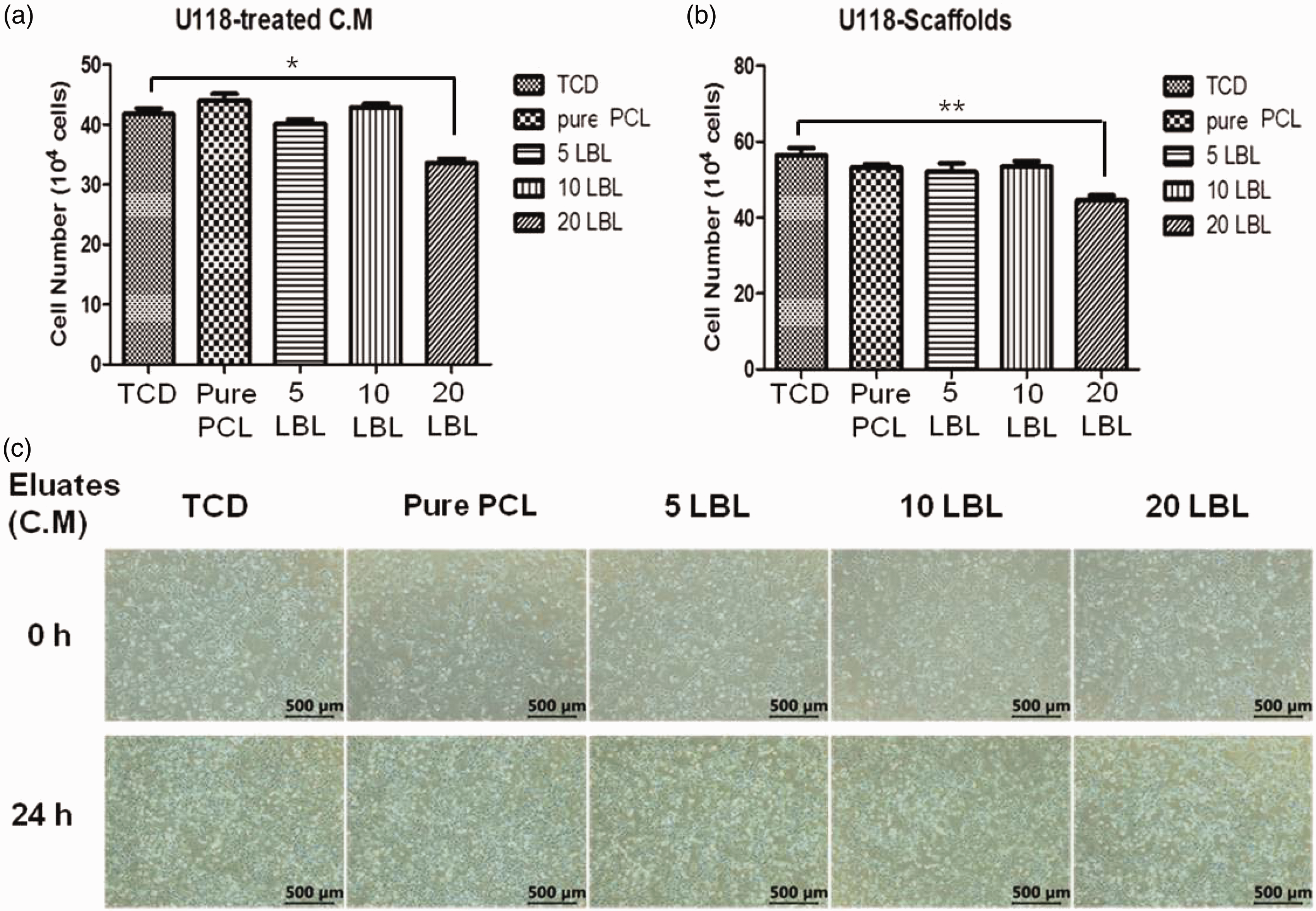
Conclusions
In this study, electroactive PCL nanofibers were fabricated using the sonication-induced LBL assembly technique. The incorporation of PPy and DNA (dopant) on the electrospun PCL nanofibers was successfully achieved by employing a LBL process for physical immobilization. The incorporation of PPy and DNA on the PCL nanofibers was confirmed using XRD, TGA, DSC, WCAs, SEM, and AFM measurements. The PCL/DNA-PPy nanomembrane (fiber diameter: 328.11 ± 8 nm, showing a smooth surface and no beads) exhibited high thermal stability (Td: 402.2℃), the highest hydrophilicity, and good conductivity (approximately 1.1 × 10⁻³ S/m). PCL/DNA-PPy 10LBL exhibited excellent cell viability and electrical conductivity. Therefore, PCL/DNA-PPy nanomembranes have immense potential in tissue engineering applications, such as cell stimulation, radiation, and regeneration, and sensor applications.
Footnotes
Declaration of conflicting interests
The authors declared no potential conflicts of interest with respect to the research, authorship, and/or publication of this article.
Funding
The authors disclosed receipt of the following financial support for the research, authorship, and/or publication of this article: This work was supported by Dankook University in 2017.