
Abstract
The present study was carried out with a new approach to evaluating the comfort of fabric contact pressure. Functional magnetic resonance imaging technology was conducted to monitor brain response when a comfortable fabric contact pressure, according to the subjective evaluation, was applied to the skin surface of the lower human chest. After a strict method of calibration with a family-wise error check analysis, the study we performed revealed the right secondary somatosensory cortex appeared prominently positively activated under a comfortable fabric pressure state. This suggested the secondary somatosensory cortex brain region, particularly the right secondary somatosensory cortex, was probably the characteristic brain region for fabric comfort perception. A reasonable explanation was that proper contact pressure facilitated the serial information flow from the primary somatosensory cortex to the secondary somatosensory cortex, and comfortable touch stimulated the firing of A-beta afferent nerves.
It is well known that there are three methods of fabric comfort evaluation: psychological (also known as subjective evaluation, linguistic assessment and organoleptic evaluation), physical, and physiological.
In 1925, Binns 1 first proposed the psychological evaluation method, using 16 pairs of adjectives including bound feeling, oppressive feeling, smooth feeling, tickling feeling, soft feeling etc. in a questionnaire survey to evaluate the hand feeling of fabric. This was the original comfort perception evaluation method. The form of the questionnaire gradually evolved from the most simple semantic differential method 2 to the more complex Hollies’s five-level interval scale evaluation method,3,4 Fritz’s seven-level semantic difference attitude scale method, 5 and eventually to a multiple attitude scale evaluation method. 6 On the whole, the questionnaires were all graded on semantic differences to arrive at the ultimate comfort level. Because this evaluation method could solve many problems that objective measurement could not, it had the advantage of combining human complex psychological activity and the fabric object itself to reflect the wearers’ real feelings about the fabric. Thus, it was still an important method of evaluating clothing pressure comfort. However, it was precisely because of the pure individual differences, randomness, and instability that this method had an irregular nature.
The physical evaluation method dates back to mechanical physical evaluation indicators on the basis of psychological evaluation and objective instrument measuring pressure proposed by Peirce in 1930, 7 which opened up a new way to represent contact perception with physical data. At the peak of researching relationships between physical fabric pressure and subjective sensation from the 1980s to 1990s, a mass of studies found the evaluation method was not only affected by subjective factors from human body including body curvature, 8 motion,9,10 soft tissue structure, and age, 11 but was also disturbed by all sorts of objective factors, such as fabric structure, fabric composition, thickness, tensile properties, 12 breaking property, 13 surface properties, bending properties, shear properties of fabrics, loose quantity, 14 and so on. Therefore, the end result of physical evaluation was more deeply indirect and unreliable.
Recently, more scientific researchers have reattributed the method of comfort assessment to the human body itself. Due to the continuous development of medical biotechnology and interdisciplinary infiltration, physiological evaluation came into being. In a comfortable state, as the maximum amplitude of P300 waves in event-related potential (ERP)15–17 and theαwave ratio in electroencephalography (EEG) 18 increases, the stronger the constraining sensation and the more theαwave is inhibited.
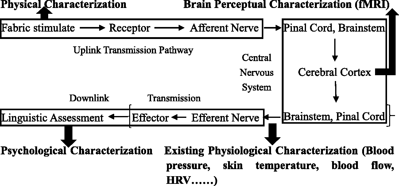
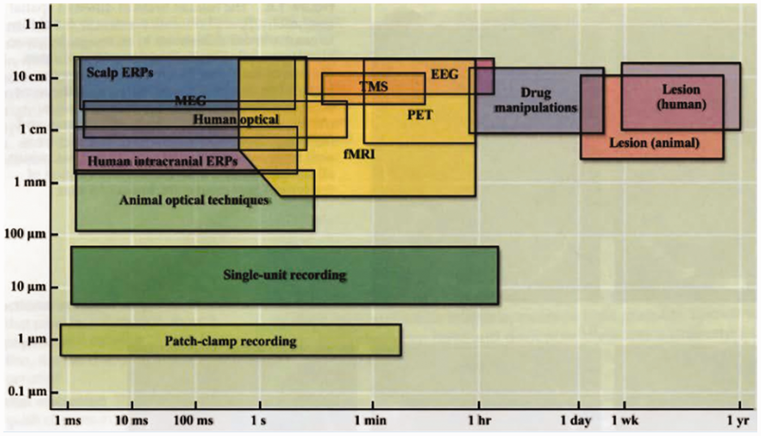
However, we now look at the mechanism of how the human body forms a fabric perception as shown in Figure 1. Perception begins in the cerebral cortex and electroneurographic signals pass through the brainstem and spinal cord and are eventually translated into speech. Apparently, the psychological evaluation is at the end of the neural circuits, so it must have a great interference and disparity to evaluate our perception. Besides, it is more indirect to express a human perception according to the physical characteristics of fabric, because the subject of perception is the human, not the fabric. Compared with existing physiological evaluation methods, such as blood pressure, skin temperature, blood flow, heart rate variability (HRV), and so on, the brain’s perceptual characterization assessment is undoubtedly better because of its more direct and closer evaluation path to the cerebral cortex, the source of tactile recognition. Therefore, if we want to explore the nature of fabric perception, we must begin at its source, the cerebral cortex. Furthermore, the best way to view the cerebral cortex is using functional magnetic resonance imaging (fMRI) technology due to its higher temporal (<1 s) and spatial resolution (<1 mm) compared with many other common techniques in neuroscience, such as positron emission tomography, EEG, and so on, as shown in Figure 2. Of most significance, in contrast to the waveform results of EEG, HRV, and ERP, the results of fMRI are brain images that can visually show which brain regions are responding to the fabric contact.
Mechanisms of various comfort evaluations of fabric contact pressure. Temporal-spatial resolution of general techniques in neuroscience.
19
Experiments
Seven healthy female volunteers of a similar size and an average body mass index of 20.6 kg/m2 were recruited as volunteers. An AMI3037 Air-pack Type Contact Pressure Measurement System was used to test clothing pressure, with an accuracy of ± 0.2–0.45 kpa. The experimental sample’s effective area was 24.5 cm × 50 cm, as shown in Figure 3. Contact pressure could be adjusted by the tightness of the magic tape. Neuroimages were obtained from fMRI equipment provided by Ruijin Hospital, as shown in Figure 4. Subjective assessment and fabric contact pressure tests were performed at the same time in a constant temperature and humidity laboratory at Donghua University, Shanghai, China. The environmental temperature and humidity were ± 20℃, ± 65% RH, respectively.
Schematic diagram of the experimental sample. Experimental functional magnetic resonance imaging scanner.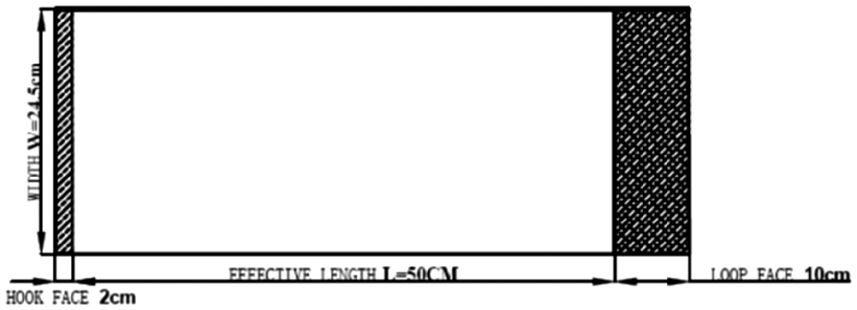

In the pressure measurement and subjective questionnaire survey, subjects were tested 30 minutes after entering the laboratory to ensure they had adapted to the temperature and humidity. As the sample pressure was mainly distributed in the chest, waist, and abdomen, similar to a large number of studies,4,20,21 the skin surface of lower chest was selected as the characteristic part to measure the clothing pressure due to its critical influence on comfort evaluation. The participants were asked to put on the sample and were given uniform earplugs and loose-fitting clothing. They were then asked to close their eyes, keep still, and remain in a state of natural relaxation. Different pressures of 0 kpa, 0.5 kpa, 0.8 kpa, 1 kpa, and 1.5 kpa were applied through tightening the magic tape. Each pressure test lasted 1 minute and the pressures were averaged after removing the maximum and minimum values. Simultaneously, a subjective evaluation questionnaire survey was conducted as shown in Figure 5. After that, we selected a comfortable contact pressure to use in the fMRI experiment to explore the brain response to comfort from a proper fabric pressure.
Z score subjective rating scale.
A block design was adopted in the fMRI experiment. In each experiment sequence, there were three sessions. In the first 30 s of each session the subject was in a resting state with no fabric pressure. In the later 30 s of each session, the fabric pressure was applied. Each session was repeated three times and the average blood oxygenation-level dependent (BOLD) signal was measured for analysis after a series of pre-treatment steps, including realignment, registration, segment, standardization, and smoothing. If the BOLD pressure signal was higher than at rest we called it a positive activation, which meant the fabric pressure increased blood flow and nerve activity in the local brain region. Conversely, if neural activity was inhibited, a negative activation had occurred.
Results and discussion
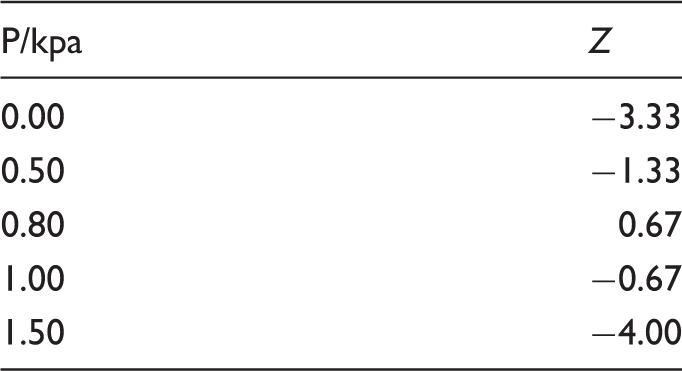
According to the subjective rating questionnaire in Figure 5, when the subjective evaluation Z value was greater than 0, the body felt comfort. When Z equalled 0, it was neither comfortable nor uncomfortable and was in a moderately comfortable scope. When Z was less than 0, it represented an uncomfortable state. The greater the Z value, the higher the degree of comfort. The subjective evaluation results were as follows.
As shown in Figure 6 and Table 1, when the contact pressure, P, was 0 and the human body made contact with the fabric without it applying any pressure, it was subjectively uncomfortable (Z = −3.33). After that, the Z value increased with the gradual increase of contact pressure, i.e., discomfort decreased gradually. When P rose to 0.77 kpa, Z reached the maximum value of 0.76, which was the most comfortable state for the sample. After that, the Z value gradually decreased with the increase in P. This meant comfort gradually disappeared and discomfort gradually increased. When the contact pressure was within the range of 0.62–0.92 kpa, it was relatively comfortable.
Result of subjective evaluation. Subjective comfort evaluation value Z under increasing pressures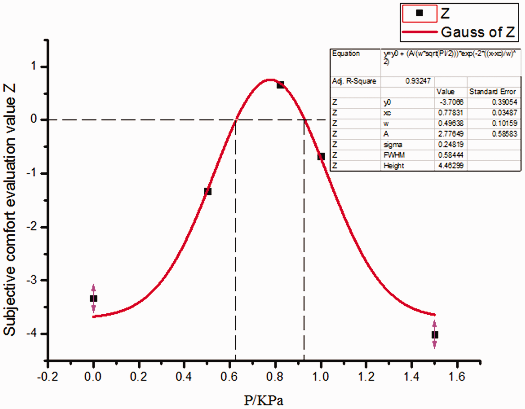
Due to the limitations of the experimental conditions, we selected a contact pressure of 0.82 kpa, which was closest to the most comfortable degree of fabric contact pressure for the fMRI scanning experiment. Before group analysis on the brain images of all subjects, we wanted to avoid various disturbance such as noise produced by fMRI scanning, the subjects' inevitable psychological activities and body-regulating muscle tension in the experiment and other interference factors that cause inaccuracy of the final results of the experiment. To do this, we used a brain mask as shown in Figure 7, only including the primary somatosensory cortex (SI), the secondary somatosensory cortex (SII), insula, amygdala, and precuneus brain regions associated with potential body perception. What should be noted was that all analyses were in the mask brain area rather than the whole brain. In group analysis, the height threshold T = 40.266253 and the extent threshold K = 0. The experimental results verified by family-wise error (FWE) (p < 0.05) were as follows.
Brain mask.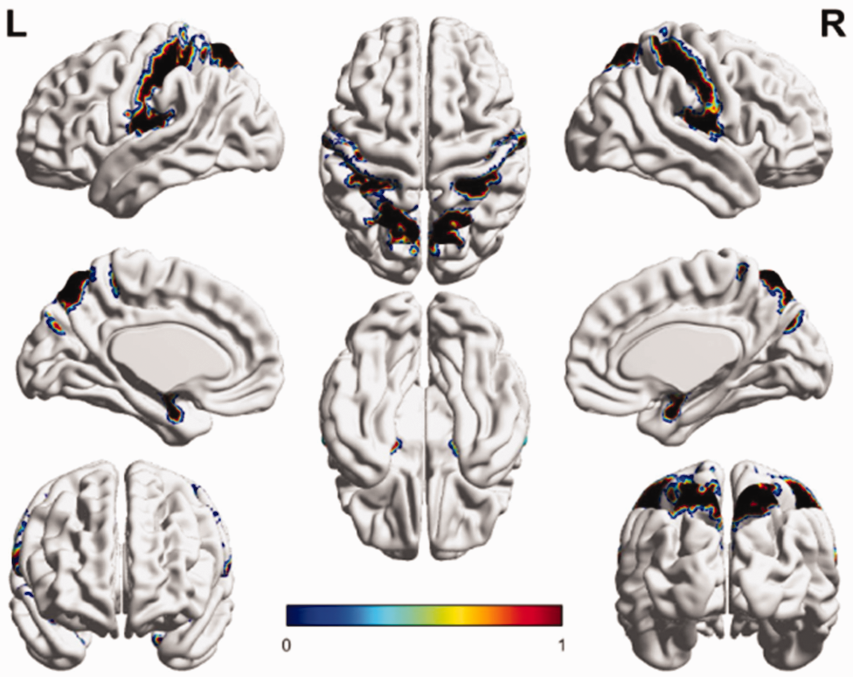
As can be seen from Figure 8, under the effect of a contact pressure of 0.82 kpa although only one positive activation point in the right SII (x = 66, y = 6, z = 15 in the Montreal Neurological Institute coordinate space) was found, the positive activation intensity in the group was up to 4.36. The result was verified by the strictest FWE check, so it was relatively reliable. In addition, there was no negative activation meeting the threshold requirements.
Full view of activation result in the brain mask regions under the stimulations of fabric contact pressure of 0.82 kpa.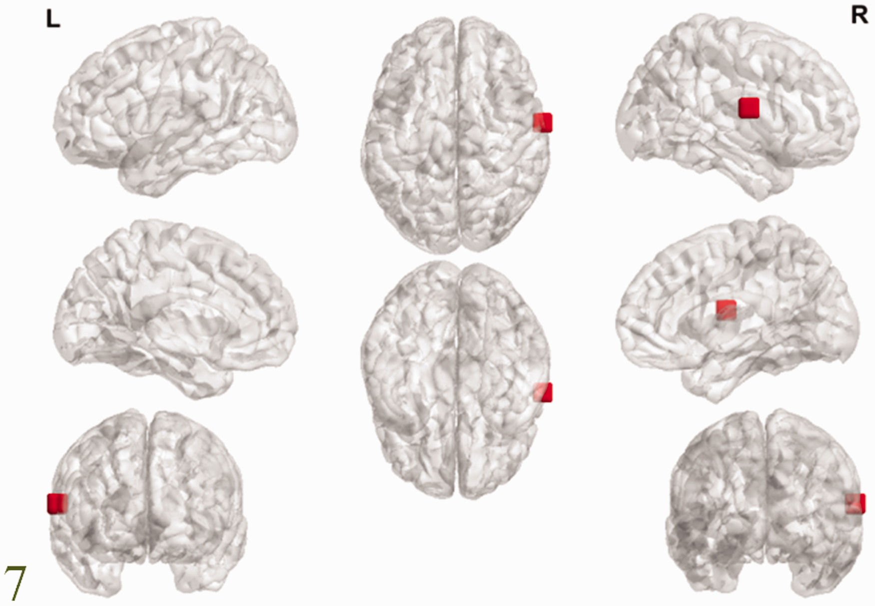
The human SII is located in deep in the parietal lobe along the upper bank of the lateral sulcus. It was first described by Adrian 22 in 1940 in the research of feeling in cats' feet. After that, Ridley and Ettlinger et al. found the SII of monkeys was involved in remembering tactile shapes and textures.23,24 Before 1992, many studies using fMRI found an essential role for the SII in normal pain (such as mechanical and temperature pain), tactile perception (such as roughness), 25 light contact, tactile attention, visceral sensation, and so on. 26 In recent years, more studies have found that the SII, particularly the right SII, was a very promising new target for treating neuropathic pain. Chronic visceral pain is a central nervous system disorder associated with a hyperexcitability of the right SII. 27 After high-frequency repetitive transcranial magnetic stimulation given to the right SII, the most significant decrease was found in thermal pain sensitivity of the face 28 and, compared with the sensorimotor cortex, the right SII had a superior analgesic effect. 29 Even more, human ability to judge pain intensity was disrupted when nociceptive transcranial magnetic stimulation was delivered over the SII, as compared to SI and vertex stimulation. 30 In contrast, Bhatia 31 also used fMRI to investigate the relationship between human perception and fabric texture, and concluded that an acceptable comfort perception was processed in the parietal lobe of high-level processing of SII. Similarly, Wang 32 found that the primary sensory cortex SI and secondary sensory cortex SII were closely related to fabric contact with the fingers. All of these findings suggested that the SII, particularly the right SII, had an effective inhibitory effect on pain, and maybe even a causal role in encoding of pain intensity. In other words, the right SII might perhaps be related to “good” feelings.
From the perspective of the transmission pathway of somatosensory information, pain and tactile information could be conveyed to the SII by an indirect pathway from the thalamus via the SI. 33 What was different was that tactile stimuli from the SI to SII was a parallel pathway, whereas pain stimuli from the SI to SII was a serial access. 34 Moreover, the flow of information from the SI to SII was reduced when the human body accepted medium or high levels of damaging and harmful input stimulus. However, at an innocuous level, an excitatory intrinsic SI-SII was found. 35 This might explain why the peak point of activation was in the SII, not the SI, under the innocuous fabric pressure of 0.82 kpa. It also suggested the fabric pressure of 0.82 kpa was a proper and comfortable contact level. The SII, particularly the right SII, was the characteristic brain region for fabric comfort perception. Moreover, it was not hard to infer that the activation peak point should be located in the SI below the comfort pressure of 0.82 kpa. With a modest increase in fabric pressure, the activation peak point transferred from the SI to SII.
From the view of neurophysiological systems mediating affective contact, there were two types of afferent nerves related to a pleasant or comfortable fabric contact. One was the C-tactile afferent nerve (CT), a type of mechanosensitive amyelinic nerve fiber only found in hairy human skin.36,37 CTs belonged to the slow-adapting type I afferent nerve group and were processed in the limbic-related cortex (including orbitofrontal cortex and posterior insula) but not in somatosensory areas such as SI and SII. Human CTs played a critical role in gentle tactile and itching sensation emotional responses (low frequency and static stimulation),38,39 and stroking of hairy skin. 40 The second type was myelinated A-beta afferent nerves, which had a fast conduction velocity related to fast sensations, and were projected to the somatosensory cortex. The present researches40,41 were consistent with the hypothesis that pleasant contact from hairy skin was mediated by CTs, which represented an innate non-learned process, but pleasant contact from glabrous skin was affected by A-beta afferent nerves, representing an analytical process dependent on previous tactile experiences. In consequence, it could be speculated that the activation location was in the right SII brain region in this project perhaps because of the excitability of A-beta afferent nerves.
Conclusion
The SII brain zone, particularly the right SII, appears prominently positively activated under a comfortable fabric pressure state. This suggests the right SII brain region is probably the characteristic brain region for fabric comfort perception. A reasonable explanation is that proper contact pressure facilitates the serial information flow from the SI to SII, and a comfort touch stimulates the firing of A-beta afferent nerves.
Footnotes
Declaration of conflicting interests
The authors declared no potential conflicts of interest with respect to the research, authorship, and/or publication of this article.
Funding
The authors disclosed receipt of the following financial support for the research, authorship, and/or publication of this article: This work was supported by Exemplary Projects of the Research on Mechanism of Body Injury in the Disaster Environment and Research and Development of Rescue Protection Technology and Equipment (Grant NO. 2016YFC0802800) and Shandong Provincial Natural Science Foundation, China (Grant No. ZR2017BEM041).