
Abstract
Coptidis Rhizoma is a medicinal plant that is well known for its high antibacterial activity against various pathogens and its anti-inflammatory, antioxidant, and hemostatic effects. Here, nanofibrous membranes containing 10, 20, and 30 wt% of Coptidis Rhizoma extracts were fabricated by electrospinning using poly(vinyl alcohol) as the drug carrier and thermally treated to increase their aqueous stability. The antibacterial properties and release characteristics of Coptidis Rhizoma nanofibrous membranes were investigated. Coptidis Rhizoma-loaded nanocomposite fibers exhibited a high drug-loading efficiency ranging from 92% to 97%. The release profile from the nanofibrous membranes of Coptidis Rhizoma showed an initial fast release followed by a gradual release for 48 h. High antibacterial activity of the Coptidis Rhizoma-loaded nanofibrous membranes was exhibited against both Staphylococcus aureus and Staphylococcus epidermidis. These results demonstrate that poly(vinyl alcohol) nanofibrous membranes containing Coptidis Rhizoma extracts have considerable potential to be effective antimicrobial wound dressings based on natural substances.
Nanofibrous materials have gained much attention in medical, protection, and hygiene applications due to their unique properties. Electrospun nanofibrous membranes have an exceptionally small pore size, large surface area-to-volume ratio, high porosity, mechanical flexibility and they are structurally similar to the extracellular matrix of natural tissues.1,2 Thus, electrospun nanofibrous membranes offer many advantages, including hemostasis, gas exchange, preventive effects against bacterial invasion, and promotion of cell proliferation and migration,1,2 which have been widely used in the field of biomedicine.
Dressings should provide an optimal environment for wound healing. An ideal dressing is expected to protect the wound from bacterial invasion, absorb and control exudates, maintain a moist environment by minimizing evaporative water loss from the wound, reduce the risk of infection, and promote healing. 3 Nanofibrous membranes possess numerous micropores and provide a moisture diffusion rate that permits respiration but is not so high as to allow drying out. Nanofibrous membranes, thus, are suitable for use in medical textiles such as wound dressings.
Interest is growing in developing advanced antimicrobial and wound healing materials for biomedical applications. An antibiotic wound dressing releases antibiotics to prevent infection and thus creates an optimal environment for healing wounded areas.1–4 In particular, fibers that contain bioactive agents or drugs within their structure can provide sustained drug release for a prolonged period of time. Incorporation of bioactive agents into fiber matrix thus facilitates the wound healing process in a more constant and stable manner than coating them on the fiber surface. Because bioactive agents can be incorporated into a nanofiber matrix via an electrospinning process in a single step, research is focused on incorporating various drugs (e.g. therapeutic or antimicrobial agents) into nanofibers.
There is considerable interest in using natural products for preventing infections and promoting wound healing in biomedical applications. Because some medicinal plants are biocompatible and have fewer side effects compared with synthetic drugs, numerous studies have focused on utilizing medicinal plants as potential antimicrobial agents for wound dressings.5–8 Coptidis Rhizoma (hereinafter referred to as C. Rhizoma) is a medicinal plant that is well known for its antibacterial properties against various pathogens, as well as its antioxidant and anti-inflammatory properties.9,10 Although there is growing evidence that C. Rhizoma is a powerful herb for treating common dermatological disorders, 9 to the best of the authors' knowledge there are no reports on incorporating C. Rhizoma into a nanofibrous matrix and investigating the potential of nanofibers containing C. Rhizoma as new wound dressing materials based on natural products.
As far as the extract is concerned, medicinal plants have various components and exhibit different medicinal effects. In a study investigating the anti-inflammatory effect of C. Rhizoma and its main alkaloid compound, berberine, in human keratinocytes, 9 a C. Rhizoma extract exhibited a robust anti-inflammatory effect, while berberine was found to have a negligible inhibitory effect. This result implied that although berberine had been reported to possess biochemical and pharmacological activities such as antitumor activities, a single active ingredient may not fully account for the overall effects of herbal extracts. Thus, whole C. Rhizoma extract rather than isolated compounds should be used. In addition, because solvents used in extracting natural substances affect the extraction efficiency as well as recovery components, the extraction solvents should be carefully chosen. Yu et al. 11 examined the antibacterial activity of medicinal herb extracts that were extracted using four different solvents (water, ethanol, methanol, and ethyl acetate). It was found that for C. Rhizoma, water extracts showed the highest antibacterial activity against skin pathogens, such as Staphylococcus aureus and Staphylococcus epidermidis. Therefore, water can be considered as the most suitable extraction solvent.
In addition, for biomedical applications such as next-to-skin wound dressings and tissue culture scaffolds, the use of organic solvents can lead to toxicity and environmental concerns. Poly(vinyl alcohol) (PVA) is a water-soluble and biodegradable synthetic polymer, which possesses non-toxicity, biocompatibility, chemical resistance, and good mechanical properties. 2 Therefore, PVA has been used for various biomedical applications, including drug delivery systems, wound dressings, dialysis membranes, implants of artificial organs, and soft contact lenses. 12 Water-soluble polymers enable environmentally friendly manufacturing processes without employing toxic chemicals. Additional treatment, however, is necessary to prevent humidity-induced structural changes. Although PVA can be chemically crosslinked using chemicals such as acetaldehyde, glutaraldehyde, or formaldehyde, the use of such organic solvents may limit its applications in biomedical applications. Heat treatment has been reported as an environmentally friendly, non-chemical method of stabilizing PVA fibrous structures in a moisture environment. 13 Therefore, heat treatment is expected to increase the aqueous stability of PVA-based membranes without using chemicals.
Therapeutic effects can be enhanced by controlling the release of drugs in polymer-based drug delivery systems. Several factors have been reported to influence the drug release from drug-loaded electrospun fibers. Drug-loading content, drug–polymer–solvent compatibility, and blend ratio in polymer blends are important parameters that can affect the release rate.8,14,15 Crosslinking has also been used to control drug release. In a study by Yang et al., 16 gelatin/PVA bicomponent nanofibers with raspberry ketone as a functional compound were prepared and then subsequently crosslinked with glutaraldehyde vapor. The authors reported that the release of raspberry ketone could be controlled by the gelatin/PVA ratio, the content of loaded raspberry ketone, and the crosslinking time of glutaraldehyde vapor. In a study on the drug (ketoprofen)-loaded PVA nanofibers, 17 methanol treatment was applied to increase their aqueous stability. The authors reported that methanol treatment effectively depressed the burst release of ketoprofen from the nanofibers. Three-layered nanofibrous mats were constructed in a study on drug release from multi-layered electrospun nanofibrous structures. 18 A gentamicin-loaded PVA layer was covered by polyurethane nanofibrous layers. The study indicated that crosslinking of the middle PVA layer by thermal treatment affected the release of gentamicin from the multi-layered structures. Although previous studies16–18 suggest that chemical and physical crosslinking of drug-loaded PVA nanofibers can influence the release rate from composite fibers, thorough research is needed to further understand how heat treatment influences the drug-release behavior and antibacterial properties of drug-loaded electrospun PVA nanofibrous membranes.
The purpose of this study was to combine the advantages of nanofibrous membranes and the pharmaceutical efficacy of medicinal plants to develop a nanofiber-based wound dressing having antibacterial properties and drug release for wound healing. Here, nanofibrous membranes containing 10, 20, and 30 wt% of C. Rhizoma extracts were developed by electrospinning using PVA as a drug carrier. The nanofibrous membranes containing C. Rhizoma extracts in various amounts were thermally treated and then assessed for their potential use as carriers for the delivery of C. Rhizoma. The antibacterial properties of both neat nanofibrous membranes and the one containing C. Rhizoma as well as the release characteristics of C. Rhizoma from the nanofibrous membranes containing C. Rhizoma were investigated. This study may provide useful results in developing medicated PVA-based membranes through ‘green’ electrospinning and post-treatments for various biomedical applications.
Materials and methods
Materials
C. Rhizoma was collected from Sichuan province in China. PVA (>99% hydrolyzed, M w = 89,000–98,000, Sigma Aldrich Co., USA) was used as a drug carrier. Distilled water was used as the solvent. Acetate buffer (pH 5.5, Abelbio, Korea) was used as the release medium for the C. Rhizoma release assay.
Extraction of C. Rhizoma
Whole C. Rhizoma extracts were used in this study and water was chosen as the extraction solvent. C. Rhizoma was ground using a grinder (HMF-3260S, Hanil Electric Co., Korea) and extracted using distilled water (1:10, w/v) through decoction for 4 h. The extracts were filtered using a Whatman no. 1 filter paper (Whatman, Japan). The collected C. Rhizoma extracts were concentrated using a rotary evaporator (Eyela, Japan) and then freeze-dried using a freeze-dryer (FDS8508, Ilshin Biobase Co., Korea).
Electrospinning
The base aqueous PVA solution was prepared at a concentration of 11 wt%. PVA solutions containing C. Rhizoma were prepared by dissolving C. Rhizoma in the PVA solution at 10, 20, and 30 wt% (based on the weight of PVA powder). Before electrospinning, the viscosity and electrical conductivity of the PVA solutions containing various amounts of C. Rhizoma were measured using a viscometer (Visco Elite-L, Fungilab, Spain) and a conductivity meter (YK-2005WA, Lutron, Taiwan), respectively. The solutions were then electrospun using a vertical electrospinning set up with a two-axis robot system (NNC-ESP200R2, NanoNC, Korea). Electrospinning was conducted under various spinning conditions to find suitable processing conditions for fabricating uniform nanocomposite fibers.
Characterization
The morphology of nanocomposite fibers containing C. Rhizoma was characterized using a field-emission scanning electron microscope (FE-SEM; Hitachi Model S 4200, Nissei Sangyo Co. Ltd., Japan) after sputter-coating each sample with platinum and palladium. Fiber diameters were measured from the SEM images (approximately 30 fibers per image) using image analysis software ImageJ (National Institutes of Health, MD, USA). All the measurements were conducted in triplicate. Infrared spectra were collected using an Fourier-transform infrared (FT-IR) spectrometer (Vertex 70, Bruker, Germany) to examine whether the chemical components of C. Rhizoma were successfully encapsulated into the fibers without any damage.
Heat treatment
Because PVA is soluble in water, heat treatment was applied to increase the aqueous stability of the membranes. PVA nanofibrous membranes containing C. Rhizoma were thermally treated at various temperatures (from 155℃ to 180℃) for different time periods (from 3 to 60 min) to determine the suitable heat treatment conditions for the membranes having varied amounts of C. Rhizoma extracts. After heat treatment, the nanofibrous membranes were immersed in water at 37℃ for 1 h. The morphology of the membranes was examined using the FE-SEM to verify the effect of heat treatment. We examined the changes in morphological structures of the membranes after immersion into water such as complete or partial disintegration, fusion of fibers into bundles.
Drug-loading efficiency
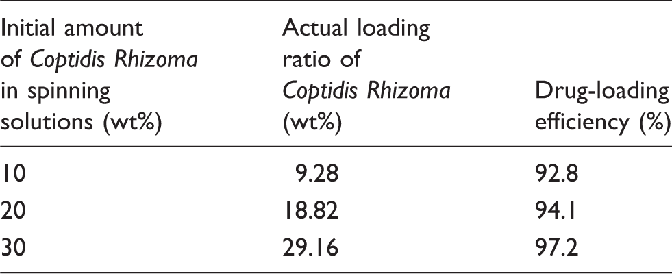
Before assessing the drug release rate, the actual amount of C. Rhizoma in the C. Rhizoma-loaded nanofibrous membranes was determined, and the drug-loading efficiency was calculated for the membranes having different concentration levels. Each specimen (40 mg) was dissolved in 10 ml of distilled water. Then 1.5 ml of the solution was used to measure the actual amount of C. Rhizoma using an ultraviolet (UV) spectrophotometer at a wavelength of 340 nm. The amount of C. Rhizoma in the C. Rhizoma-loaded nanofibrous membranes was calculated from the obtained data against a predetermined calibration curve for C. Rhizoma.
The drug-loading efficiency was calculated using the following equation:

In vitro drug release assay
C. Rhizoma release studies were carried out on nanofibrous membranes having different concentration levels to find out how much C. Rhizoma was released from each membrane in certain periods. A total immersion method was used, and an acetate buffer solution (pH 5.5) was chosen as the release medium to create a similar environment as human skin. The total immersion method is widely used for characterizing bioactive agent-loaded textiles in terms of their drug release and drug-loading efficiency.
19
In this method, the release of bioactive agents is conducted in vitro in simulated body fluids at physiological temperatures and the amount of released drug is determined using an appropriate analytical technique. In this study, each specimen (40 mg) was immersed in 20 ml of the release medium at 37℃, and the solution was stirred at 100 rpm. After a specified immersion period (e.g. 0.5, 1, 3, 5, 8, 11, 24, 48 h), 1.5 ml of the sample solution was collected, while an equal amount of fresh medium was added. The amount of C. Rhizoma in each sample solution was determined using a UV spectrophotometer at a wavelength of 340 nm. The cumulative amount of C. Rhizoma released from the specimens was calculated using the following equation:

Antibacterial activity
Antibacterial activity of the nanofibrous membranes containing C. Rhizoma was examined in accordance with ISO 20645:2004 (Determination of antibacterial activity – agar diffusion plate test). This method allows the diffusion of antimicrobial agents in the sample and creates an inhibition zone around the sample where virus growth is restrained by the concentration gradient of the antimicrobial agent.
20
The susceptibility of the test strain against the antimicrobial agent can be evaluated by examining the extent of bacterial growth in the contact zone under the specimen and, if present, measuring the extent of an inhibition zone around the specimen. Inhibition zones were calculated using the following equation:

Results and discussion
Fiber morphology
In preliminary experiments, electrospinning was conducted with an 11 wt% PVA solution under various spinning conditions, including feed rates of 0.2–0.3 ml h−1, needle gauges of 26–30, voltages of 18–23 kV, and tip-to-collector distances of 11–15 cm to find a suitable processing condition. The suitable spinning conditions for fabricating uniform PVA nanofibers were found to include a solution feed rate of 0.2 ml h−1, a voltage of 19 kV, and a tip-to-collector distance of 13 cm through a 27-gauge needle (0.20 mm inner diameter). The neat PVA nanofibers produced under such spinning conditions are shown in Figure 1(a). Cylindrical fibers with diameters ranging from 150 to 200 nm were obtained. The PVA solutions with varying amounts of C. Rhizoma extracts were then electrospun under the same spinning conditions to fabricate C. Rhizoma-loaded nanocomposite fibers.
SEM images of electrospun PVA nanofibers and nanocomposite fibers containing Coptidis Rhizoma (30,000 × magnification): (a) 11 wt% PVA, (b) 10 wt% Coptidis Rhizoma, 11 wt% PVA, (c) 20 wt% Coptidis Rhizoma, 11 wt% PVA, and (d) 30 wt% Coptidis Rhizoma, 11 wt% PVA.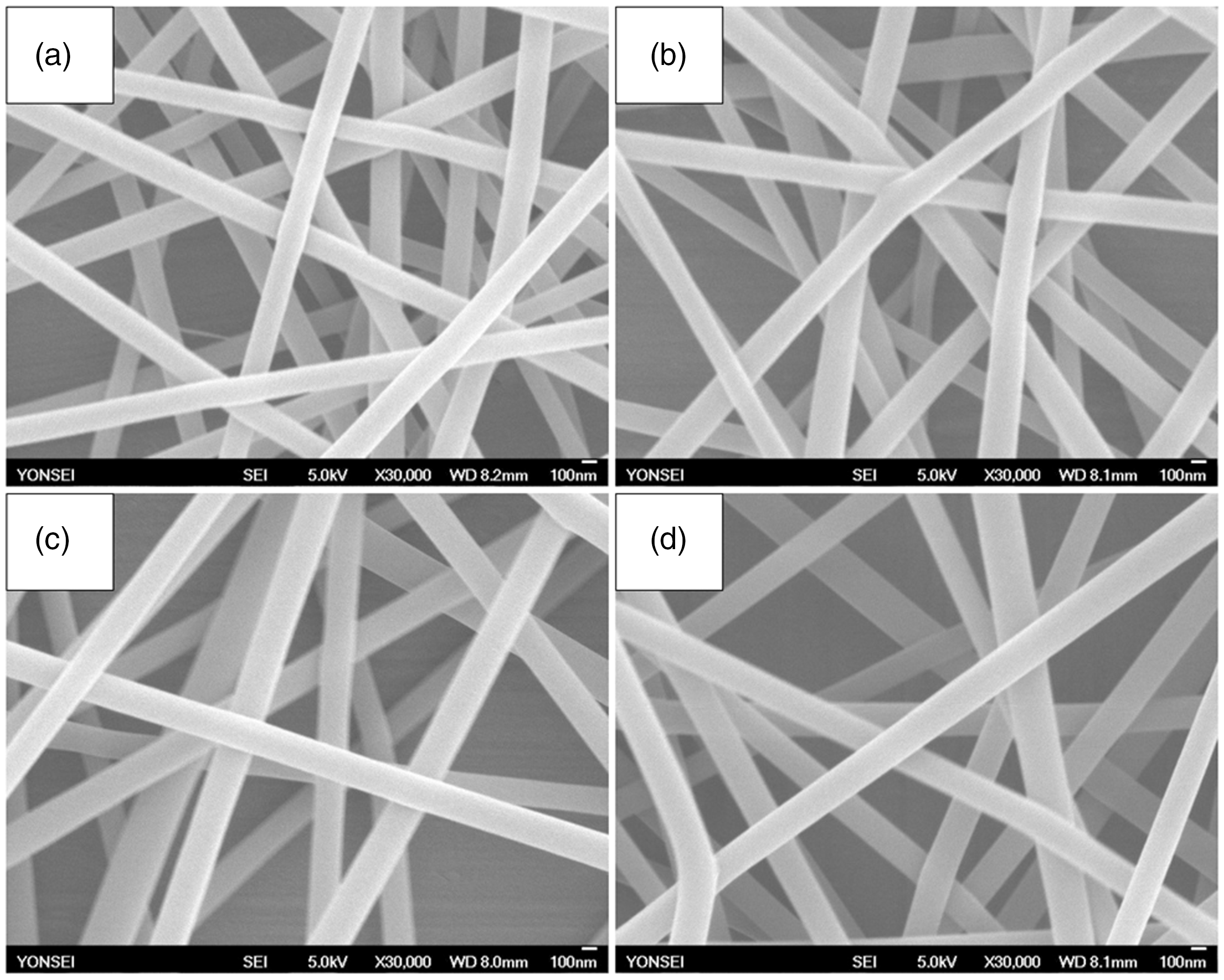
Figure 1(b)–(d) shows PVA nanocomposite fibers containing 10, 20, and 30 wt% of C. Rhizoma, respectively. Bead-free composite fibers were observed, which had smooth surfaces and diameters ranging from 170 to 280 nm. Negligible C. Rhizoma aggregates were observed on the surface of C. Rhizoma-loaded nanocomposite fibers, indicating that C. Rhizoma was well blended and encapsulated within the fibers. Phase separation was not observed between the polymer and drug in the spinning solutions. It has been reported that well-blended composite fibers with smooth surfaces can be formed at a proper polymer concentration, with the aid of increased intermolecular interactions and viscoelastic force. 21 Our SEM images confirm that, as a drug carrier polymer, a PVA concentration of 11 wt% was appropriate for forming uniform nanocomposite fibers containing C. Rhizoma extracts for our experimental conditions.
Viscosity and electrical conductivity of neat and Coptidis Rhizoma-containing PVA solutions, and fiber diameters of the resulting electrospun fiber webs
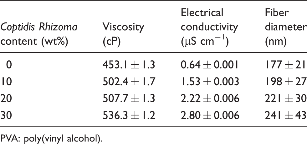
PVA: poly(vinyl alcohol).
For drug release applications, it is highly critical to produce fibers without any beads, because beads can inhibit the release of drug by trapping portions of the drug in the fibers. 26 As shown in Figure 1, bead-free composite fibers having uniform diameters along the fiber were obtained in all examined C. Rhizoma concentrations. Producing fibers having small diameters is also important for use in drug release systems because small diameter of fiber increases the surface area-to-volume ratio and can maximize the effect of drug delivery. 26 Relatively small fiber diameters ranging from 170 to 280 nm were obtained for our experimental conditions (Table 1). These results ensured that the produced composite fibers met the requirement of drug-releasing nanofibers in terms of fiber morphology.
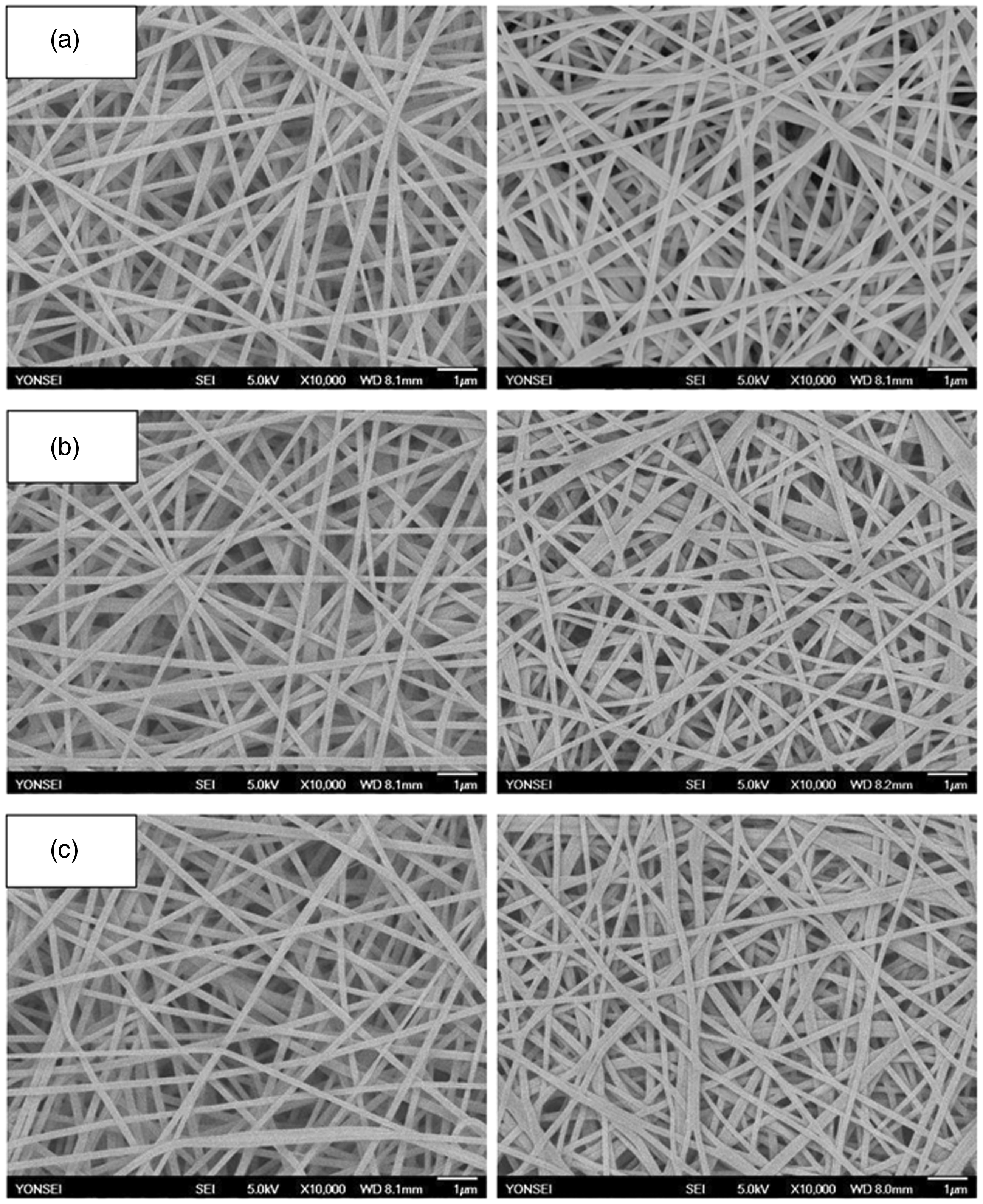
A major advantage of electrospun nanofibrous structure for wound care applications is that the morphology, porosity, and composition of nanostructures can be tailored to meet the needs for a specific biomedical application. Figure 2 depicts low-magnification SEM images of the pristine PVA nanofibrous membrane and composite membranes containing 10, 20, and 30 wt% of C. Rhizoma. The porous structure of nanofibrous membranes was demonstrated. Such a highly porous structure having interconnected pores is expected to allow fluid absorption and gas exchange, while maintaining a moist wound healing environment and accordingly accelerating wound healing.
SEM images of electrospun PVA and Coptidis Rhizoma-loaded PVA nanofibrous membranes (5000 × magnification): (a) neat PVA membrane, (b) 10 wt% Coptidis Rhizoma composite membrane, (c) 20 wt% Coptidis Rhizoma composite membrane, and (d) 30 wt% Coptidis Rhizoma composite membrane.
Heat treatment effect
C. Rhizoma-loaded PVA nanofibrous membranes were thermally treated at various temperatures (from 155℃ to 180℃) for various time periods (from 3 to 60 min) to determine suitable heat treatment conditions. It is ideal to carry out heat treatment at the lowest possible temperature and for the shortest time period to maintain fiber morphology.
PVA nanofibrous membranes containing 10, 20, and 30 wt% of C. Rhizoma were heated first at 155℃ with the time varying between 3 and 15 min, and then immersed in water at 37℃ for 1 h to examine the effect of heat treatment. In all cases, the fiber structure disappeared and most fibers were dissolved in water regardless of the treatment time (results not shown). The heat treatment temperature was then increased to 165℃, and the samples were also heated for 3 to 15 min. The samples which were heated for 10 min and then immersed in water are shown in Figure 3. Partial dissolution of the fibrous membrane was achieved. However, in this case, the degree of dissolution was more prominent for the samples containing a greater content of C. Rhizoma when heated for the same duration, as shown in Figure 3(b) and (c). Some individual fibers in the samples containing 20 wt% and 30 wt% of C. Rhizoma were fused into bundles (as indicated by the arrows) due to aqueous instability. This result indicated that an increase in the concentration of C. Rhizoma increased the required time of heat treatment for stabilizing fiber morphology. The heat treatment temperature was further raised to 180℃, and the samples were treated for different durations depending on concentrations of C. Rhizoma (i.e., 10–25, 15–45, and 35–60 min for the membranes containing 10, 20, and 30 wt% of C. Rhizoma, respectively). Figure 4 presents the selected SEM images of C. Rhizoma-loaded nanofibrous membranes heated at 180℃ for different durations before and after immersion in water. It was found that the suitable heat treatment conditions for PVA nanofibrous membranes containing 10, 20, and 30 wt% of C. Rhizoma were at 180℃ for 25, 35, and 55 min, respectively. Figure 4 confirms that the thermally treated nanofibrous membranes containing C. Rhizoma maintained the fibrous structure after exposure to an aqueous environment.
SEM images of PVA nanofibrous membranes containing Coptidis Rhizoma thermally treated at 165℃ for 10 min: (a) 10 wt% Coptidis Rhizoma composite membrane, (b) 20 wt% Coptidis Rhizoma composite membrane, and (c) 30 wt% Coptidis Rhizoma composite membrane. (The images were taken after immersion in water.) SEM images of PVA nanofibrous membranes containing Coptidis Rhizoma before (left) and after (right) immersion in water: (a) 10 wt% Coptidis Rhizoma composite membrane heated at 180℃ for 25 min, (b) 20 wt% Coptidis Rhizoma composite membrane heated at 180℃ for 35 min, and (c) 30 wt% Coptidis Rhizoma composite membrane heated at 180℃ for 55 min.
Chemical component analysis
The structure of C. Rhizoma-loaded PVA composite fibers was further characterized using FT-IR spectroscopy. FT-IR spectra of PVA nanofiber, C. Rhizoma powder, and PVA nanofibers containing 30 wt% C. Rhizoma are shown in Figure 5. As shown in Figure 5(a), the spectrum of PVA had characteristic peaks assigned to stretching vibrations of –OH at 3338 cm−1, stretching vibrations of CH2 and CH groups at 2941 and 2910 cm−1, bending vibrations of CH at 1449 cm−1, together with C–O stretching vibrations at 1144 and 1098 cm−1. The spectrum of C. Rhizoma powder is shown in Figure 5(b). Featured peaks from –OH at 3396 cm−1 and C = O stretching vibrations at 1674 cm−1 were found. Moreover, bands associated with aromatic rings in C. Rhizoma were observed at 1604, 1507, and 1480 cm−1 due to C = C stretching of aromatic rings.
FT-IR spectra of (a) PVA nanofibers, (b) Coptidis Rhizoma, and (c) PVA nanofibers containing 30 wt% Coptidis Rhizoma.
The spectrum of PVA nanofibers containing 30 wt% C. Rhizoma is given in Figure 5(c). It comprises a composite of PVA and C. Rhizoma spectra, displaying the characteristic bands of both PVA and C. Rhizoma with some extra features. The absorption band of PVA at 3338 cm−1 which was attributed to –OH stretching vibrations not only broadened but also shifted to 3325 cm−1 in the composite fibers possibly due to the formation of hydrogen bonds between C. Rhizoma and PVA, indicating intermolecular interactions. The characteristic peak of C. Rhizoma at 1674 cm−1 associated with C = O stretching vibrations shifted to 1635 cm−1 in the composite fiber possibly due to hydrogen bonds between C. Rhizoma and PVA as well. The typical absorbance bands of C. Rhizoma at 1604 and 1507 cm−1 attributed to C = C stretching of aromatic rings were observed in C. Rhizoma-loaded PVA nanofibers, suggesting that C. Rhizoma was successfully incorporated in electrospun nanofibers and maintained an intact molecular structure.
Drug-loading determination
Actual amounts of Coptidis Rhizoma incorporated in Coptidis Rhizoma-containing nanofibrous membranes and drug-loading efficiency
In vitro drug release studies
All samples were heat-treated at the suitable heat treatment conditions (i.e. 25, 35, and 55 min at 180℃ for membranes loaded with 10, 20, and 30 wt% C. Rhizoma, respectively) before the drug-release studies. Release profiles of C. Rhizoma from the nanofibrous membranes having different C. Rhizoma concentration levels are illustrated in Figure 6. The drug release curves exhibited a similar trend where an initial rapid release was followed by a gradual release throughout the remaining testing period. In the case of 10 wt% C. Rhizoma-loaded fibers, about 54% was released within the initial 5 h, and then an additional 12% was released during the remaining testing period. The release of C. Rhizoma reached 66% after 48 h. Such an initial burst release may be attributed to a release of the drug molecules near the surface of fibers or those adsorbed or loosely bound on the surface. The drug molecules encapsulated deep in the interior would need to diffuse through a longer distance and thus take a longer time to be released.8,15
Coptidis Rhizoma release profiles from nanofibrous membranes containing different Coptidis Rhizoma concentration levels.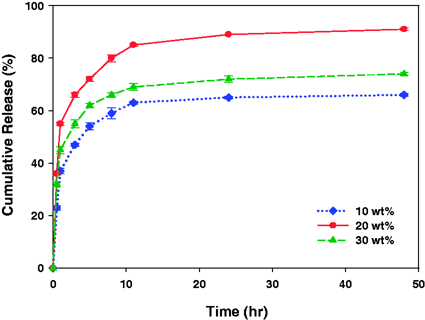
The amount of C. Rhizoma released in a given amount of time varied with the C. Rhizoma concentration levels. After a 5 h release, about 54%, 72%, and 62% were released from the 10, 20, and 30 wt% C. Rhizoma-loaded fibers, respectively. The release rates of C. Rhizoma after 48 h were 66%, 91%, and 74% for the fibers loaded with 10, 20, and 30 wt% C. Rhizoma, respectively. Therefore, the release rates of C. Rhizoma followed the order of 20 wt% > 30 wt% > 10 wt%. It is interesting to note that the fibers loaded with 30 wt% C. Rhizoma exhibited slower release and thus a lesser extent of release than the ones loaded with 20 wt% C. Rhizoma. Generally, a greater extent of release and fast release should be observed with high drug loadings, but an opposite tendency was observed here. This result could be related to the long-term heat treatment applied to the fibers loaded with 30 wt% C. Rhizoma. The fibers loaded with 30 wt% C. Rhizoma were heated at 180℃ for 55 min, whereas the ones loaded with 20 wt% C. Rhizoma were thermally treated at the same temperature for 35 min. Such a long-term heat treatment on the fibers loaded with 30 wt% C. Rhizoma may cause physical crosslinks between drug molecules within the interior fibers and the polymer, resulting in a slow drug release. Similar results were reported in a study on the sodium salicylate-loaded electrospun PVA fiber mats. 27 In that case, chemical crosslinking was applied to the drug-loaded PVA fiber mats, which slowed down the release of sodium salicylate from fiber mats. The release rate decreased with increasing crosslinking time, which was similar to our findings. Our results implied that heat treatment applied to impart water resistance to the PVA could be a key factor affecting the release of the drug incorporated in PVA nanofibers. In other words, the release behavior of drug-containing PVA-based nanofibers can be controlled by heat treatment conditions such as the duration of heat treatment in addition to the drug-loading content.
In drug delivery systems, a high initial release of antibiotic agents is beneficial at the wounded area to quickly eliminate bacteria, while a subsequent gradual release of drug helps preventing an infection and accelerates wound healing. 4 The in vitro release studies of the nanofibrous membranes loaded with C. Rhizoma demonstrated an initial fast release of C. Rhizoma followed by a sustained release over 48 h. In particular, the PVA nanofibrous membranes loaded with 20 wt% C. Rhizoma released nearly 55% of the embedded drug within the initial 1 h, followed by a sustained release and 91% was released after 48 h. Such a drug release behavior can be beneficial for rapid bacterial elimination for a short term as well as a long-term treating effect in preventing an infection.
Antibacterial properties
C. Rhizoma has been reported to show outstanding antibacterial properties against various pathogens including Staphylococcus aureus, Escherichia coli, Pseudomonas aeruginosa, Propionibacterium acnes, and Vibrio cholerae. 9 In this study, the antibacterial effect of nanofibrous membranes containing C. Rhizoma extracts was examined to investigate whether the antimicrobial properties of C. Rhizoma were retained after being incorporated into the fibers with subsequent heat treatment. The antibacterial activity of PVA nanofibrous membranes containing 10, 20, and 30 wt% of C. Rhizoma was evaluated against two kinds of strains, Staphylococcus aureus (ATCC 6538P), and Staphylococcus epidermidis (ATCC 12228), which are the most common causes of wound infection. Because PVA is water-soluble, all samples were thermally treated at the suitable heat treatment conditions before antibacterial assessment. The neat PVA nanofibrous membranes without C. Rhizoma were used as a control.
Antibacterial activity of electrospun PVA nanofibrous membranes containing Coptidis Rhizoma against Staphylococcus aureus and Staphylococcus epidermidis

PVA: poly(vinyl alcohol).
Antibacterial effect of the antibacterial treatment 20
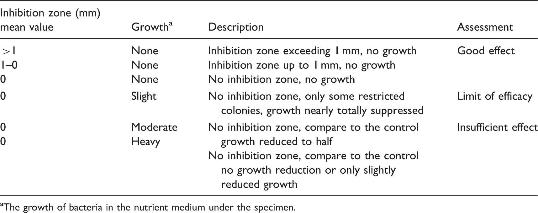
The growth of bacteria in the nutrient medium under the specimen.
The level of antibacterial activity can be further assessed by examining the extent of the inhibition zone around the specimen. As shown in Table 3, the extent of the inhibition zone varied depending on the concentration of C. Rhizoma, exhibiting higher antibacterial activity in the order of 20 wt% > 30 wt% > 10 wt% of C. Rhizoma. This tendency was in agreement with the results of C. Rhizoma release studies. The membranes containing 20 wt% of C. Rhizoma had a stronger antibacterial effect than the ones containing 30 wt% of C. Rhizoma, because C. Rhizoma may have become immobilized within the fibers by a long-term heat treatment, resulting in low C. Rhizoma release and thus low antibacterial effects. Our observations indicate that the antibacterial properties as well as the release of C. Rhizoma extracts loaded in the PVA nanofibers were affected by both the thermal treatment and the amount of loaded drug. These results also demonstrate that the level of antibacterial activity of PVA nanofibrous membranes containing C. Rhizoma extracts is determined by the released amount of C. Rhizoma extracts from the composite membranes.
Our findings show promise for developing medicated nanotextile materials having tunable drug release by varying drug loading and thermal treatment. Ultrathin and light-weight PVA nanofibrous membranes comprising bioactive agents, which show a biphasic release profile (an initial burst release followed by a sustained release), would serve as good candidates for use in advanced wound dressings. Although our findings revealed that drug-loaded PVA nanofibrous membranes have considerable potential as effective antimicrobial wound dressings, several issues need to be further addressed in future research. Additional studies are required to investigate the release behavior of the drug-loaded dressings during and/or after long-term storage under varying storage conditions with the aim of enabling their use in real-life conditions. In addition, efforts must be made to produce drug-loaded membranes on large scales with improved production rate for real practical biomedical applications. Herein, we implemented uniaxial electrospinning of drug–PVA polymer blend solutions and subsequently applied thermal treatment to fabricate drug-loaded PVA-based nanofibers. This technique may be useful for the scale-up production of drug-loaded nanofibrous membranes having a controlled drug release because it is relatively simple and scalable in comparison with coaxial electrospinning. Thus, this technique may be applicable to needleless production-scale electrospinning instruments.
Conclusions
Due to the recent interest in natural products, medicinal plants and their extracts have gained much attention as therapeutic or antimicrobial agents in the field of biomedicine. In this study, Coptidis Rhizoma, a medicinal plant well known for its high antibacterial and anti-inflammatory properties, was incorporated into a nanofiber matrix to investigate the potential for biomedical applications such as antimicrobial wound dressings. Antibacterial properties of the composite nanofibrous membranes as well as the release characteristics of C. Rhizoma from the composite fibers were examined.
PVA nanofibrous membranes containing 10, 20, and 30 wt% of C. Rhizoma were successfully fabricated by electrospinning. Bead-free composite fibers with diameters ranging from 170 to 280 nm were obtained. The PVA-based nanofibrous membranes containing C. Rhizoma were thermally treated to attain stability in aqueous environments, and it was found that increasing the concentration of C. Rhizoma in the fibers increased the time of heat treatment needed for stabilizing fiber morphology. The suitable heat treatment conditions for PVA nanofibrous membranes containing 10, 20, and 30 wt% of C. Rhizoma were 25, 35, and 55 min at 180℃, respectively. FT-IR spectra suggested that C. Rhizoma was successfully incorporated into electrospun nanofibers and the main chemical aspects of C. Rhizoma were retained after electrospinning. High drug-loading efficiencies ranging from 92% to 97% were obtained for nanofibrous membranes containing C. Rhizoma. The release rates of C. Rhizoma varied with its concentration levels in the order of 20 wt% > 30 wt% > 10 wt%, which may be related to the combined effects of heat treatment conditions and drug-loading content. The PVA nanofibrous membranes containing 20 wt% C. Rhizoma released the embedded drug most efficiently among the tested samples, resulting in 91% release after 48 h. Overall, the PVA nanofibrous membranes loaded with C. Rhizoma showed an initial fast release of C. Rhizoma followed by a sustained release over 48 h. High antibacterial activity was exhibited for the PVA nanofibrous membranes containing C. Rhizoma against both Staphylococcus aureus and Staphylococcus epidermidis, indicating that the C. Rhizoma-loaded in the fibers retained its antibacterial properties even after it had been subjected to a high electrical voltage and heat treatment. The results of this study demonstrated that the applied thermal treatment as well as drug-loading content greatly affected the drug release behavior and antibacterial properties of the membranes. This study suggests the possibility of controlling drug release rates via post-spinning thermal treatment on drug-containing PVA-based nanofibers.
In this study, for possible biomedical applications, the use of chemicals such as organic solvents for electrospinning media or crosslinking agents was avoided throughout the manufacturing process by using PVA, a water-soluble polymer, as a drug carrier polymer, water as an electrospinning medium, and subsequent heat treatment rather than chemical crosslinking of PVA-based membranes. Our findings demonstrated that PVA-based nanofibrous membranes containing C. Rhizoma exhibited strong antibacterial properties and a sustained drug release. Thus, they have the potential to serve as effective antimicrobial wound dressings based on natural substances. Future efforts need to focus on examining the performance of the bioactive agent-loaded membranes in vivo to demonstrate their effectiveness in real-use situations.
Footnotes
Declaration of conflicting interests
The authors declared no potential conflicts of interest with respect to the research, authorship, and/or publication of this article.
Funding
The authors disclosed receipt of the following financial support for the research, authorship, and/or publication of this article: This work was supported by the Basic Science Research Program through the National Research Foundation of Korea (NRF) funded by the Ministry of Education (NRF-2013R1A1A2009730); and the Brain Korea 21 Plus Project of Dept. of Clothing and Textiles, Yonsei University in 2018.