
Abstract
Acupoint catgut embedding therapy (ACET) has become the most promising method for treatment of juvenile pseudomyopia due to its advantages of lasting effect, high efficiency, safety and no side effects. However, poor hydrophilicity and biocompatibility are the main disadvantages, preventing ACET materials from having wide applications. This work fabricated four types of polylactic acid (PLA) and polyglycolic acid (PGA) monofilaments from their polymer chips, and then the prepared monofilaments were treated ultrasonically by dipping them in a mixed solution composed of ethyl alcohol and H2O2 (volume ratio 1:1) at 250 W ultrasonic power for 30 min. Afterwards, the PLA and PGA groups were fully characterized with respect to structure characterizations, mechanical properties and in vitro properties. The results showed that the surface roughness of the monofilaments had been greatly enhanced by ultrasonic modification. The PLA groups’ molecular structures changed little, while those of the PGA group emerged with some polar hydrophilic bonds. By deionized water measurement of contact angle values, the ultrasound modified PLA monofilaments (UMPLA1 = 87.2 ± 2.5°, UMPLA2 = 83.6 ± 3.5°) presented a decrease compared to that of untreated PLA ones (PLA1 =103.5 ± 3.4°, PLA2 = 108.4 ± 1.2°), while that of ultrasound modified PGA monofilaments (UMPGA1 = 75.6 ± 4.3°, UMPGA2 =70.5 ± 3.1°) was smaller than untreated PGA ones (PGA1 = 97.3 ± 1.7°, PGA2 = 95.8 ± 2.6°). Based on the measurement of the mechanical properties, the tensile properties and bending stiffness of the PLA and PGA groups changed little, and their swelling ratios were greatly improved after modification. All the prepared monofilaments presented non-toxicity with good cell viability (more than 75%), and samples UMPGA2 (81.4 ± 3.1%) and UMPLA2 (65.8 ± 0.8%) exhibited the largest cell attachment ratio values among their groups. In conclusion, these findings present important clinical implications regarding the ACET materials manufacturing process, which warrant further study.
Keywords
In recent past decades, pseudomyopia has become a serious threat to the adolescent health owing to its fundamental properties, including stubbornness, recurrent attacks, high morbidity rate, etc.1,2 The incidence of pseudomyopia among the senior high school students and college students is over 70%, and has a high upward trend. 3 More than that, the prevalence rate of myopia in children in China over the age of 5 will increase to about 51% by 2020, and the overall population will reach 700 million. 4 In comparison with the other methods of treating juvenile pseudomyopia, acupoint catgut embedding therapy (ACET) has emerged as a very attractive approach for its advantages of lasting effect, high efficiency and easy operation. 5 These attractive characteristics make ACET the most promising method to replace the traditional acupuncture therapy.6,7
Embedding materials is one of the potential risks of ACET in its application in the treatment of juvenile pseudo myopia, especially biodegradable aliphatic polyester embedding materials such as polylactic acid (PLA) and polyglycolic acid (PGA).8–10 As foreign bodies, the surface structures of PLA and PGA monofilaments are relatively smooth, and suffer from poor hydrophilicity and cell attachment ability, which increases the potential risk of surgical site infections, thus limiting their applications in the ACET field.11,12
Recently, attempts have been made to modify the surface structures of biomedical materials to enable the expected functions, including chemical vapor deposition, 13 surface graft polymerization 14 and ion beam injection, 15 etc. Among the various surface modification methods, ultrasound treatment has several important advantages such as being rapid and convenient, non-polluting, safe in operation and highly efficiency.16–18 Hence, the application of ultrasound treatment to surface modification is a hot topic. Shimpi et al. 19 studied and analyzed the effects of ultrasound on calcium carbonate nanoparticles; the results showed that the contact angle of modified materials became smaller, and that the resulting surface structures were rougher according to scanning electron microscopy (SEM) and transmission electron microscopy measurements. Akindoyo et al. 20 applied the ultrasound surface modification on PLA-hydroxyapatite composites, and rougher surfaces and better thermal properties were observed. Moreover, properties such as modulus, tensile strength and impact were enhanced by about 20, 25 and 42%, respectively. Wang et al. 21 attempted to use ultrasound treatment to modify PGA and poly lactic-coglycolic acid (PLGA) fibers, and the results suggested that both PGA and PLGA fibers achieved better hydrophilicity and cytocompatibility, while the tensile strength of PLGA increased and that of PGA changed little. Basto et al. 22 adopted laccase as the modified agent in the ultrasound modifications. The results showed that ultrasonic power of 7 W and a processing time of 30 min were optimized parameters for the cotton fibers. Aimin et al. 23 studied the influence of ultrasound treatment on cellulose fibers using sodium periodate solutions, and the morphology roughness, accessibility and oxidation reactivity of cellulose were improved. Khajavi et al. 24 studied the effect of ultrasound- sodium hypochlorite modification on wool fibers, and better wettability and rougher surface structures were observed. Even through much research has been conducted on ultrasonic modification, with existing studies tending to target materials such as films, stents and composites, few reports have focused on PLA and PGA monofilaments, and their surface modification process has not yet been discussed and analyzed in detail.
In the present work, we aimed to improve the properties of PLA and PGA monofilaments, so that they can be used for ACET materials in our future studies. As biomedical materials, the surface structure influences the hydrophilicity and cell attachment ability. Meanwhile, maintaining adequate mechanical properties after modification is worth consideration, as it is a benefit if embedding materials can bear the pressure from the peripheral tissue. However, strong oxidizing medium such as hydrochloric acid 25 and sodium hydroxide, 26 which have been used in other ultrasonic modification studies, cannot be used on PLA and PGA because they are biodegradable materials, which will cause a big loss of the mechanical properties in the modification process. Considering the above factors and analysis, we attempted to surface modify PLA and PGA monofilaments via an ultrasonic method, with the help of mixed absolute ethyl alcohol and H2O2 solution as an appropriate medium. Afterwards, the characteristics such as structure, and mechanical and in vitro properties were characterized.
Materials and methods
Materials
PLA polymer chips (density = 1.25 g/cm3; intrinsic viscosity = 4.2 dL/g; glass transition temperature =54℃ melting temperature = 150℃) were provided by Shenzhen Esun Industrial Co., Ltd, China. PGA polymer chips (density = 1.63 g/cm3; intrinsic viscosity =1.3 dL/g; glass transition temperature = 36℃ melting temperature = 213℃) were purchased from PURAC Co., Ltd, Holland (Gorinchem, Holland). The drawing and winding devices were provided by Shanghai Tianqing Biomaterial Company (Shanghai, China). The absolute ethyl alcohol with a purity of 95% was purchased from Lianyu Chemistry industry Co., Ltd, Shanghai, China. H2O2 solution at a concentration of 30% was provided from Sinopharm Chemical Reagent Co., Ltd, Shanghai, China. Self-made phosphate-buffered saline (PBS), consisted of NaCl 8 g/L, KClO 2 g/L, Na2HPO4•12H2O 2.9 g/L and KH2PO4 0.2 g/L. All chemicals were analytically pure and used without further purification.
Preparation of PLA and PGA monofilaments
The basic properties of polyglycolic acid and polylactic acid monofilaments
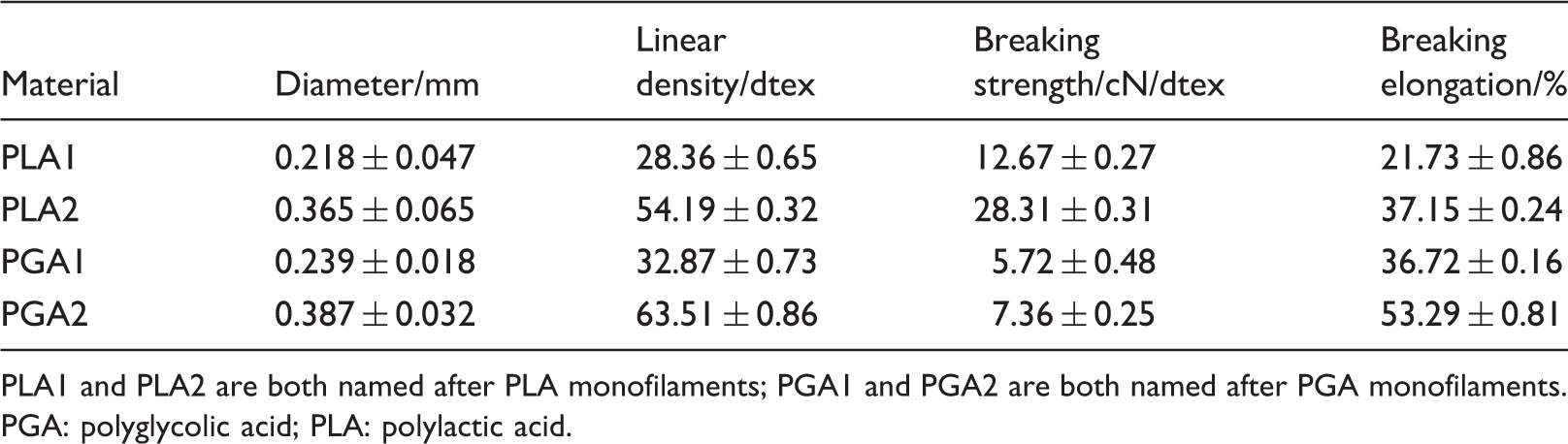
PLA1 and PLA2 are both named after PLA monofilaments; PGA1 and PGA2 are both named after PGA monofilaments.
PGA: polyglycolic acid; PLA: polylactic acid.
Ultrasonic modification of PLA and PGA monofilaments
Figure 1 shows the ultrasonic modification process of PLA and PGA monofilaments. To achieve a fully mixed and homogeneously modified solutions (H2O2:Alcohol = 1:1 (V/V)), the mixture was pretreated with an ultrasound instrument (KQ500VDE, Ultrasound instruments Co., Ltd, Kunshan, China) at 250 W power and a frequency of 40 KHz for 15 min. Subsequently, the prepared PLA and PGA monofilaments went through the clearing tank filled with mixed solutions (power = 250 W; time = 30 min; frequency =75 KHz). Finally, these modified monofilaments were taken out and washed with deionized water several times, and freeze-dried in a vacuum freeze-drying dryer (FD-1 A-50; Bilang instruments Co., Ltd, Shanghai, China) for 12 h. In addition, the ultrasound modified PLA and PGA monofilaments were coded as UM-PLA1, UM-PLA2, UM-PGA1 and UM-PGA1, respectively. Then, their properties were tested and compared to evaluate the efficiency of this ultrasonic modification method.
Schematic drawing of the ultrasonic modification process of the polylactic acid and polyglycolic acid monofilaments.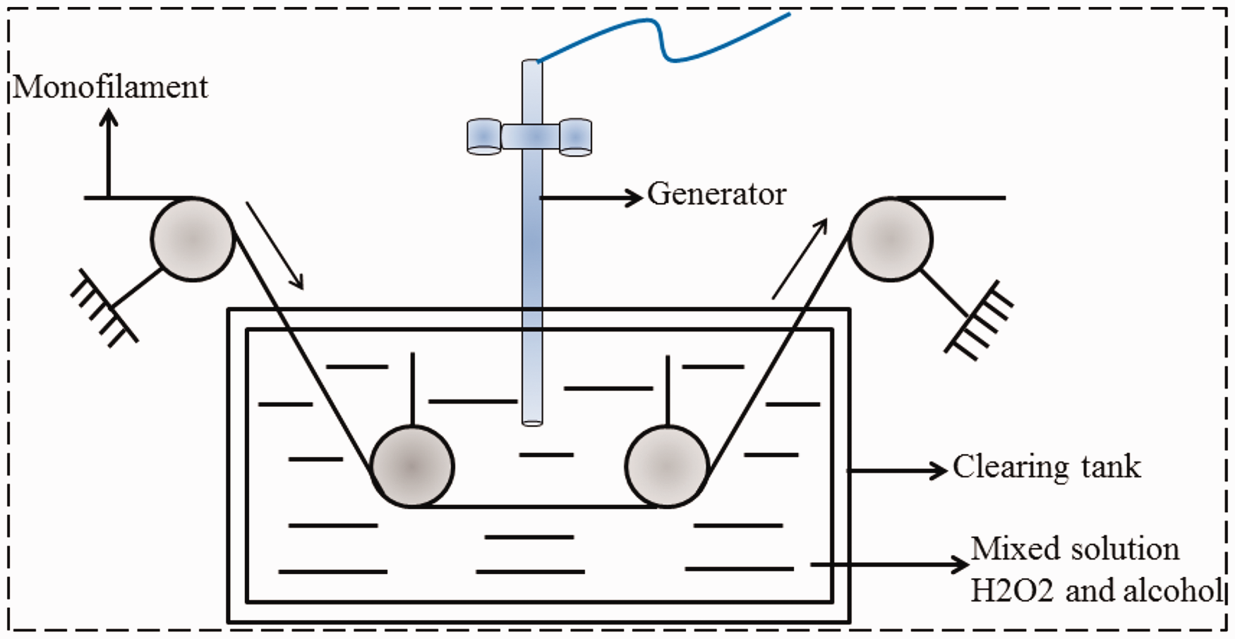
Characterization
Structural characterization
The surface morphologies of the PLA and PGA monofilaments before and after ultrasonic modification were examined by SEM (DXS-10ACKT, low vacuum secondary detection, 15 kV, Shanghai Tianjing Electronic Optics Co. Ltd, Shanghai, China). Before observation, these samples were set on round stainless steel holders with double-sided adhesive conductive tapes and then coated with gold. To check the chemical structures of the prepared PLA and PGA monofilaments, they were detected using Fourier transform infrared spectroscopy (FT-IR) (Spectrum Two, PerkinElmer, UK) with a resolution of 4 cm−1 at room temperature, with 64 cumulative scans in the range of 500–4000 cm−1. To analyze the surface hydrophilicity of modified and unmodified monofilaments, a contact angle goniometer (JC2000A, Shanghai Zhongchen Instrument Co. Ltd, Shanghai, China) was used to measure the contact angle values with deionized water in this experiment.
Mechanical properties
All the samples were conditioned for 24 h at standard atmospheric conditions according to the Chinese National Standard GB/T 6529 before testing. After conditioning, all the tests were also conducted at the standard atmospheric condition (temperature = 20 ± 2°, relative humidity = 65 ± 5%).
29
Based on the testing standard GB/T14344, an electronic single-yarn tensile tester (YG061f, Shandong Laizhou electron instrument Co., Ltd, Shanghai, China) was used to measure tensile properties, including tensile strength and breaking elongation. The final result was expressed as an average value and an SD of 10 replicates. To evaluate the flexibility of prepared monofilaments, bending stiffness values of samples were measured according to the cantilever beam method;
30
the detail operation process was described in previous literature.
31
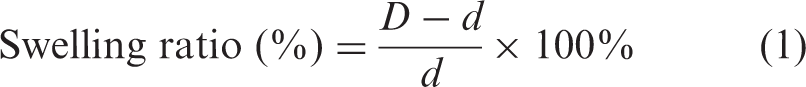
To test the swelling ratio of monofilaments, samples were immersed in 10 mL of PBS (pH = 7.4), and incubated at 37℃ in a shaker bath at 100 rpm for a certain period of time. Afterwards, samples were removed from the excess solution covering the surface, and diameters of samples were obtained via an optical microscope and processed with Image J software. Swelling ratios were calculated using equation (1) as follows:
In vitro properties
Cytotoxicity
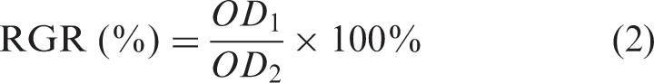
The cytotoxicities of prepared PLA and PGA monofilaments were evaluated via the cell counting kit-8 (CCK-8) cell proliferation method. To sterilize and remove impurities in measured samples, monofilaments were immersed in alcohol solution (75% (v/v)) for 3 h, and then washed with PBS. In detail, samples were first placed into Dulbecco’s modified Eagle’s medium at a certain ratio and cultured under standard culture conditions at 37℃ for 48 h in the incubator to achieve leach liquor. Then, second generation rat fibroblasts (1 × 10⁴ mL) were seeded into a 96-well plate, and the leach liquor was added to each well after good adherence of the cells. Finally, a CCK-8 assay was carried out to measure the cell viability of the samples. Moreover, a microplate reader at a wavelength of 450 nm was adopted to test the optical density (OD) value, and the relative growth rate could be calculated by the equation (2) as follows:
Cell attachment and cell morphology
The sterilized samples were placed in the wells of 24-well plates, with the bottom surfaces of the wells fully covered by the disk, and then the C2C12 cells (provided by the China Center for Type Culture Collection, Shanghai, China) were harvested and seeded in the wells, and each well was inoculated with a total of 1 mL medium of 2.0 × 10⁶ cells/mL. Afterwards, the unattached cells were removed by washing with PBS three times and the attached cells were fixed for 10 min in 3% glutaraldehyde at room temperature, after culturing at 24 h in a humidified incubator (37℃, 5% CO2). Finally, samples were rinsed in PBS, and then the cells were dyed with one drop of Giemsa stain (Sigma) for 30 min and washed with distilled water. An inverted fluorescence microscope (Nikon, Shanghai, China) was used to observe the cell attachment and morphology of prepared samples, and the percentage of C2C12 coverage on modified and unmodified monofilament surfaces was calculated and analyzed by Image J 6.0. The final results were expressed with average values (repeated three times).
Statistical analysis
Statistical analysis was performed using Origin 9.0 (Origin lab, USA). Data results were averaged and expressed as means ± SD. Student’s t-tests were used to determine the statistical difference between samples. The data in the figures were marked by (*) for P < 0.05.
Results and discussion
Structural characterization of prepared samples
Surface morphologies of modified and unmodified PLA and PGA monofilaments
SEM images of the PLA and PGA groups are shown in Figure 2. The PGA group was observed to have a relatively smooth surface, while that of the PLA group had some impurities that may have been caused during the preparation process. In the case of modified embedding monofilaments, all the samples presented relatively rough surfaces, with some long grooves emerging for both the PLA and PGA groups. Moreover, the surfaces of the PGA group monofilaments were found to be coarser than those of the PLA group.
Scanning electron microscope images of surfaces of polylactic acid and polyglycolic acid groups. polylactic acid group: PLA1, PLA2, UMPLA1 and UMPLA2; polyglycolic acid group: PGA1, PGA2, UMPGA1 and UMPGA2; UMPLA: ultrasound modified polylactic acid; UMPGA: ultrasound modified polyglycolic acid.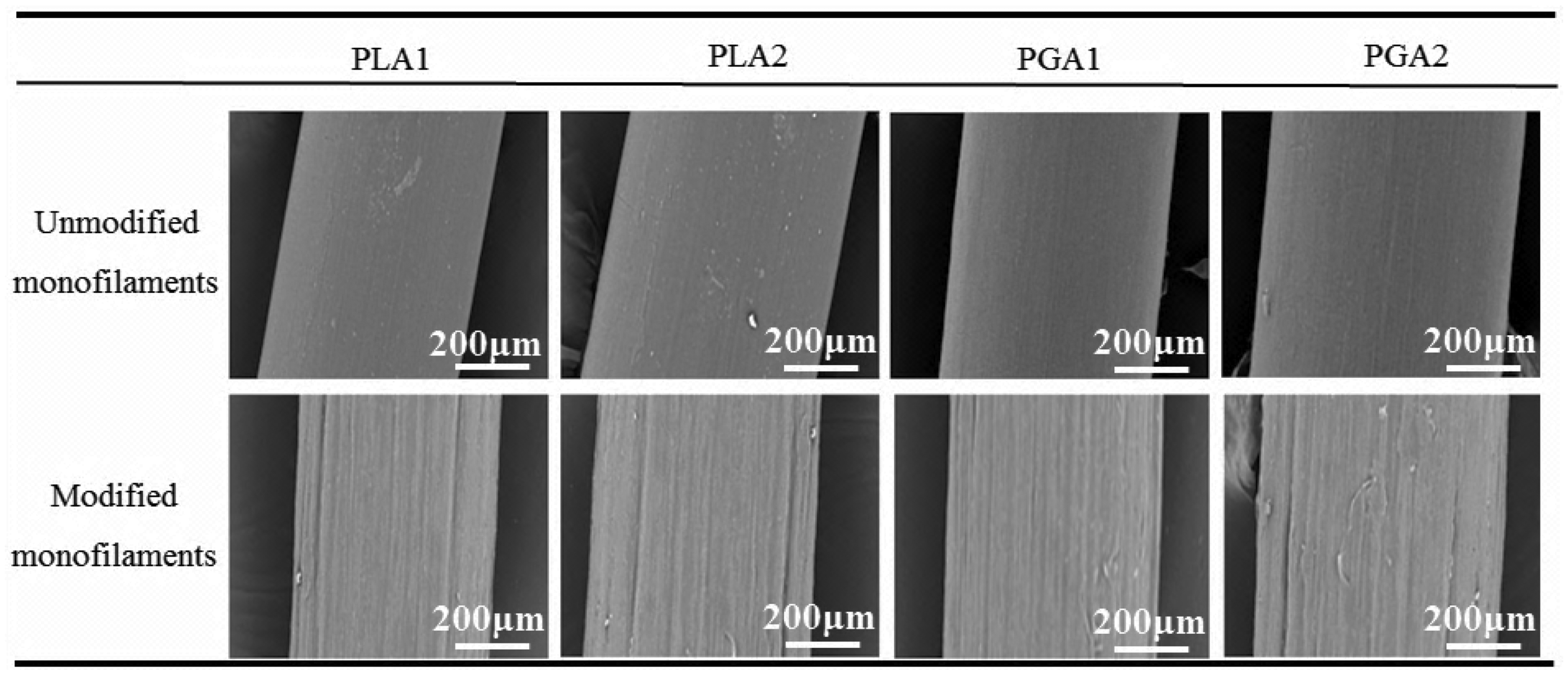
FT-IR analysis of modified and unmodified PLA and PGA monofilaments
Figure 3 shows the FT-IR analysis curves of the PLA and PGA groups. PLA monofilaments presented similarly changing curves, and they had four absorption bands at 3480.4, 2962.1, 1764.5 and 1186.3 cm−1, respectively. The sharp band at 1186.3 cm−1 was attributed to the bending vibration of C-O, and the absorption bands at 1764.5, 2962.1 and 3480.4 cm−1 confirmed the presence of C = O, C-H and O-H. Therefore, it was concluded that the PLA monofilaments had the typical structure of aliphatic polyester, and few new chemical bands or groups were observed. In the case of the PGA group, sharp peaks were presented at 3515.6, 2960.1, 1436.8, 1149.4 and 1083.7 cm−1, respectively. Among them, the absorption bands at 1436.8 and 1149.4 cm−1 confirmed the presence of -CH2 and -CH2-COO-.
32
Moreover, the modified PGA group (UMPGA1 and UMPGA2) exhibited a new sharp peak at 3515.6 cm−1, which may be caused by a hydroxyl dimer. These findings could be explained according to inner changes in the molecular structures of chemical bonds caused by the ultrasound modification.33,34 As such, some ester bonds of PGA monofilaments may be partially hydrolyzed under the effects of ultrasound cavitation and hydrogen peroxide corrosion.
35
Fourier transform infrared analysis curves of the polylactic acid and polyglycolic acid groups. (a) wavenumbers at 4000 and ∼2000 cm−1; (b) wavenumbers at 2000 and ∼500 cm−1. Polylactic acid group: PLA1, PLA2, UMPLA1 and UMPLA2; polyglycolic acid group: PGA1, PGA2, UMPGA1 and UMPGA2; UMPLA: ultrasound modified polylactic acid; UMPGA: ultrasound modified polyglycolic acid.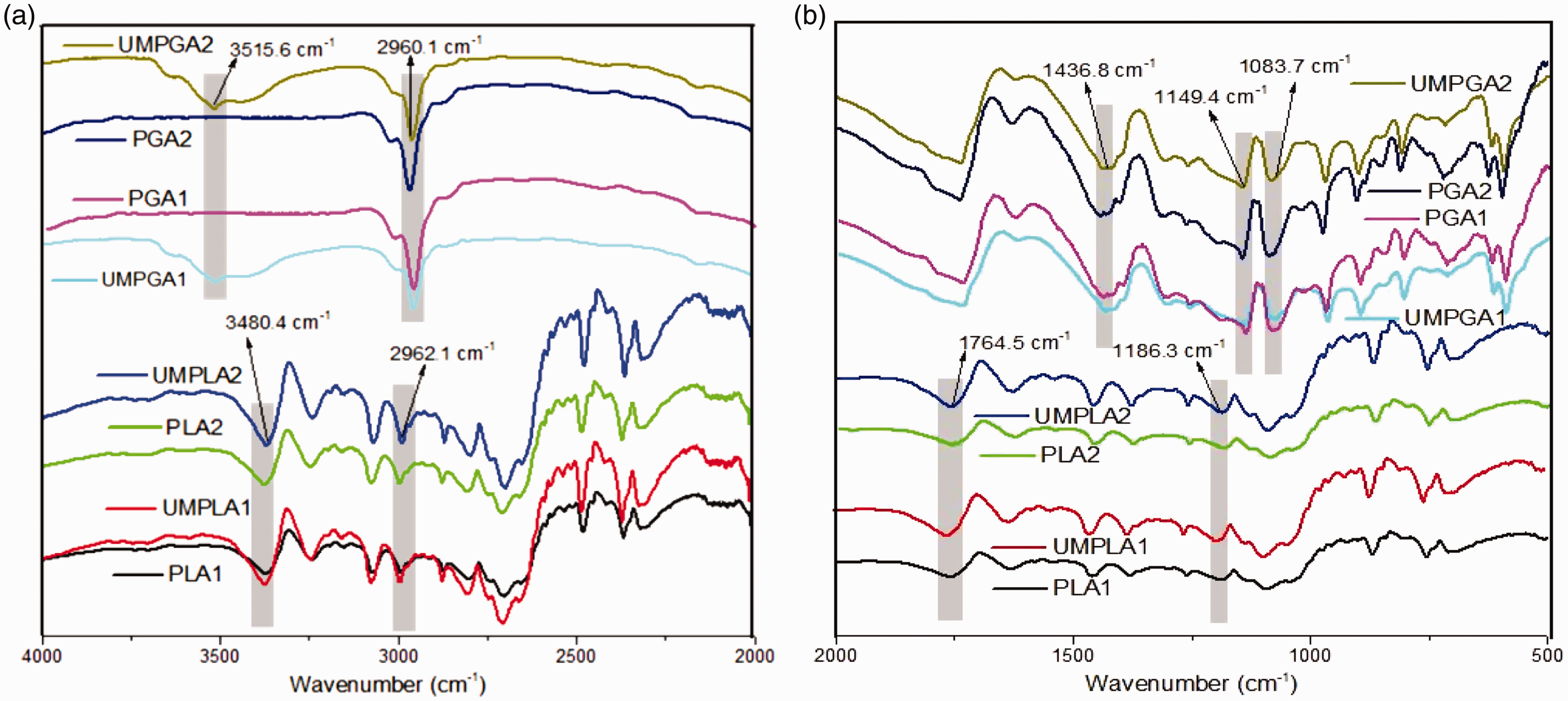
Surface hydrophilicity of modified and unmodified PLA and PGA monofilaments
Figure 4 shows the contact angle values of the PLA and PGA groups. The modified monofilaments presented were observed to have smaller contact angle values than those of the unmodified ones, which indicated that they were hydrophobic materials (more than 90°), and that the ultrasound treatment could greatly improve the hydrophilicity of both the PLA and PGA groups. As for the PLA group, samples PLA1 (103.5 ± 3.4°) and PLA2 (108.4 ± 1.2°) showed significant difference from samples UMPLA1 (87.2 ± 2.5°, P < 0.05) and UMPLA2 (83.6 ± 3.5°, P < 0.05), respectively. A similar result was observed among the PGA group; sample PGA2 (95.8 ± 2.6°) presented a significantly larger contact angle value compared with that of sample UMPGA2 (70.5 ± 3.1°, P < 0.05). Meanwhile, the contact angles of PGA1 (97.3 ± 1.7°) were larger than those of UMPGA1 (75.6 ± 4.3°, P < 0.05). In conclusion, these findings illustrate that the contact angle values of the PLA and PGA groups became smaller after modification, and that the PGA group showed better hydrophilicity than the PLA group.
Contact angle values of the polylactic acid and polyglycolic acid groups. Polylactic acid group: PLA1, PLA2, UMPLA1 and UMPLA2; polyglycolic acid group: PGA1, PGA2, UMPGA1 and UMPGA2; UMPLA: ultrasound modified polylactic acid; UMPGA: ultrasound modified polyglycolic acid.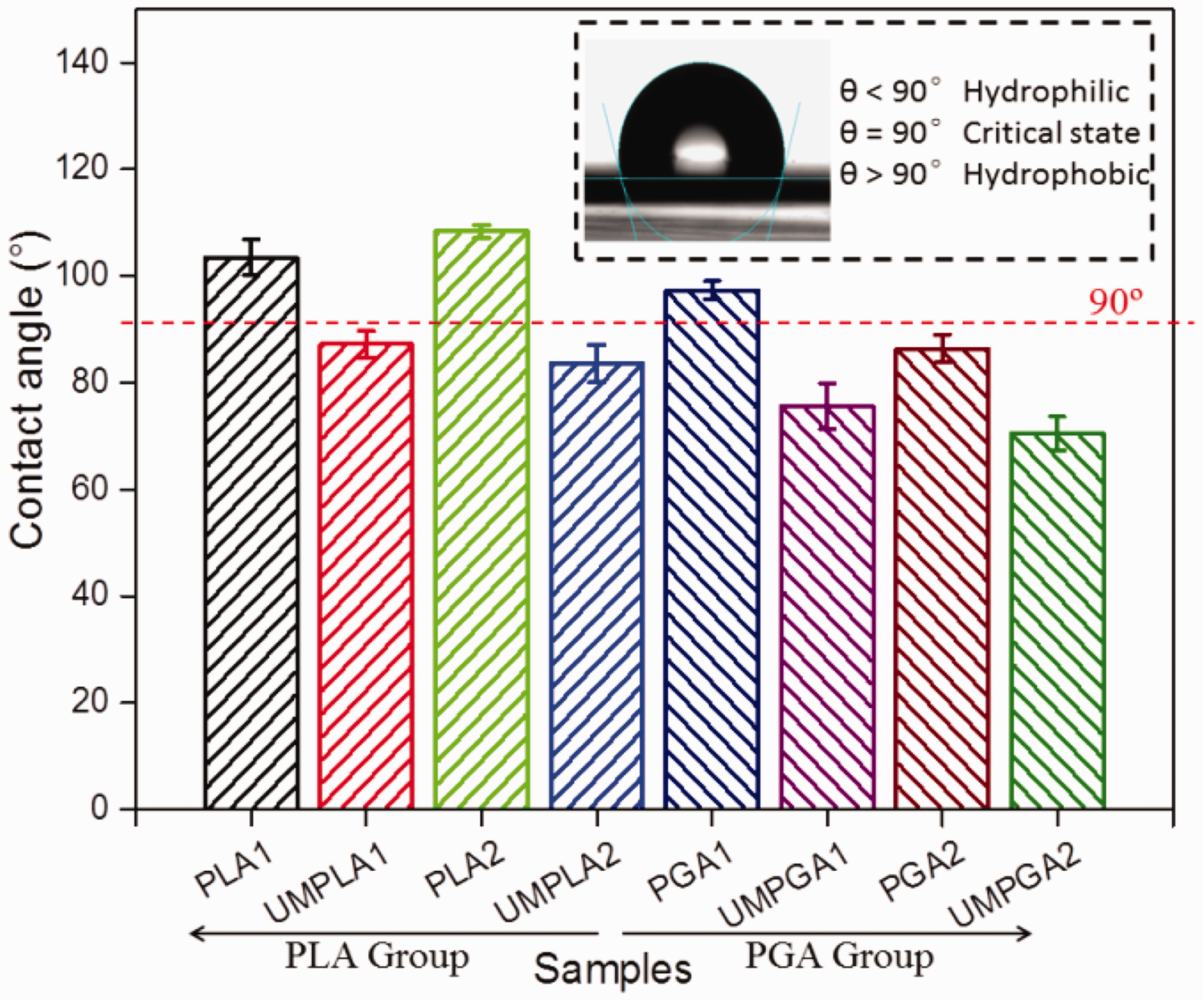
Mechanical properties of prepared samples
Tensile properties of modified and unmodified PLA and PGA monofilaments
The mechanical properties of PLA and PGA monofilaments before and after ultrasound modification were examined and compared, as shown in Figure 5. In the case of tensile strength, sample PLA2 (58.61 ± 4.35 cN/tex) exhibited a significant difference from that of sample PGA2 (28.35 ± 4.33 cN/tex, P < 0.05), and sample PLA1 (36.47 ± 3.18 cN/tex) had the larger tensile strength value than sample PGA1 (21.83 ± 1.39 cN/tex, P < 0.05). Moreover, the tensile strength value of sample PLA2 was similar to that of the ultrasound modified one (sample UMPLA2, 56.34 ± 2.86 cN/tex), and sample PLA1 had a tensile strength value that was similar to that of sample UMPLA1 (33.82 ± 1.03 cN/tex). A similar result was found for the breaking elongation values of the PLA and PGA groups. For instance, the breaking elongation of sample PLA2 (52.24 ± 1.26%) was larger than that of sample PGA2 (27.63 ± 2.15%), while it was similar to the ultrasound modified monofilament (UMPLA2, 50.48 ± 2.85%). In addition, sample UMPLA1 (43.16 ± 3.06%) presented a breaking elongation value that was similar to that of sample PLA1 (46.28 ± 5.62%).
Tensile properties of polylactic acid and polyglycolic acid groups. Polylactic acid group: PLA1, PLA2, UMPLA1 and UMPLA2; polyglycolic acid group: PGA1, PGA2, UMPGA1 and UMPGA2; UMPLA: ultrasound modified polylactic acid; UMPGA: ultrasound modified polyglycolic acid.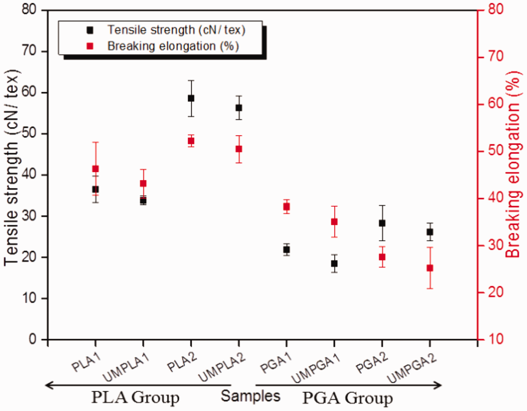
These findings illustrate the fact that ultrasonic modification had little influence on the tensile properties of both the PLA and PGA groups, and that the PLA group presented larger tensile strength and breaking elongation values than those of the PGA group. This may be explained by the theory of molecular motion of textiles. Ultrasonic modification would enhance the surface roughness of monofilaments via its erosive effect, causing a loss of tensile properties for monofilaments, while ultrasonic modification could also result in the rearrangement of molecular chains, further eliminating some shape and interstress defects of monofilaments.26,36 Hence, the tensile properties of the PLA and PGA groups changed little after the ultrasonic modification.
Flexibility properties of modified and unmodified PLA and PGA monofilaments
Bending stiffness was adopted to evaluate the flexibility of PLA and PGA groups according to the cantilever beam method and calculation formula. As shown in Figure 6, sample PLA2 (17.32 ±3.29 cN/cm2) exhibited a similar bending stiffness value to that of sample UMPLA2 (16.58 ±2.25 cN/cm2), and sample PLA1 (12.83 ± 2.18 cN/cm2) showed little difference from sample UMPLA (14.56 ± 1.15 cN/cm2) regarding flexibility. In the case of the PGA group, the bending stiffness values of modified samples (UMPGA1 = 7.04 ± 3.26 cN/cm2; UMPGA2 = 5.08 ± 1.63 cN/cm2) changed little compared to the unmodified ones (PGA1 = 6.81 ±1.93 cN/cm2, PGA2 = 4.73 ± 0.87 cN/cm2).
Bending stiffness of the polylactic acid and polyglycolic acid groups. Polylactic acid group: PLA1, PLA2, UMPLA1 and UMPLA2; polyglycolic acid group: PGA1, PGA2, UMPGA1 and UMPGA2; UMPLA: ultrasound modified polylactic acid; UMPGA: ultrasound modified polyglycolic acid.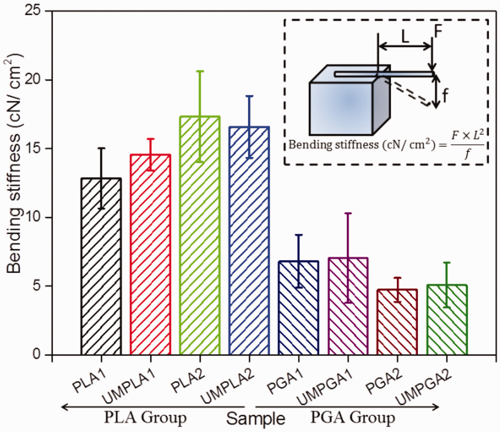
The reason may be that the ultrasound modification only changed the surface structures of monofilaments, without causing further damage to the PLA and PGA groups. In sum, both the PLA and PGA groups changed little regarding their bending stiffness values after ultrasonic modification, and the PLA group presented greater rigidity than the PGA group.
Swelling behavior of modified and unmodified PLA and PGA monofilaments
The swelling behaviors of the PLA and PGA groups are shown in Figure 7. Obviously, the PGA group exhibited larger swelling ratio values than the PGA group. For example, sample PGA2 (11.49 ± 2.15%) had a larger swelling ratio compared to that of sample PLA2 (3.85 ± 0.53%), and the swelling ratio value of sample UMPGA2 (23.82 ± 3.16%) was larger than that of UMPLA2 (7.64 ± 1.05%). Moreover, sample UMPLA2 exhibited a larger swelling ratio value than that of sample PLA2, which revealed that the ultrasonic modification had a beneficial effect on the swelling behavior of the PLA group. This result was more obvious for the PGA group. Considering the statistical difference of the swelling behavior among all the samples, sample UMPGA1 (17.38 ±3.07%) presented a significant difference from that of sample PGA1 (9.36 ± 2.44%, P < 0.05), and sample UMPGA2 (23.82 ± 3.16%) presented a significant difference from that of sample PGA2 (11.49 ± 2.15%, P < 0.05).
Swelling ratios of the polylactic acid and polyglycolic acid groups. Polylactic acid group: PLA1, PLA2, UMPLA1 and UMPLA2; polyglycolic acid group: PGA1, PGA2, UMPGA1 and UMPGA2; UMPLA: ultrasound modified polylactic acid; UMPGA: ultrasound modified polyglycolic acid.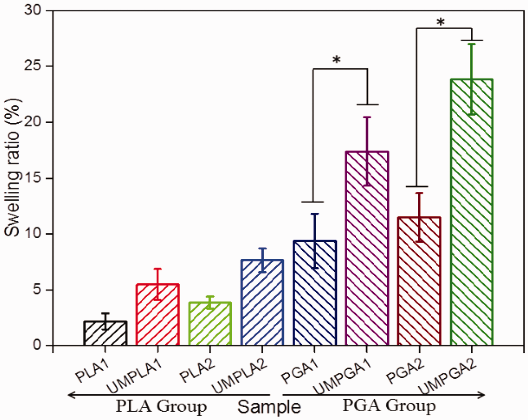
This phenomenon could be explained by the molecular chains of the modified samples being rearranged, increasing the distance between the molecular chains, and further enhancing the swelling properties of the PLA and PGA groups.37,38 In total, this ultrasonic modification could improve the swelling behavior of both the PLA and PGA groups, and the modified PGA monofilaments showed better swelling behaviors than the modified PLA monofilaments.
In vitro properties of prepared samples
Cell viability
The cell viability was determined by incubating the C2C12 rat skeletal muscle cells in the media of the PLA and PGA groups’ leach liquors for 48 h. As shown in Figure 8, all the samples were observed to present no toxicity, with more than 75% of cells being viable. For both the PLA and PGA groups, the cell viability values changed little before and after ultrasonic modification. In detail, sample UMPLA1 (90.2 ± 0.9%) exhibited a similar cell viability value to that of sample PLA1 (87.5 ± 1.3%), while sample PGA2 (92.3 ± 2.8%) had little effect on cell viability compared to that of sample UMPGA2 (95.6 ± 1.9%).
Cell viability of polylactic acid and polyglycolic acid groups. Polylactic acid group: PLA1, PLA2, UMPLA1 and UMPLA2; Polyglycolic acid group: PGA1, PGA2, UMPGA1 and UMPGA2; UMPLA: ultrasound modified polylactic acid; UMPGA: ultrasound modified polyglycolic acid.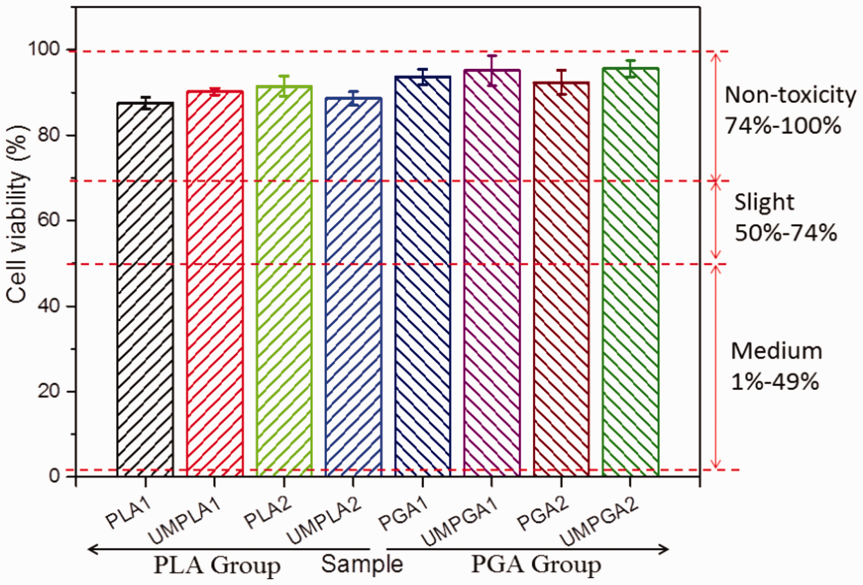
Cell attachment and cell morphology
In this part of the work, the attachment characteristics of ultrasound modified and unmodified PLA and PGA groups were studied. The cell attachment ratios of samples were measured by fluorescence staining and expressed as fluorescence area proportion values of the whole area. As shown in Figure 9, considering the statistical difference among all the samples, sample UMPLA2 (65.8 ± 0.8%) exhibited significantly greater cell attachment than sample PLA2 (32.7 ± 1.6%, P < 0.05), while the cell attachment of sample UMPLA1 (53.4 ± 3.2%) was greater than that of sample PLA1 (28.6 ± 1.8%, P < 0.05). In addition, a similar result was found for the PGA group; sample UMPGA2 presented the largest cell attachment of 81.4 ± 3.1%, which was significantly different to that of sample PGA2 (50.2 ± 2.6%, P < 0.05), and the cell attachment of sample UMPGA1 (74.6 ± 1.5%) was much larger compared to that of sample PGA1 (47.3 ± 3.8%, P < 0.05). These trends are in agreement with the regular of surface morphology and hydrophilicity indicated by the water contact angle, which revealed that ultrasonic modification had effectively enhanced the surface roughness of both the PLA and PGA groups, and further promoted the cell attachment ability of the monofilaments.
Cell attachment of polylactic acid and polyglycolic acid groups. Polylactic acid group: PLA1, PLA2, UMPLA1 and UMPLA2; polyglycolic acid group: PGA1, PGA2, UMPGA1 and UMPGA2; UMPLA: ultrasound modified polylactic acid; UMPGA: ultrasound modified polyglycolic acid.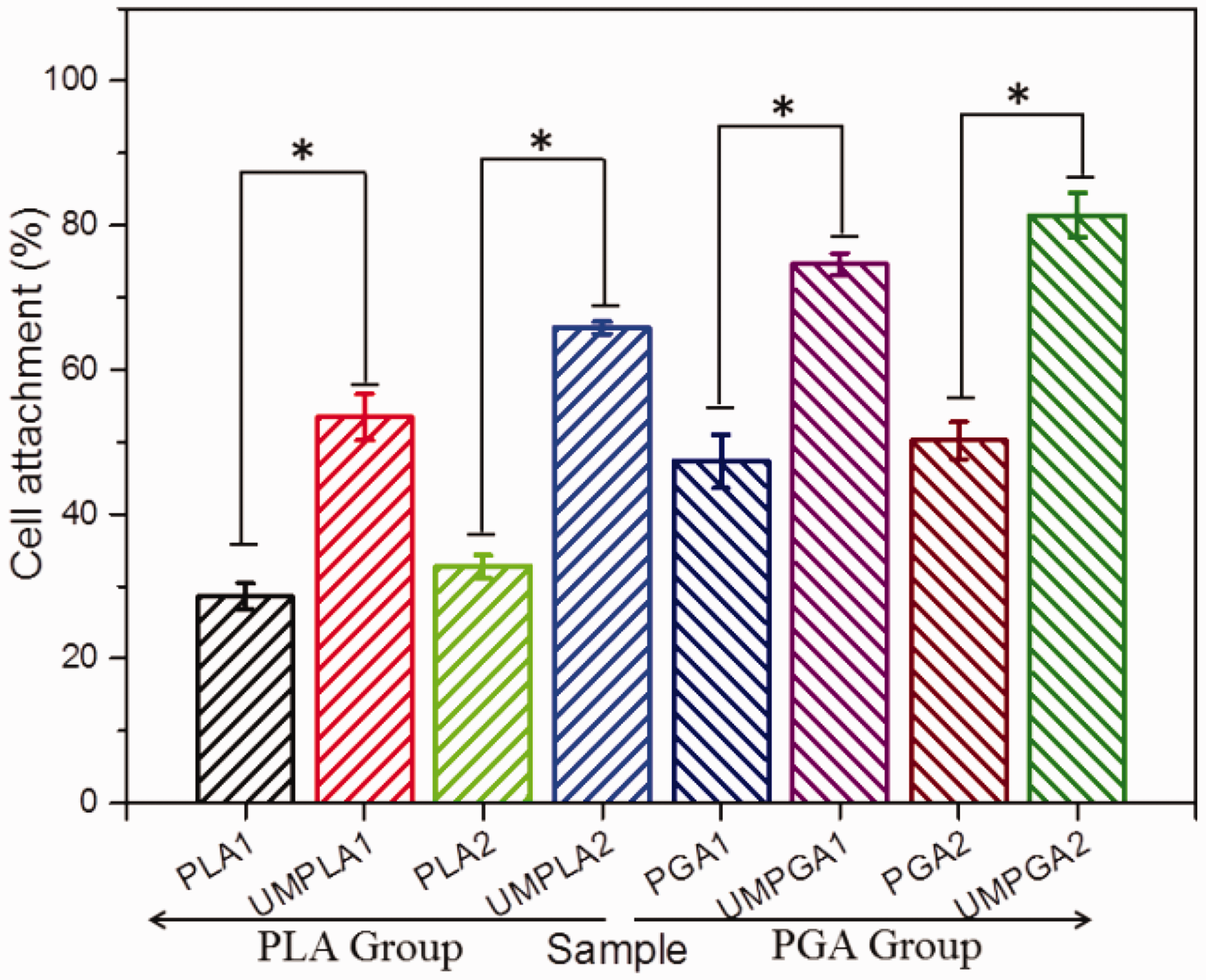
The optical micrographs of the C2C12 cells attached to the PLA and PGA groups are shown in Figure 10. The unmodified monofilaments were observed to attach relatively small numbers of cells, and there existed little difference between the PLA and PGA monofilaments cultured for 48 h. As for the modified monofilaments, the cells attached onto the surfaces for both the PLA and PGA modified samples were obviously increased, and the cell growth around the modified monofilaments became better than that of the unmodified ones. These findings revealed that the ultrasonic modification could improve the cytocompatibility of both the PLA and PGA monofilaments, and the PGA sample exhibited a better cell attachment ratio than that of the PLA sample.
Cell morphology of modified and unmodified polylactic acid and polyglycolic acid monofilaments.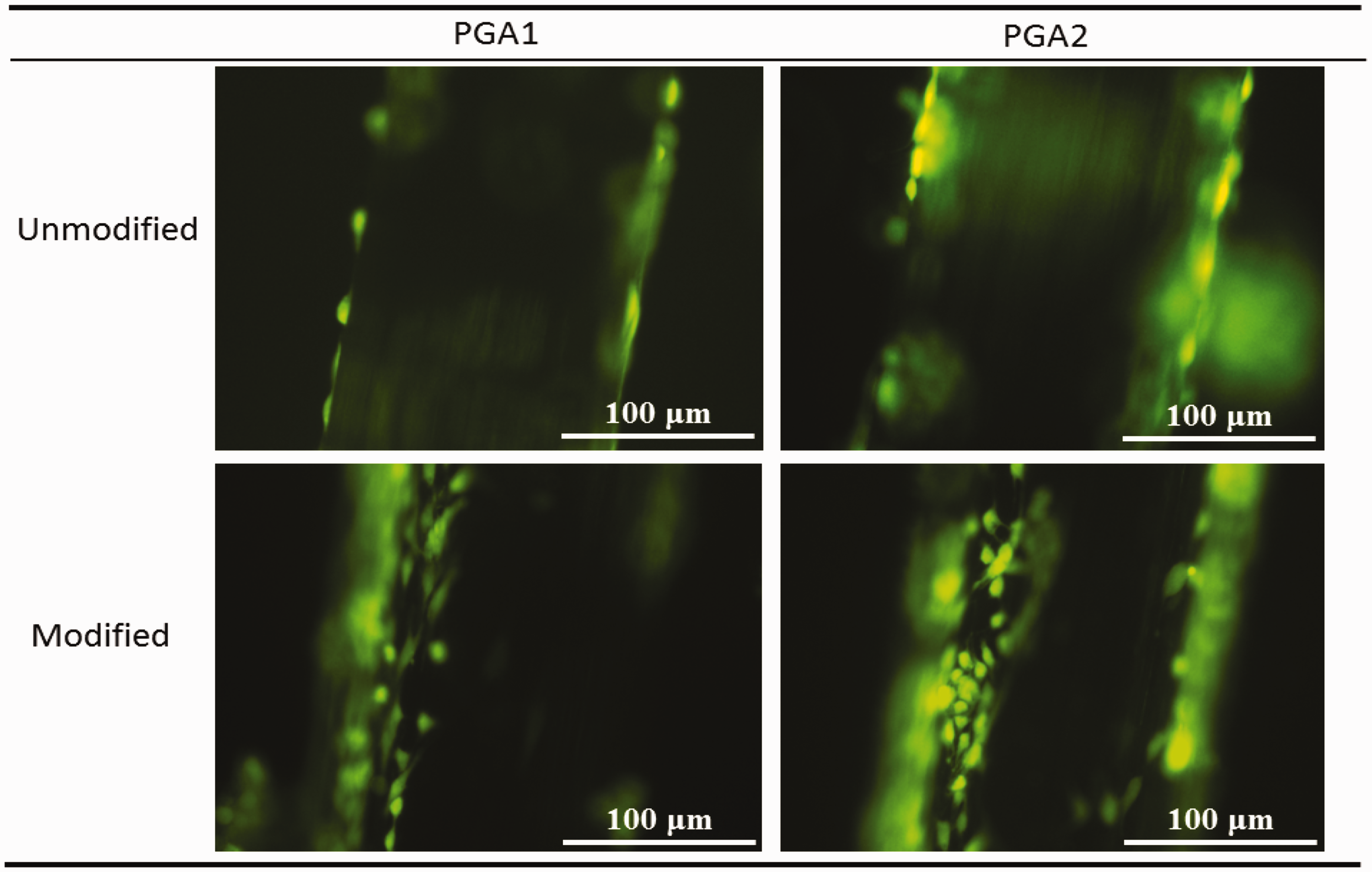
Conclusion
In this work, four types of PLA and PGA monofilaments were first fabricated from their polymer chips, and were then subjected to surface modification using ultrasonic technology. Meanwhile, other characteristics such as surface morphology, FT-IR analysis, cytotoxicity, cell attachment and cell morphology were also tested and analyzed, and the feasibility of using the ultrasonic technology to surface modify ACET materials was evaluated according to these experiment results. The following conclusions were established through this research:
Via the analysis of structural characteristics, the modified monofilaments were observed to have rougher surfaces than the unmodified ones. FT-IR analysis indicated that there were few new chemical bonds for the PLA group, while some hydrophilic hydroxyl and carboxyl groups on the termini of molecules for the PGA group increased after modification. The surface hydrophilicity of both the PLA and PGA groups had been greatly improved via ultrasonic modification according to the analysis of contact angle values. The mechanical properties showed that both the PLA and PGA groups retained their tensile performance and bending stiffness after ultrasonic modification, and that the PLA group exhibited better tensile properties than the PGA group. Moreover, the swelling behaviors of all the samples were greatly enhanced after modification due to the rearrangement and increasing distance of the molecular chains. UMPGA2 (23.82 ± 3.16%) and UMPLA2 (7.64 ± 1.05%) presented the largest swelling ratio values among their groups. Through the evaluation of in vitro experiments, all the prepared monofilaments were proven to be non-toxic with more than 75% cell viability, and the ultrasonic modification had little influence on the cell viability of the PLA and PGA groups. In addition, ultrasonic modification effectively enhanced the cell attachment ability of both the PLA and PGA groups, which was caused by the increasing surface roughness and surface hydrophilicity after modification. The PGA group had a larger cell attachment ratio than the PLA group, which exhibited more outstanding potentiality in clinical applications. These findings suggest that ultrasonic modification could be selected as a novel approach to improve the hydrophilicity and cytocompatibility of both the PLA and PGA groups, while retaining their other excellent characteristics after modification. Moreover, the ultrasound modified PLA and PGA groups presented better prospects for application in the ACET field.
In sum, this study may inspire advances in the design and manufacture of novel PLA and PGA monofilaments to satisfy clinical requirements.
Footnotes
Declaration of conflicting interests
The authors declared no potential conflicts of interest with respect to the research, authorship and/or publication of this article.
Funding
The authors disclosed receipt of the following financial support for the research, authorship, and/or publication of this article: This work is supported by “111 Project” - Biomedical Textile Materials Science and Technology (B07024) and “The Fundamental Research Funds for the Central Universities” (BCZD2018008). The first author is grateful for scholarship support from the China Scholarship Council (CSC).