
Abstract
Development of novel antibacterial fibers with mass production is urgently required in the technical textiles industry. In this paper, a series of segmented-pie composite microfibers based on polyamide 6 (PA6) and different amounts of copper–carbon nanoparticles (CuCNPs) were fabricated by utilizing a melt-spinning apparatus with twin-screw extruders. The encapsulation of CuCNPs and the formation of segmented-pie structure of as-prepared PA6/CuCNP microfibers were confirmed. The CuCNPs or their agglomeration with an average diameter of approximately 200 nm exhibited a uniform distribution in PA6/CuCNP segmented-pie microfibers. Compared with the pure PA6 microfibers, the PA6/CuCNP segmented-pie microfibers showed obviously enhanced crystallinity, thermal stability as well as UV resistance. As the CuCNP content increased to 1.0 wt%, the tensile strength and initial modulus increased to 3.79 cN/dtex and 22.4 cN/dtex, respectively. Importantly, the PA6/CuCNP segmented-pie microfibers presented excellent antimicrobial activities to both Escherichia coli and Staphylococcus aureus (antimicrobial efficiency around 99%) and great antifungal activity to Candida albicans (antimicrobial efficiency around 82%). Taken together, our present study demonstrated that the PA6/CuCNP segmented-pie microfibers show great prospects in the fabrication of technical textiles for healthcare applications.
Keywords
Public health emergencies caused by emerging infectious diseases have become the forefront of global security concerns. 1 Fibers with antibacterial functions are favorable for preventing the reproduction and spread of pathogens, as well as manufacturing reusable antibacterial textiles such as sportswear, socks, shoe linings and lingerie. 2 Therefore, the design and construction of composite fibers with efficient antibacterial properties have gained widespread attention within the technical textile industry with the aim of improving human survival and health.3,4
The ideal antimicrobial textiles are recognized as possessing effective, broad-spectrum and durable antimicrobial activity, but exhibit significantly low toxicity to humans.2,3 Although a wide variety of textile techniques have been implemented for preparing antimicrobial fibers, the melt-spinning technique provides the simplest and most effective way to encapsulate the antimicrobial agents.2,4 In addition, the melt-spinning processing imparts fibers with antimicrobial durability as the antimicrobial agents are physically embedded in the fiber structure and present a slowly released behavior during use.2,4 Compared with natural and other organic antibacterial agents,5–7 inorganic antibacterial agents, i.e., metals and their oxides, have drawn much attention owing to their good thermal resistance and antibacterial effect.8–11 Previous studies showed that copper exhibited broad-spectrum biocide effects and cost-effectiveness, which expanded the application in the fields of clothing, decoration, and medical care.10–15 Other studies indicated that copper nanoparticles possessed notably increased physicochemical and antibacterial properties in comparison with copper micro particles, which was attributed to their higher surface-to-volume ratio.12–14 Jiao et al. reported that carbon fibers loaded with nano-copper showed good antibacterial activity and an electromagnetic interference-shielding property. 14 Marković et al. found that cotton fabrics modified with copper nanoparticles exhibited excellent antibacterial activity of about 99.9%. 12 Swar et al. developed an approach to immobilize copper nanoparticles on an mPEG-grafted PA6 surface by physisorption, which improved the stability for long-term antibacterial efficacy. 15 However, the color of the samples was transformed through the process of drying from dark brown to green shades due to the instability of the copper surface in the air.12,15 Copper nanoparticles without a protective coating often have a high oxidation tendency, resulting in an obviously shortened shelf-life.12,15 Lian et al. developed CuCNPs by loading copper ions into fibers of biological origin, and then carbonizing the fibers. 16 In addition, the as-prepared copper nanoparticles coated with a thin carbon layer displayed excellent antimicrobial performance and exhibited promising application in the fabrication of antimicrobial fibers.16,17 Thus, CuCNP-loaded fibers/textiles are thought to have great potential for possessing effective, broad-spectrum and durable antimicrobial uses.
Polyamide 6 (PA6) is one of the most common commodity polymers with a high resistance to elongation and abrasion, appropriate mechanical strength as well as low wrinkling, which could be employed to fabricate the nanocomposite antibacterial fibers by the melt-spinning technique.18–21 Zhang et al. incorporated functionalized graphene (FGO) with PA6 by melt-spinning and found that the FGO/PA6 nanocomposite fibers exhibited multi-functional characteristics, such as high strength, UV resistance, and good antibacterial properties. 19 In addition, polyamide composite fibers with segmented-pie cross-section have been used previously to obtain splittable ultrafine fibers and nonwovens,21–23 which could also load functional nanoparticles to impart antibacterial property.
In the present paper, we employed PA6 as a fiber matrix material and CuCNPs as a multi-functional additive to manufacture segmented-pie shaped composite microfibers by using a melt-spinning apparatus with twin-screw extruders. We hypothesized that the incorporation of a low concentration of CuCNPs could increase the structural stability, mechanical properties, and thermal performance of PA6/CuCNP segmented-pie microfibers, as well as impart the as-obtained fibers’ excellent antimicrobial activities. The structural properties, mechanical properties, and thermodynamic properties of as-prepared PA6/CuCNP segmented-pie microfibers were studied systematically. Gram-positive bacteria Staphylococcus aureus, Gram-negative bacteria Escherichia coli, and fungi Candida albicans were employed as model strains to explore the antimicrobial properties of different PA6/CuCNP segmented-pie microfibers.
Experimental methods
Materials
CuCNPs (COPPWARE®, spherically shaped nanoparticles with a diameter less than 60 nm) were supplied by Suzhou Guanjie Nano Materials Technology Co., Ltd (China). PA6 chips were supplied by Shandong CRC Atsugi Nylon Co., Ltd (China). Nutrient agar, nutrient broth, ethanol, and deionized water were purchased from Sinopharm Chemical Reagent Co., Ltd (China).
Fabrication of PA6/CuCNP antimicrobial master batch
The master batch of PA6/CuCNP composite was prepared according to the procedure of Zhang et al. 19 Briefly, 1 g of CuCNPs was uniformly mixed with 100 g of neat PA6 chips in a high-speed mixer for 5 minutes. Then, the premixed samples were oven-dried at 100°C for 24 hours and added to a lab tech twin-screw extruder (SHJ-20, Nanjing Giant Machinery Co., Ltd, China) with an L/D of 40 for melting and extrusion, at a barrel temperature of 240–245–247–250–247–245°C. Dried PA6 granules were mechanically premixed with the CuCNPs for 5 minutes and then extruded and granulated. A dried master batch was obtained with the same drying process. Melt flow index (MFI) measurements were carried out using an XNR-400a melt flow indexer (China), at 235°C with 2.16 kg loading. MFI of PA6 and PA/CuCNP master batches were 44.8 and 47.4 g/10 min, respectively.
Preparation of PA6/CuCNP segmented-pie microfibers
The PA6 fibers with different CuCNP concentrations were prepared by melt-spinning with twin-screw extruders. A schematic illustration of the fabrication of PA6/CuCNP composite fibers is shown in Figure 1. The dried PA6/CuCNP master batch and neat PA6 granules (100°C, 24 hours in a vacuum oven) were added to the twin-screw extruders A and B, respectively, and then extruded through capillary holes of the specific spinneret for fabrication of 16 segmented-pie fibers. The PA6/CuCNP master batch and pure PA6 granules were melt-spun at a barrel temperature of 252–257–262–267°C controlled by an FDY spinning machine (Dalian Longsheng Machinery Co., Ltd, China). Different loadings of CuCNPs such as 0.5 and 1.0 wt% of COPPWARE were utilized. The as-prepared microfibers were heated to 100°C for thermal stretch, and the stretch multiple was three times to obtain the final draw-textured microfibers. Pure PA6 melt-spun microfibers were prepared as control. The as-prepared multifilaments containing different amounts of PA6/CuCNPs were denoted as PA-CuCNP1 and PA-CuCNP2 (Table 1).
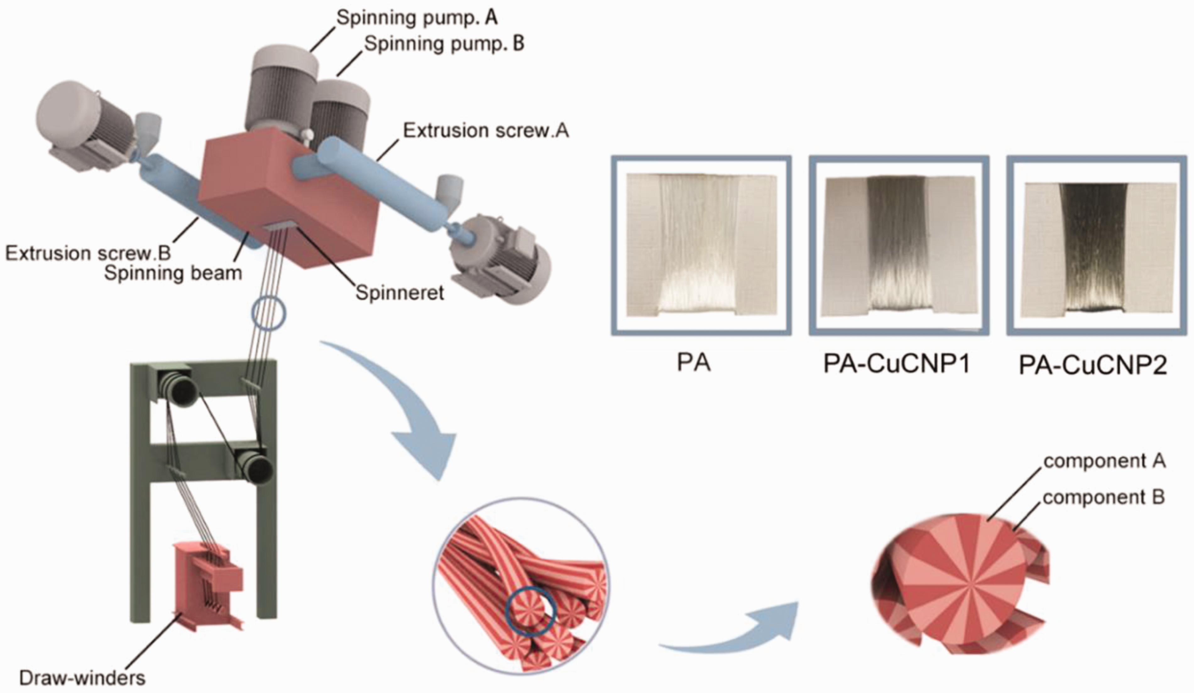
Schematic illustration of the preparation of PA/CuCNP segmented-pie microfibers by melt-spinning.
The composition and element amounts of PA/CuCNP microfibers

aPA6/CuCNPs represents the as-prepared copper-contained PA6 master batches.
bTheoretical addition of CuCNPs.
cElement amounts in PA-CuCNP composite fibers measured by SEM–EDX.
Characterization of PA/CuCNP segmented-pie microfibers
Morphology observation
The microstructure of PA/CuCNP fibrous samples was observed by utilizing scanning electron microscopy (SEM; TESCAN VEGA3) with an energy dispersive X-ray (EDX) spectrometer. The fibrous samples were sputter-coated with gold to avoid negative-charge accumulation. A 5 kV acceleration voltage was employed during the test.
FTIR analysis
A Fourier transform infrared (FTIR) spectrometer (TSN-iS50, ThermoFisher, USA) was used to determine the chemical groups of PA6/CuCNP segmented-pie microfibers. The scan range was from 4000/cm to 400/cm with a resolution of 5/cm.
Mechanical test
The mechanical properties of the segmented-pie microfibers, including PA, PA-CuCNP1, and PA-CuCNP2, were characterized using a universal testing machine (INSTRON 5965, USA) equipped with a 5 N loaded cell. The stress–strain curves of the fibers were calculated from the load–deformation curves recorded at a cross-head speed of 100 mm/min and the gage length of 100 mm. 19 The tensile testing was performed five times for all the samples.
XRD analysis
The crystal morphology of PA6/CuCNP segmented-pie microfibers was investigated using wide-angle X-ray diffraction (XRD; Rigaku Ultima IV, Japan) using a diffractometer equipped with Ni-filtered CuKα radiation, which was operated at a voltage of 40 kV and a current of 40 mA. The intensity versus 2θ values were recorded at a step size of 3°/min.
Thermal analysis
The thermal properties of PA6/CuCNP segmented-pie microfibers were studied using a simultaneous thermal analyzer (DSC/DTA-TG, NETZSCH, German) in an N2 atmosphere. PA6/CuCNP segmented-pie microfibers were scanned from 25°C to 310°C at a heating rate of 10°C/min and maintained at a temperature for 5 minutes to remove the thermal history. After that, samples were cooled from 310°C to 25°C at a rate of 10°C/min, and then heated from 120°C to 550°C at a rate of 10°C/min. The data obtained from the first cooling scan and the second heating scan were used for the study.
Data color analysis
The K/S values were measured to evaluate color strength of PA6/CuCNP segmented-pie microfibers. The K/S values of PA6/CuCNP segmented-pie microfibers before and after being laundered 10 times were determined using a color measurement system (Datacolor 850, USA).
UV transmittance
Weft-knitted fabrics with weft plain stitch (170 × 90) were knitted by different PA6/CuCNP segmented-pie microfibers. The UV transmittance of the fabrics (diameter 60 mm) was assessed before and after being laundered 10 times by Textile Anti-Ultraviolet Performance Tester (YG912E, Meibang Instrument Co., Ltd, China) in the wavelength range of 280–400 nm. The thickness of the fabric was around 0.12 mm. All the tests were repeated five times.
Assessment of antimicrobial activity
Antimicrobial activities of PA6/CuCNP segmented-pie microfibers before and after being laundered 10 times were determined using three different bacterial strains, namely Gram-negative E. coli (ATCC 29522), Gram-positive S. aureus (ATCC 6538), and fungi C. albicans (ATCC 10231). The inhibition zone tests for antimicrobial activities of PA6/CuCNP fabrics after being laundered 10 times were also conducted using two bacterial strains, namely S. aureus and E. coli, according to the agar diffusion plate test (GB/T 20944.1-2007). In this procedure, fabric samples were cut into discs with diameter of 10 mm and sterilized under UV for 30 minutes. Then 200 μL bacterial culture prepared previously was evenly plated over the solidified agar Petri dish and then incubated at 37°C for 24 hours. The inhibition zone surrounding the fabric disc was recorded at the predetermined incubation period.
The quantitative procedures for antimicrobial activity were conducted according to the shake-flask method (GB/T 20944.3-2008). Briefly, E. coli, S. aureus, and fungi C. albicans were cultured in the nutritive agar medium for 24 hours at 37°C. Fresh bacteria cultures were added separately by a sterilized loop into 10 mL of sterile culture broth until it reached the required standard concentration (3.0 × 10⁹ CFU/mL). Then 0.75 g sterilized samples were shaken for 18 hours in an incubator with 70 mL culture broth at 25°C. After incubation, a series of dilutions was carried out to obtain a concentration of 30–300 colony forming units (CFU)/mL. Then 1 mL of the last diluted solution was scrubbed onto a Luria agar Petri dish and placed in an incubator for 18 hours at 37°C. The growing colonies of microbes on the agar Petri dish were counted, and the percentage of cell-growth reduction was calculated using the equation Y = (W – Q)/W × 100%, where W is the number of CFU of bacteria from the control sample after 18 hours and Q is the number of CFU of bacteria from PA-CuCNP samples after 18 hours. All the tests were repeated three times.
Results and discussion
Morphology of PA6/CuCNP segmented-pie microfibers
The morphology of the segmented-pie microfibers is shown in Figure 2(a,b). Two components, i.e., PA and PA/CuCNP matrix, melt together in PA-CuCNP samples during the spinning process. In order to visualize the orange petal shape of PA/CuCNP 16-segmented pie, another component, a polyethylene terephthalate (PET) matrix, was employed to fabricate PA/PET 16-segmented pie by melting PET and PA/CuCNP matrix together and then extrusion through capillary holes of the specific spinneret with the same processing parameters of PA-CuCNP fibers. As shown in Figure 2(a), the prepared samples showed aligned fiber morphology and uniform dispersal. The diameter of PA-CuCNP1, PA-CuCNP2, and pure PA was about 13.4 ± 0.4 μm, 12.2 ± 0.3 μm, and 12.6 ± 0.2 μm, respectively. Simultaneously, the cross-sectional images of the fibers indicated that CuCNPs or their agglomeration with average diameter of approximately 200 nm were uniformly dispersed in PA-CuCNP1 and PA-CuCNP2 fibers (Figure 2(b)). The agglomerated morphology was probably attributable to the interactive forces between different copper nanoparticles. 26
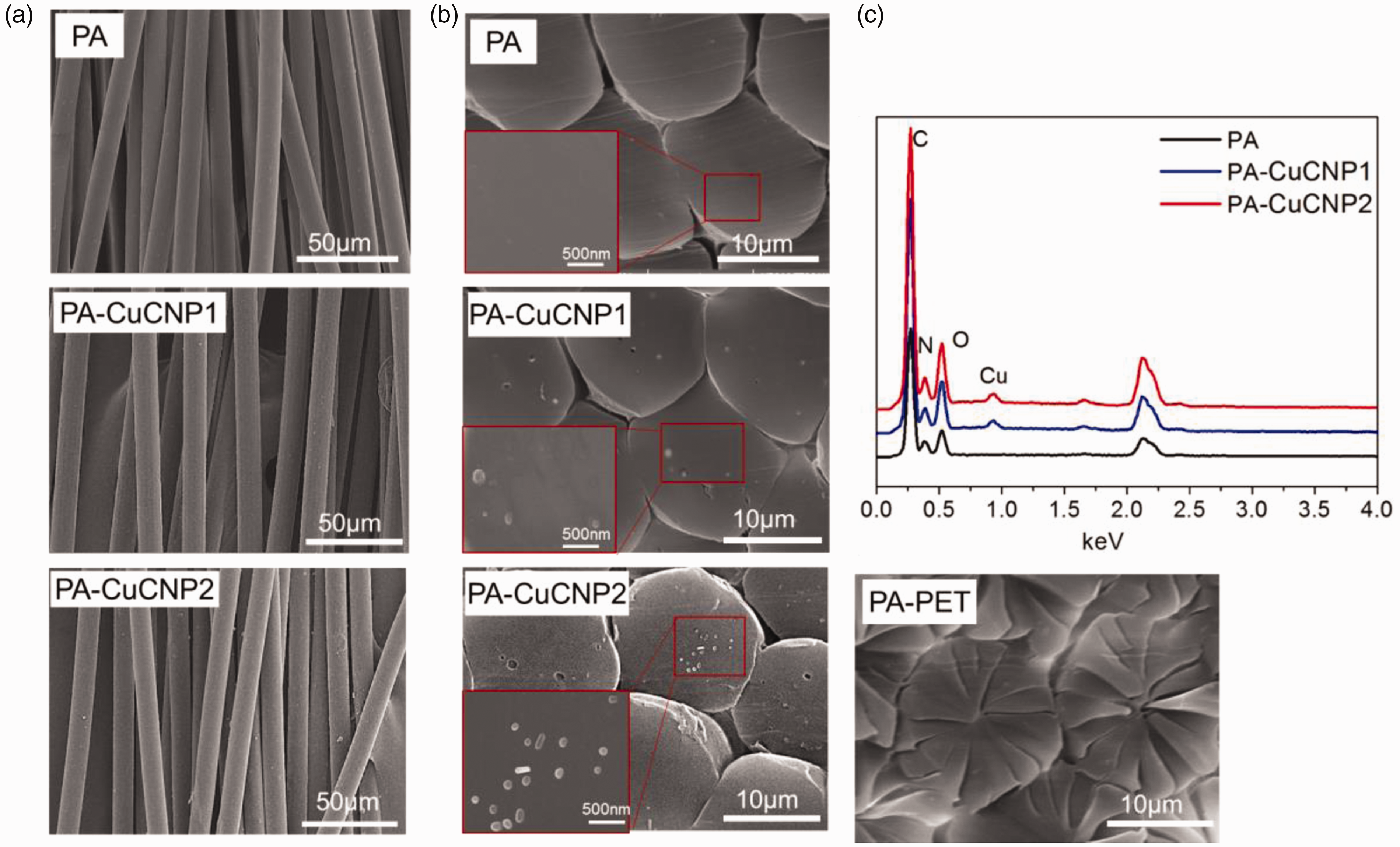
SEM images of the PA6/CuCNP segmented-pie microfibers: (a) the surface morphology, (b) cross-section morphology, and (c) the EDX spectra of cross-sections of various segmented-pie microfibers.
EDX analysis was performed on the cross-section of the segmented-pie microfibers to analyze the presence of CuCNPs loaded in the PA-CuCNP samples. Table 1 lists the content of different elements in PA/CuCNP composite fibers determined by EDX. Specifically, the copper content of the PA-CuCNP2 sample was approximately 0.6%, which was less than the actual amounts added to fibers owing to the low penetration depth of EDX (around 500 nm). The cross-section morphology of PA-CuCNP fibrous samples demonstrated that CuCNPs dispersed uniformly in the microfibers.
Mechanical property of PA6/CuCNP segmented-pie microfibers
The mechanical property of PA6/CuCNP segmented-pie microfibers with different amounts of CuCNPs was explored with a tensile tester. The representative stress–strain curves and as-calculated mechanical properties of PA, PA-CuCNP1, and PA-CuCNP2 samples are presented in Figure 3. Compared with pure PA fiber, the tensile strength of PA-CuCNP composite fibers increased slightly from 3.45 to 3.79 cN/dtex with the addition of CuCNPs from 0.5 to 1.0 wt% (Figure 3(b)). At the same time, according to the variation tendency of strength, the initial modulus increased as the CuCNP content increased to 1.0 wt%, and reached a maximum value at 22.4 cN/dtex, with an enhancement of 48.3% (Figure 3(c)). In addition, the elongation at break of PA composite fibers decreased slightly from 26.2% to 23.9% with the addition of CuCNPs from 0 to 1.0 wt% (Figure 3d). Thus, the incorporation of CuCNPs into the PA segmented fibers presented a positive effect on enhancing the strength and the stiffness of the samples.
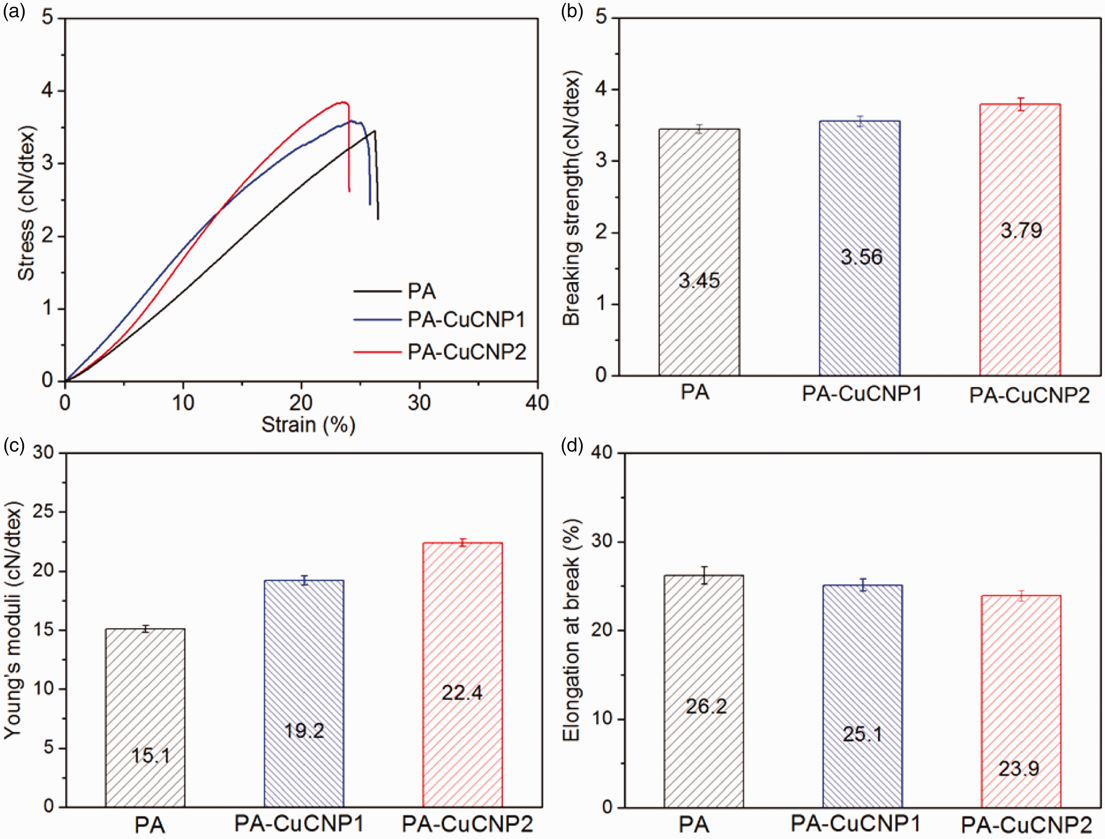
(a) Stress–strain curve of PA6/CuCNP segmented-pie microfibers with different CuCNP amounts, (b) Tensile strength, (c) Young’s moduli, and (d) elongation at break.
The enhancement of the mechanical property of the composite fibers could be attributed to CuCNPs dispersed well in fibers and randomly oriented in PA matrix19,26. Zhang et al. 19 fabricated melt-spinning PA6 fiber and found that the tensile strength and tensile modulus reached a maximum value at 4.2 and 33.9 cN/dtex, respectively, with a loading of 0.5 wt% antibacterial agent. Compared with Zhang et al., 19 the antibacterial agent dispersed well in PA6/CuCNP fibers, even with a high content (i.e., 1.0 wt%) of CuCNPs, and the tensile strength and tensile modulus reached a maximum value at 3.79 and 22.4 cN/dtex, respectively. CuCNPs dispersed in the segmented-pie microfibers uniformly, which was also validated by morphological study as mentioned previously. This indicates that proper addition of CuCNP agent could promote the tensile strength of the PA segmented-pie microfibers.
XRD analysis
Figure 4 illustrates XRD spectra of PA6/CuCNP segmented-pie microfibers, and shows that the α-crystal diffraction peaks of PA6 appear when 2θ values are 20.2° and 23.3°, and that the γ-crystal diffraction peak appears when the 2θ value is 21.3°. The diffraction peak of Cu(1,1,1) at 42° did not show up in the spectra owing to the low content (1.0 wt%) of CuCNPs loaded in PA matrix. 27 PA/CuCNP composite fibers exhibited both α-phase and γ-phase, two different crystal structures, owing to the high-speed draft during the melt-spinning. 28 In addition, the γ-crystal form increased with an increase in the content of CuCNPs. The phenomenon might be due to the addition of CuCNPs destroying PA6 α-crystal hydrogen bonding, inducing α-phase conversion into γ-phase. 4 The results indicated that added copper nanoparticles acted as nucleating agents in the PA matrix and promoted the crystallinity rate of the PA microfiber 29 .
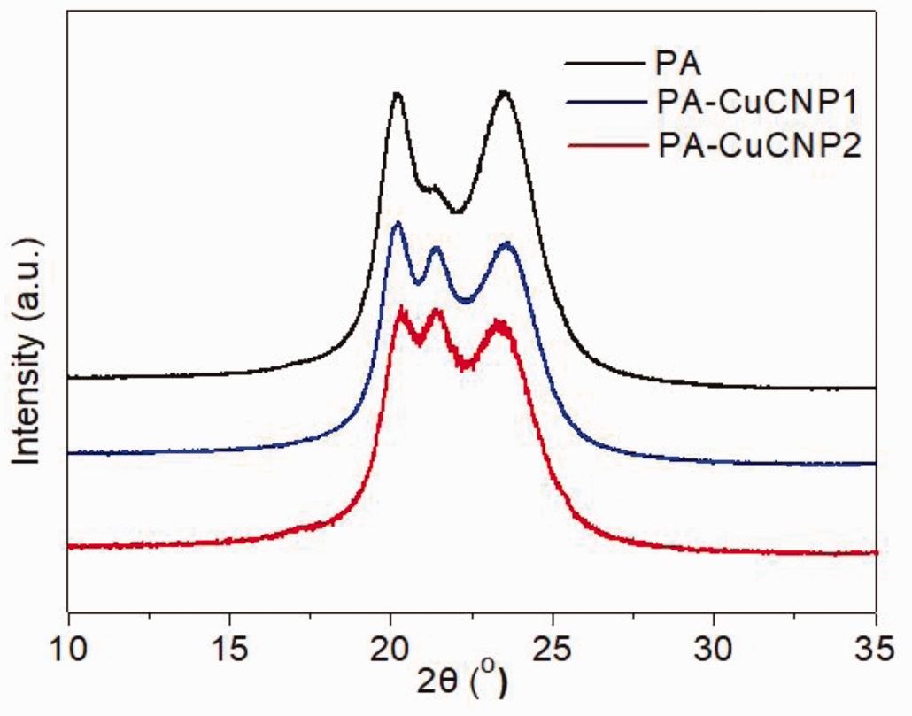
XRD curves of PA6/CuCNP segmented-pie microfibers.
FTIR analysis
The characteristic absorption bands of PA6/CuCNP segmented-pie microfibers are shown in Figure 5. Characteristic vibration frequency peaks of PA6 are found at 3287/cm (N–H stretching), 2927/cm (CH2 stretching), 1630/cm (C=O stretching, amide I), 1540/cm (C–N stretching vibration), and 689/cm (N–H bending vibration) (Figure 5(a)). The absorption vibration peak at 2350/cm is attributed to carbon dioxide. In addition, the presence of CuCNPs in the PA6 fibers did not change the vibration frequency of the carbonyl group and amide group of PA6 in the high-wavenumber region (4000–1600/cm), but slightly varied the intensity of characteristic peaks of the amine group in the low-wavenumber region (1600–400/cm). As shown in Figure 5(b), the characteristic peaks of the α-crystal phase at 1462, 1265, and 1120/cm can be easily discerned. The peak at 620/cm was assigned to the r-crystal form of the amide IV due to the wagging vibration of N–H, 30 and its intensity became stronger with the increasing addition of CuCNPs in PA samples. Furthermore, the intensity of the characteristic peaks of the α-phase peak at 1265/cm (N–H stretch bend) 31 became weaker in the PA-CuCNP2 sample. The variation of peak at 620 and 1265/cm clearly indicates that CuCNPs suppress the arrangement of hydrogen bond and induce the perfect arrangement of molecular chains.30,31
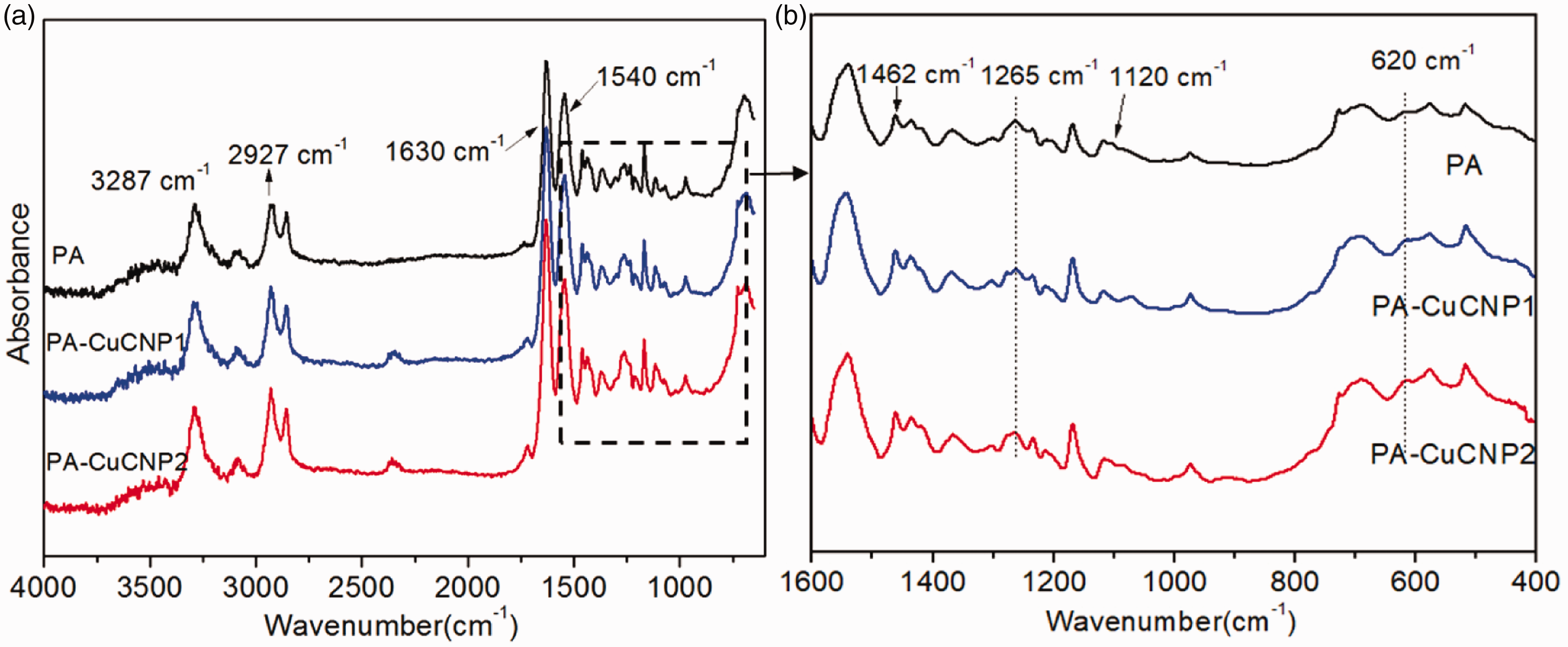
FTIR curves of PA6/CuCNP segmented-pie microfibers.
Thermal analysis
The melting and crystallization behavior of the PA/CuCNP composite fibers were observed based on the differential scanning calorimetry (DSC) thermograph. The first heating scan was conducted to remove previous thermal history. DSC curves of the prepared PA/CuCNP composite fibers were determined from the second heating scan as shown in Figure 6(a) and the glass transition temperature (Tg), the melting temperature (Tm), and melting enthalpy (ΔHm) are listed in Table 2. In Figure 6(a), a series of PA/CuCNPs show similar melting behavior. Specifically, with the increasing content of CuCNPs, melting points increased from 214.3 to 219.7°C, while at the same time, melting enthalpy increased from 38.8 to 43.4 J/g, respectively. The percentages of PA crystallinity for composites fibers were calculated according to the following equation, Xc =
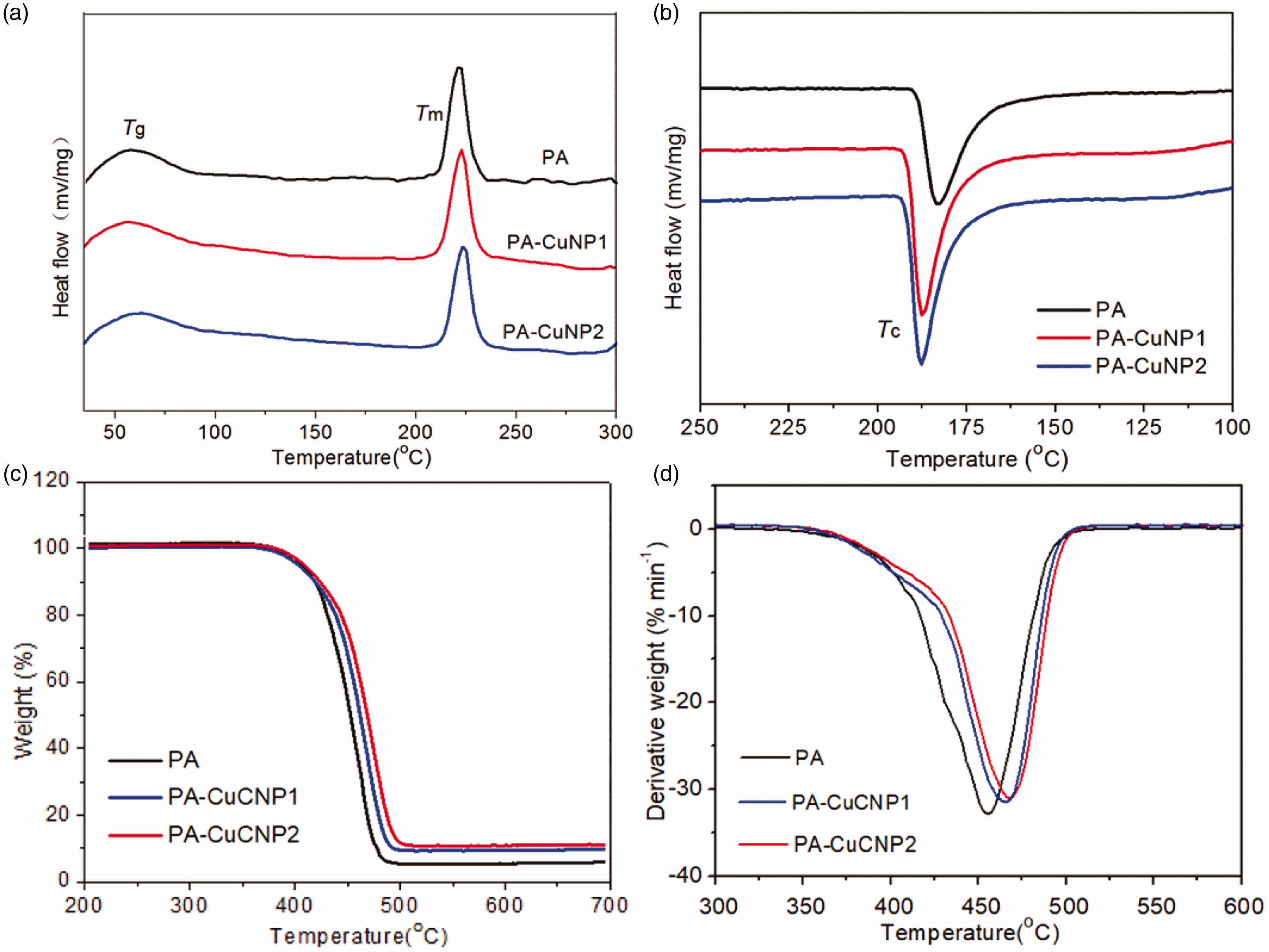
DSC curve of (a) second heating and (b) cooling scan of PA6-CuCNP segmented-pie microfibers; (c) TG and (d) DTG curves of PA6-CuCNP segmented-pie microfibers.
Thermal characteristics of PA6/CuCNP segmented-pie microfibers

In Figure 6b, the crystallization temperature increased from 183.7 to 187.8°C. It was possible that CuCNPs loaded in PA6 played heterogeneous nucleation catalyst in crystallization, resulting in acceleration of the crystallization of composites.4,21 Thermogravimetric analysis (TGA) was employed to test the thermal stability of the PA6/CuCNP segmented-pie microfibers. Figure 6(c,d) shows the TGA and differential thermogravimetric (DTG) curves of pure PA and composites. As is shown in Figure 6(c,d) and Table 2, all of the thermogravimetric curves exhibited one weight-loss stage and the initial decomposition temperature (Tonset) as well as temperature of 50% weight loss (T50) increased gradually with the increasing addition of CuCNPs. This was because the reinforcing effect of the CuCNPs reduced the PA6 chain segmental mobility and hence the enhancement in the maximum decomposition temperature (Tmax) was observed with increasing CuCNP content (Table 2). This indicates that the range of thermal degradation temperature was wider with the increase of CuCNPs from 0.5 to 1.0 wt%. Thus, the thermal stability of segmented-pie PA microfibers was improved after the incorporation of CuCNPs.
UV resistance of the PA/CuCNP segmented-pie microfibers
CuCNPs loaded in segmented-pie PA fibers improved the UV resistance significantly. As listed in Table 3, the UV protection factor (UPF) increased significantly from around 3.2 to 16.0 with the addition of CuCNPs before being laundered 10 times. Meanwhile, with the increased content of CuCNPs, the transmittance of solar UVA (with the wavelengths 315–400 nm) and UVB (with the wavelengths 280–315 nm) decreased significantly from approximately 63.2% and 59.6% to approximately 14.5% and 10.9%, respectively. After being laundered 10 times, the UPF value of PA/CuCNP microfibers decreased less than 25% as listed in Table 3. The improvement of anti-UV property before or after being laundered 10 times is contributed to by the gradual deepening color of PA6/CuCNP segmented-pie microfibers. 33
UV-resistance characteristics of PA/CuCNP segmented-pie microfibers

PA6/CuCNP segmented-pie microfibers exhibited various depths of gray (Figure 1). In order to record the color in a quantitative manner, the color data of fibers is determined. The color strength (K/S value) indicates the color depth of fibers or fabrics, and a higher K/S value indicates a deeper color of the fibers or fabrics.24,25 The K/S value of the two different PA6/CuCNP segmented-pie microfibers is about 0.65 and 1.11 before washing, and decreased slightly to 0.64 and 0.97, respectively, after being laundered 10 times. As a control, the K/S value of pure PA6 segmented-pie microfibers was only 0.04 before or after being laundered 10 times. The PA6/CuCNP segmented-pie microfibers exhibited a gradual deepening color owing to the increase of CuCNPs in the microfibers. Corresponding with the K/S value of the prepared microfibers, the UPF value of PA/CuCNP microfibers increased with an increasing amount of CuCNPs. Thus, CuCNP-loaded PA composite microfibers exhibited good color stability and facilitated the improvement of UV resistance.
Antimicrobial activity of PA6/CuCNP segmented-pie microfibers
Previous studies demonstrated that copper ions released from the samples could destroy the permeability of the bacterial membranes, resulting in the leakage of sugars and proteins from the cells. 34 Copper-loaded PA6/CuCNP microfibers were expected to exhibit excellent antibacterial activity in the inhibition zone test.8,11 However, the results showed that the CuCNP-containing PA6 fabrics had no obvious inhibition zone against S. aureus and E. coli before or after being laundered 10 times (Figure 7). This can possibly be ascribed to the low copper content at the fiber surface and the low solubility and diffusion rate of CuCNPs.16,17
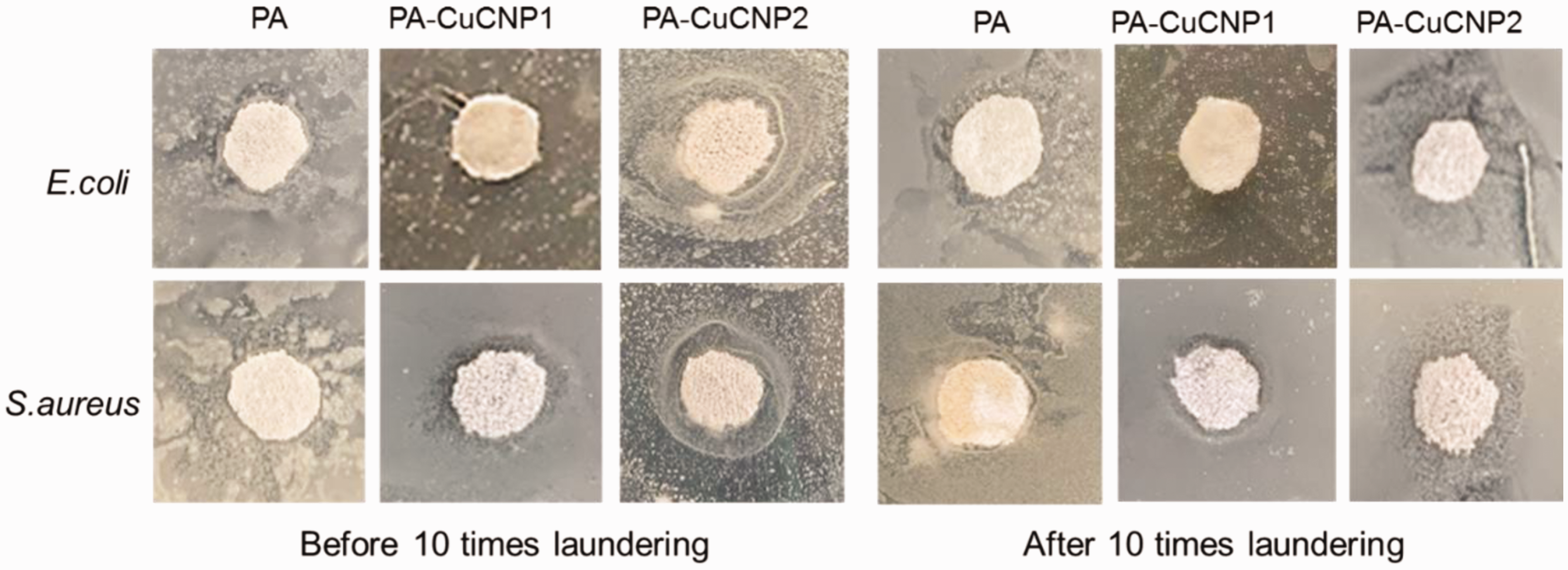
Inhibition zone tests against E. coli and S. aureus of PA-CuCNP fibers before and after being laundered 10 times.
A shake-flask method was further employed to determine the antimicrobial behaviors of the PA6/CuCNP segmented-pie microfibers after being laundered 10 times. In addition, Gram-negative E. coli (ATCC 29522), Gram-positive S. aureus (ATCC 6538), and fungi C. albicans (ATCC 10231) were all utilized. The reduction in bacterial colonies of different compositions are shown in Figure 8(a). Compared with the PA control sample, PA-CuCNPs showed excellent antimicrobial activity against all the strains of bacteria and fungi, and the antibacterial activity of PA-CuCNP2 was obviously higher than that of PA-CuCNP1 for Gram-negative E. coli and Gram-positive S. aureus. With the increasing content of CuCNPs, the inhibition rate of E. coli and S. aureus increased from 91.5% and 99.0% to 99.6% and 99.9%, respectively. Simultaneously, PA-CuCNP2 composite fibers demonstrated a better inhibition rate against C. albicans of approximately 82.6% compared with PA/CuCNP1 with the content of 0.5 wt% CuCNPs. At 1.0 wt% copper loading, the PA6/CuCNP segmented-pie microfibers exhibited approximately 99.6%, 99.9%, and 87.5% reduction of bacterial colonies against Gram-negative E. coli, Gram-positive S. aureus, and C. albicans, respectively, which is better than copper–montmorillonite reinforced nanocomposite filaments (approximately 93.1% and 94.5%) as reported by Roy et al. 8 The antimicrobial effect of PA/CuCNP composite fibers after being laundered 10 times was also analyzed, and the inhibition rate of microbes in PA/CuCNP composites is shown in Figure 8(b). As can be expected, PA6/CuCNPs prepared via melt-spinning preserved good antimicrobial property after being laundered 10 times. Consistent with the antimicrobial effect measured before washing, the antimicrobial activity rose with increasing content of CuCNPs after being laundered 10 times.
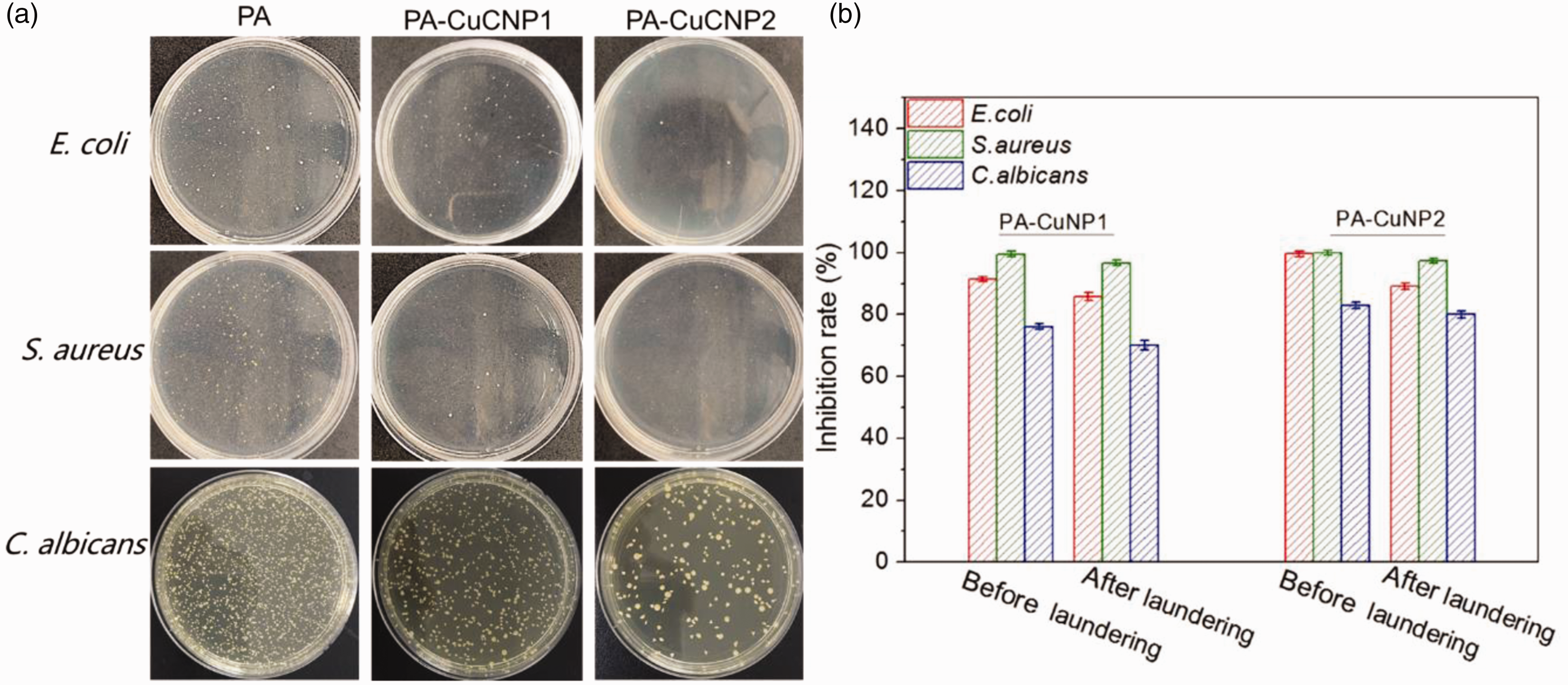
(a) Bactericidal photographs of PA6/CuCNP segmented-pie microfibers after being laundered 10 times against E. coli, S. aureus, and C. albicans. (b) Antimicrobial activity assay of PA/CuCNP composite fibers before and after being laundered 10 times.
CuCNPs exhibited good antimicrobial property before and after being laundered 10 times, which was attributed to active copper-catalyzed reactions that promote the generation of bacteria-killing reactive oxygen species (ROS).16,17 With the increasing addition of CuCNPs, the concentration of ROS increased, which contributed to the lethal damage of microbes. Copper-catalyzed reactions produce ROS and bind to essential amino acids and DNA leading to denaturation, ultimately leading to cell death.35,36
Combining excellent antimicrobial properties with enhanced mechanical property, the prepared PA-CuCNP segmented-pie fibers are expected to be a suitable candidate for the production of medical textiles. For instance, they could be used as medical sequential decompression socks for the prevention and treatment of varicose veins, as well as for effectively reducing the spread of infection.
Conclusion
A series of PA6/CuCNP segmented-pie microfibers was prepared via the melt-compounding and fiber-spinning routes. With the increase in CuCNPs from 0.5 to 1.0 wt%, the tensile strength of as-obtained fibers improved to 3.79 cN/dtex and the crystallinity of as-manufactured fibers increased significantly from 16.9% to 18.9%. In the presence of CuCNPs, PA6 composite fibers exhibited good UV resistance with a UPF value of approximately 16.0. With the addition of 1.0 wt% CuCNPs, PA6/CuCNP microfibers exhibited maximum antimicrobial activity against E. coli, S. aureus, and C. albicans. Combining the characteristics of excellent antimicrobial properties, good UV resistance, and enhanced mechanical property, the CuCNP functionalized PA segmented-pie fibers are expected to be a suitable candidate for the production of medical textiles, which could be potentially utilized in healthcare sectors to effectively reduce the spread of infection and contamination.
Footnotes
Declaration of conflicting interests
The author(s) declared no potential conflicts of interest with respect to the research, authorship and/or publication of this article.
Funding
The author(s) received no financial support for the research, authorship, and/or publication of this article.