
Abstract
The hairy skin of the human body is the main receptor of fabric contact. In order to evaluate clothing comfort objectively, electroneurophysiological tests with nine subjects were carried out to determine their cognitive differences in fabric static and dynamic contact. Electroencephalography and electrocardiography signals were collected when the subjects statically and dynamically touched a piece of polyester fabric with their forearms. According to their electroneurophysiological responses to fabric contact, the subjects could be categorized into three different types, namely, extraordinary, ordinary and nonsensitive ones. Their different sensitivity could be observed from the activities of α waves, sympathetic nervous system and pneumogastric nervous system. The extraordinarily sensitive subject responded to the fabric–skin static contact with an intensive α rhythm reaction followed by a suppressed α wave, an enhanced activity of the sympathetic nervous system and a mild participation of the pneumogastric nervous system. During dynamic contact, the pneumogastric nervous system activity increased greatly to balance the nervous system. The ordinarily sensitive subjects responded gently to the fabric–skin static contact, with a gradually suppressed α wave, an enhanced activity of the sympathetic nervous system and the near absence of the pneumogastric nervous system. Fabric dynamic contact induced a strong α rhythm reaction followed by a suppressed α wave combined with a joint effort of the sympathetic nervous system and pneumogastric nervous system. The nonsensitive subjects responded with almost no difference between the fabric–skin static and dynamic contact in both electroencephalography and electrocardiography tests. This preliminary study could provide an efficient way to identify different types of subjects and lay a fair ground for comparison purposes in future electroneurophysiological studies of clothing comfort.
When the human body receives external stimulus, spontaneous physiological response will take place to adapt with corresponding physiological needs. 1 This instinctive and involuntary physiological response is of great research value because it reflects the nature of external stimulus and the sensory information it brings. 2 As is known to all, three different approaches have been adopted to measure clothing comfort, namely, physical, psychophysical and electroneurophysiological approaches. 3 The physical approach is to relate the physical properties of fabrics to clothing comfort objectively without participation of human subjects. 4 The psychophysical approach is to investigate the feelings of the subjects to evaluate clothing comfort subjectively, lacking scientific precision. 5 The electroneurophysiological approach is to combine objective and subjective evaluation of human comfort, which intuitively reflects the interaction between stimulus and the human body, filling the gap between physical and psychophysical methods. 6
The studies of clothing tactile comfort based on electroneurophysiology have continued for years since the 1990s. 7 In the early years, the capability of the data acquisition and analysis was relatively low due to insufficient computing power. These technical barriers restricted the output of the meaningful results for electroneurophysiological testing, 8 discouraging researchers to sustain this type of research. In recent years, the cost of the electroneurophysiological device has come down significantly and the computer capability has been enhanced greatly, which makes this kind of research more feasible. However, in more recent publications, the subjects have been assumed to have similar electroneurophysiological characteristics, which brought in large variation in testing results and inconclusive outcomes. 9 Therefore, in the current study, we utilized the latest electroneurophysiological system with high precision, and tried to come up with more meaningful and discriminative results.
Electroneurophysiological measurement consists of electromyography (EMG), electrocardiography (ECG), electroencephalography (EEG), event-related potential (ERP) and functional magnetic resonance imaging (fMRI). For years, clothing tactile comfort evaluation by electroneurophysiological measurements has been studied continuously and essential progress has been made. 10
EEG is a medical technique that measures the electrical activity generated by the human brain. There are normally four kinds of brainwaves categorized by frequency bands. 11 When people are awake, α and β waves take charge of the main brain activity. 12 The relationship between EEG and tactile comfort of the human body has been reported. Yoshiaki et al. 13 found that α waves were related to tactile neural activity. This was proved by Park and Watanuki, 14 who reported that α waves from 10 to 13 Hz had certain relations to human tactile comfort based on the study of sanitary napkins. Pan and Li 15 concluded that when a subject was relaxed and comfortable, the number of α waves increased, while a subject felt nervous and uncomfortable, the number of α waves declined. On the other hand, the number of β waves increased if the subject was focused. Zhang 16 proved that α waves were positively and β waves were negatively related to the smoothness and softness of fabrics. In other words, a rougher surface could enhance the arousal level of the subjects and brought discomfort. 17 Therefore, fabric surface roughness has appeared to be one of the most study-worthy haptic properties. 18
ECG is another medical technique that records the electrical activity generated by the human heart periodically, and the key parameter is heart rate variability (HRV). HRV is related to the activity of the sympathetic nervous system (SNS) and pneumogastric nervous system (PNS). 19 A certain amount of effort has been paid to the relation between ECG and comfort of the human body. Yu et al. 20 found that when human skin touched wood, metal and rock, the activity of the sympathetic nerve was strengthened while that of the pneumogastric nerve was reduced. However, these activities varied less for skin-wood contact than those of skin-metal and skin-rock contact, showing that wood brought better tactile comfort than metal and rock. Yin and Zhang 21 picked three time-domain indices and three frequency-domain indices to study the relationship between HRV and pressure comfort of the human body. The results showed that increasing the pressure on the abdomen (no more than 12 mmHg) induced an enhanced activity of the pneumogastric nerve and reduced activity of the sympathetic nerve. When the pressure on the waist increased (from 12 to 18 mmHg), the autonomic nervous system (ANS) was suddenly dominated by the sympathetic nerve with fast increasing activity, while the activity of the pneumogastric nerve was greatly suppressed.
The signals of EEG and ECG are also influenced by the method of skin-material contact. There are mainly two types of contact, namely, static contact and dynamic contact. Wang et al. 9 reported that when the subjects were wearing the shirts made of various fabric samples (static contact), the rougher the fabrics, the lower the energy percentage of the α waves at the left occipitalia. Wang et al. 22 chose the palms and forearms of the subjects to perform fabric–skin dynamic contact experiments by moving a piece of silk fabric back and forth at a frequency of 1 Hz. The authors concluded that it was better to choose glabrous skin, such as fingers, to access fabric hand and to choose hairy skin, such as forearms, to evaluate fabric comfort. However, unlike the comparison between different types of fabrics, 23 little has been reported about the comparison between fabric–skin static contact and dynamic contact for the same fabric, which could be important for choosing a proper method of fabric–skin contact to study the tactile comfort because either way represents a wide range of applications. For example, fabric–skin static contact is a better choice for sleepwear and home textile tactile comfort study, while fabric–skin dynamic contact could be preferable for sportswear tactile comfort study. In addition, for developing artificial intelligence for human comfort response simulation, there would be different comfort responses even if the subject wears the same cloth because of the different body status, namely, keeping still or making movements.
In most published literature, time-domain signals of EEG and ECG are often transformed into frequency-domain patterns to facilitate data analysis. However, human physiological and psychological activities often vary over time. 24 Therefore, analysis of time-domain signals should also be important. In the present study, EEG and ECG signals were collected from the subjects to analyze the human electroneurophysiological responses to fabric–skin contact in both time and frequency domains, aiming to provide more in-depth information for understanding fabric tactile comfort.
Materials and methods
Subjects
Nine healthy right-handed subjects (five women and four men), aged 24–30 years, with normal body mass index (BMI) (18.5 < BMI < 24), participated in the experiment. They were recruited from Hangzhou City, Zhejiang Province, China, with different professions, namely: doctor, computer engineer and office clerk. Their consents to participate in the study were acquired. They had no history of taking medicine for at least 1 year and were told to wash their hair and have enough sleep the day before the experiment. Before the experiment, they were trained in detail about the whole procedure and signed the informed consent. The experiment was carried out in an artificial climate chamber with 25 ± 1.5°C and 31 ± 4.5% relative humidity. The subjects were asked to get used to the environment for 30 min after they came into the experiment chamber. The protocol of the study was approved by the ethics committee of Donghua University.
Materials and stimuli
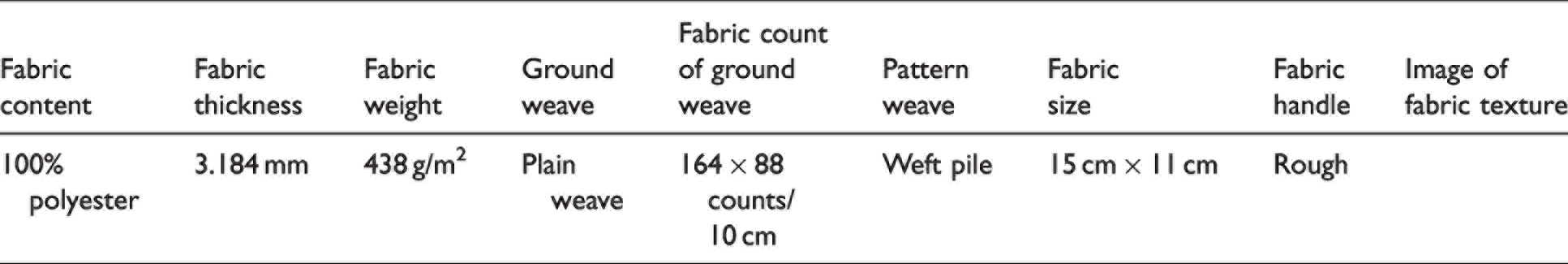
A piece of polyester fabric with ground weave and pattern weave was chosen, the detailed parameters are shown in Table 1. Due to its weft pile pattern, it had a rough surface which was supposed to bring more tactile stimulation 18 of fabric prickle. In the static contact experiment, the fabric was placed directly on the forearm with no gap, as shown in Figure 1. Because of the stiffness of the fabric, the fabric–skin contact area excluded the side of the forearm. As the contact area between fabric and skin was about 67.5 cm2 and the contact force was the fabric weight, the contact pressure was calculated as 10 Pa. While in the dynamic contact experiment, a stimulus applicator was applied. As shown in Figure 2, the stimulus applicator was a 4.5-cm-diameter plastic cylinder with one end covered by a sponge. The testing fabric was laminated on top of the sponge. In the dynamic contact test, the applicator was moved back and forth within 7 cm distance along the arm at a frequency of 0.5 Hz. The operator held the applicator at the uncovered end and used the covered end to rub against the subject’s forearm with an average pressure of 387 Pa. This was calculated by dividing the contact area by the average force measured with an electrical balance when the applicator was rubbing against its surface in the same way as in the test. The force was not a constant due to the dynamic nature of the test. We tried to record the maximum and the minimum values and calculate the mean of the two values. According to Yin et al., 25 a pressure below 1.0 kPa, which is the case in the current study, would bring no discomfort to the human body, and a pressure of approximately 1.0–2.0 kPa may even improve the attention level. 26
Details of the testing fabric for the experiment
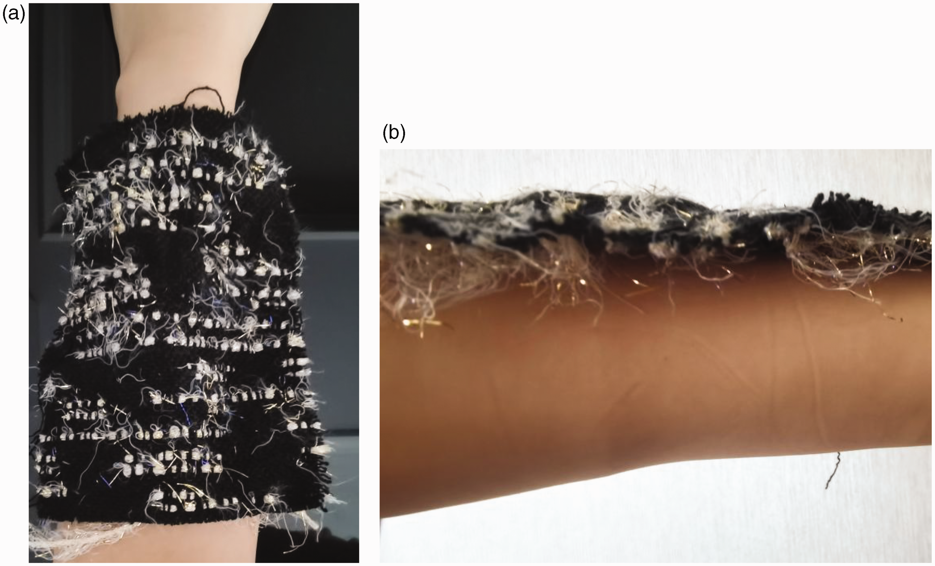
The fabric placement for the fabric–skin static contact experiment: (a) top view of the fabric placement and (b) side view of the fabric placement.
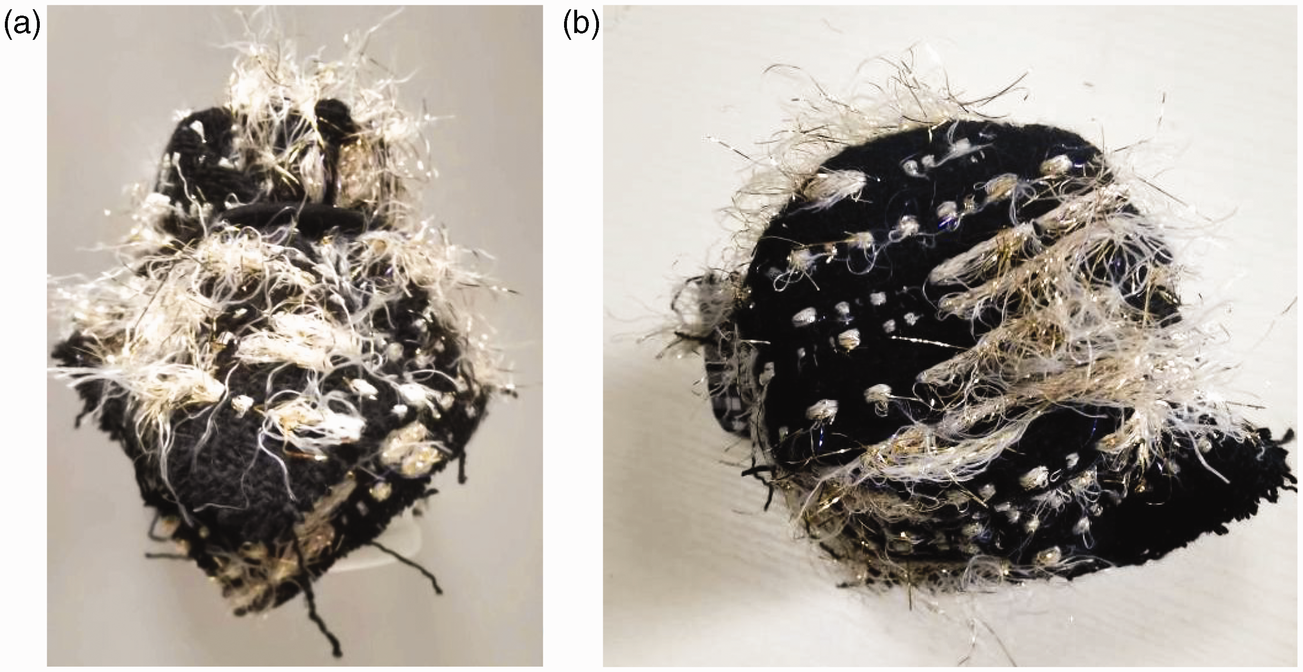
Stimulus applicator with one end covered by a piece of polyester fabric. (a) the side of the applicator and (b) the top of the applicator.
EEG and ECG experiments
The EEG and ECG tests were performed simultaneously using physiological signal acquisition equipment (MindAngel UBR08; MindAngel Intelligent Technology Corporation, Hangzhou, China) with eight channels, 250 Hz sampling speed, 125 db common-mode rejection ratio, 24 amplification and less than 0.1 µV background noise.
The trial protocol included the physiological signals acquisition positions on the human head and the corresponding block design. According to the International 10–20 system, 27 anterior and posterior brain areas (Fp1, Fp2, O1, O2) were chosen as the positions for electrodes to be attached. Then the subjects were equipped with the physiological signal acquisition equipment and straightened their two arms naturally onto the table top in front of them so that both the EEG and ECG signals were recorded throughout the experiment simultaneously. For EEG signals, the four different kinds of brainwaves 28 were recorded, as shown in Table 2. And for ECG signals, time-domain indices were chosen as HRV indicators,29,30 as shown in Table 3. To build the block design, two sessions of the experiment were carried out, namely, static and dynamic contact sessions, each of which contained three 300 s phases, that is, rest, stimulus, rest phases as shown in Figure 3.
Basic characteristics of four kinds of brainwaves 21

Introductions of the HRV indicators 23
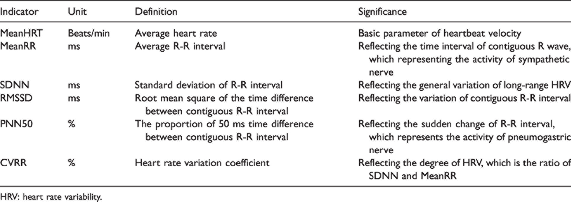
HRV: heart rate variability.
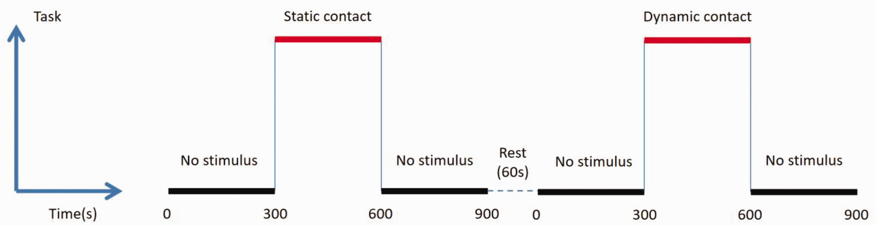
Block design of the experiment with the testing fabric.
During the whole experimental process, the subjects were asked to close their eyes but keep awake, remain relaxed while sitting quietly and focusing their attention only on the stimulation of fabric touch. There was a 60 s rest interval between each experiment session in which the physiological signal acquisition equipment was stopped working for a moment.
Electroneurophysiological data acquisition and processing
To determine the physiological response of different subjects for fabric–skin static contact and dynamic contact, the EEG and ECG signals were processed by signal purification, wave filtering and feature extraction. 31
Results and discussion
EEG analysis
After the EEG signals were preprocessed, the time domain features of δ, θ, α and β rhythm were obtained by band filtering (as shown in Figure 4). 32 Then the frequency domain features (as shown in Figure 5) of α and β rhythm were extracted by a fast Fourier transformation.
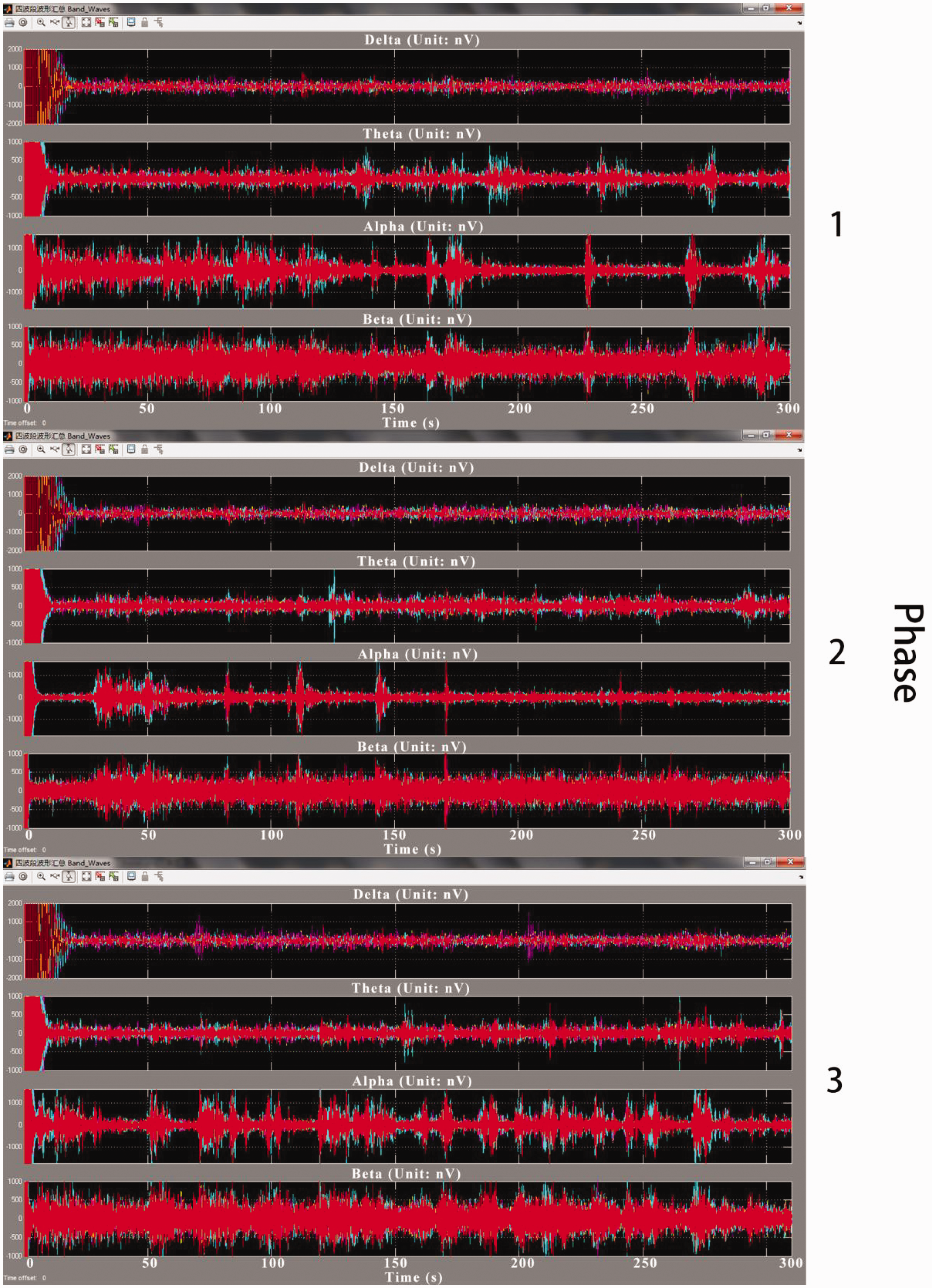
Subject no. 1: time domain features of δ, θ, α and β rhythm for three phases of fabric–skin dynamic contact trial using the testing fabric.
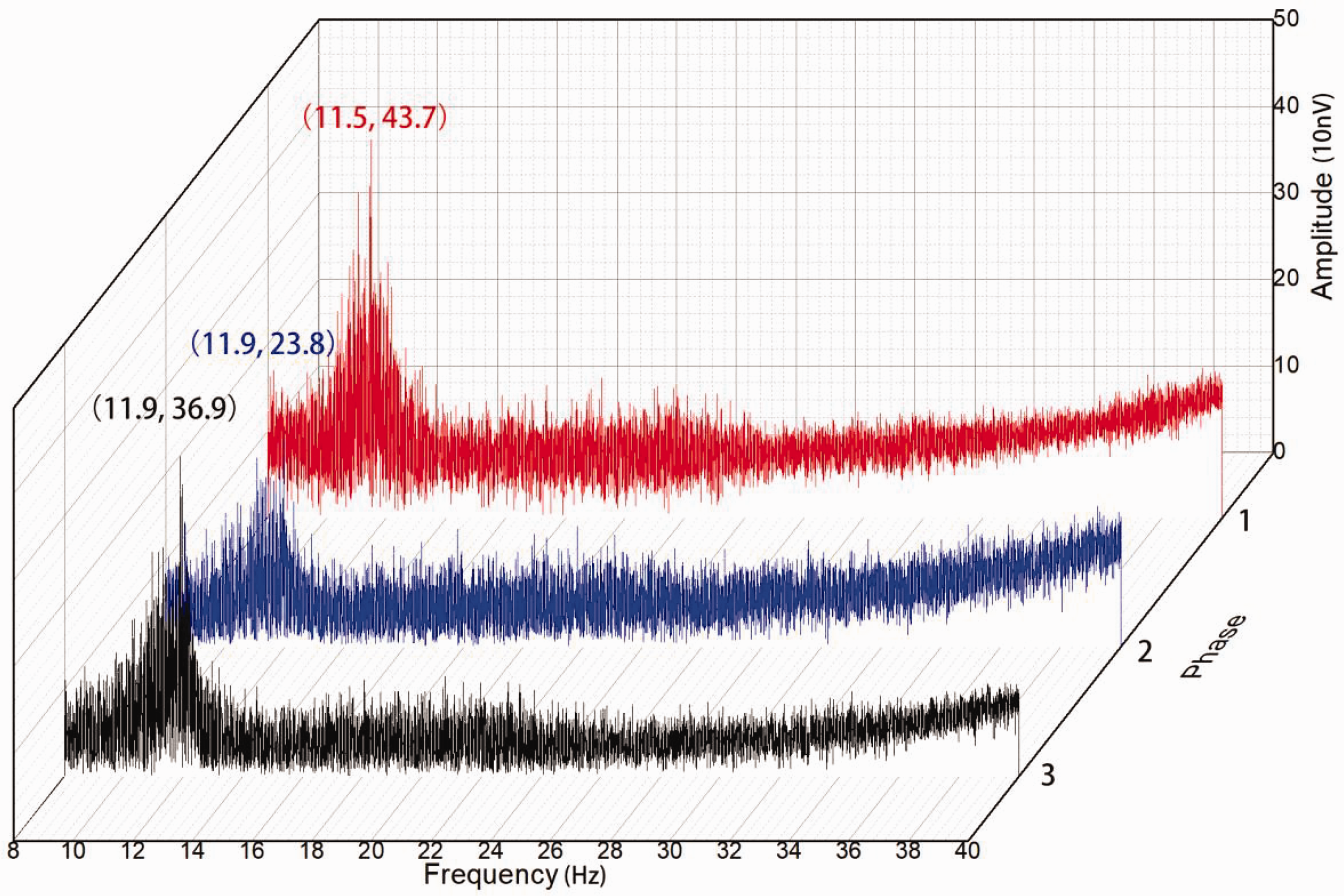
Subject no. 1: frequency domain features of α and β rhythm for three phases of fabric–skin dynamic contact trial using the testing fabric.
Based on the α rhythm analysis of EEG signals, it was found that a huge variation of brain wave neural response existed among different subjects, in terms of signal pattern, total energy in the time domain and peak value in the frequency domain. No consistent characteristics in the frequency domain were observed and even no dominant frequencies existed for almost half of the subjects. In some published literature, time-domain signals were often transformed into frequency-domain patterns to facilitate data analysis. In contrast, more attention was paid to time-domain signals in our study because they reflected the human physiological activities which varied over time. Indeed, three typical patterns were observed among nine subjects, as shown in Figure 6(a). The first type of pattern had a group of large signals followed by signals with relatively small and stable amplitudes during the fabric–skin static contact. This means that the subject was extraordinarily sensitive to the fabric touch and only one subject reacted in that way. The second type of pattern was composed of signals with steadily declining amplitude, indicating that the subject gradually felt discomfort over time. This type of subject was ordinarily sensitive to the fabric touch and three subjects had this kind of sensitivity. The third type of pattern had no particular features, that is, the amplitudes of the major peaks were roughly constant. This means that the subject was not sensitive to the fabric touch and there were five subjects who belonged to this type.

Electroencephalography (EEG) patterns in time domain of three different types of subjects for fabric–skin static contact: (a) the time-domain signals of α rhythm at O1; (b) the cumulative energy curve of the α rhythm and (c) the first derivative curve of the cumulative energy curve.
Therefore, three types of people could be categorized by tactile sensitivity according to the patterns of α rhythm, namely, extraordinarily sensitive, ordinarily sensitive and nonsensitive. In order to understand the patterns better, the cumulative energy of the signals and their first derivative in the time domain were calculated while the corresponding curves were also plotted, as shown in Figures 6 and 7. For the extraordinarily sensitive subject, the cumulative energy curve appeared with two distinctive slopes and its first derivative curve showed a large peak followed by a plateau (peak-plateau) during the fabric–skin static contact period. When it turned to dynamic contact, the cumulative energy curve became straight and its first derivative curve showed a multipeak shape. For the ordinarily sensitive subjects, the cumulative energy curve was convex and its first derivative curve appeared to be a declining multipeak (multipeak declining) shape for static contact. When it came to dynamic contact, the cumulative energy curve showed two distinctive slopes and its first derivative curve showed a peak-plateau shape. For the nonsensitive subjects, the cumulative energy curves showed almost a straight line for both fabric–skin static contact and dynamic contact, while the corresponding first derivative curve remained a multipeak shape.

Electroencephalography (EEG) patterns in time domain of three different types of subjects for fabric–skin dynamic contact: (a) the time-domain signals of α rhythm at O1, (b) the cumulative energy curve of the α rhythm and (c) the first derivative curve of the cumulative energy curve.
ECG analysis
ECG signals were filtered from the raw electroneurophysiological signals and six time-domain HRV indicators were extracted, 33 namely, mean heart rate (meanHRT), mean R-R interval (meanRR), standard deviation of R-R interval (SDNN) and root mean square of the time difference between consecutive R-R intervals (RMSSD). The proportion of 50 ms time differences between consecutive R-R intervals (PNN50) and heart rate variation coefficient (CVRR) are shown in Table 3. Besides, an instantaneous heart rate chart (IHRC), R-R histogram (RRH) and Poincare plot (PP), as shown in Figures 8 and 9, were adopted to understand better the three different types of people, namely, extraordinarily sensitive, ordinarily sensitive and nonsensitive.

Electrocardiography (ECG) patterns of three different types of subjects for fabric–skin static contact: (a) instantaneous heart rate chart (IHRC); (b) R-R histogram (RRH) and (c) Poincare plot (PP).

Electrocardiography (ECG) patterns of three different types of subjects for fabric–skin dynamic contact: (a) instantaneous heart rate chart (IHRC); (b) R-R histogram (RRH) and (c) Poincare plot (PP).
For the extraordinarily sensitive subject, when fabric–skin static contact took place, SDNN and CVRR decreased, while meanRR, RMSSD and PNN50 increased. It suggested a declined HRV level and increased activity of the SNS. Meanwhile, the PNS took a part in maintaining the balance of the ANS, but it seemed to be not so intensively active. On the other hand, when it turned to fabric–skin dynamic contact, meanRR, SDNN, RMSSD, PNN50 and CVRR all increased. It meant the enhanced HRV level, the suppressed activity of the SNS and the extremely active participation of the PNS helped to lower the heart rate and maintain the system balance. This trend can be visually observed in IHRC, RRH and PP. Comparing IHRC for static contact, dynamic fabric contact led to a heart rate that declined quickly initially and leveled off afterwards, indicating active intervention by the PNS, resulting in a closer to normal distribution of RRH and more concentrated and somewhat elongated PP.
For the ordinarily sensitive subjects, during the fabric–skin static contact period, meanRR, SDNN, RMSSD, PNN50 and CVRR all decreased, suggesting a declined HRV level, increased activity of the SNS and near absence of the PNS. When it came to fabric–skin dynamic contact, meanRR decreased again while SDNN, RMSSD, PNN50 and CVRR all largely increased. It meant that the HRV level was obviously enhanced, indicating the participation of both the SNS and PNS. However, the PNS was not able to maintain the balance of the ANS. This phenomenon can also be observed in IHRC, RRH and PP. Compared with IHRC for fabric–skin static contact, that for dynamic contact showed visually obvious premature beats, leading to sudden promotion of the heart rate and suggesting active participation of the SNS. According to the similar distribution and a wider range of RRH, the active intervention by the PNS can be seen, along with the much enlarged tail of the comet-shaped PP pattern as a result of premature beats.
For the nonsensitive subjects, during the fabric–skin static contact period, meanRR, SDNN, RMSSD and PNN50 all decreased while CVRR remained almost the same, indicating the stabilization of the HRV level and the joint effort of the SNS and PNS. During the dynamic contact period, meanRR, SDNN, RMSSD and CVRR slightly increased while PNN50 slightly decreased. It suggested that the PNS made more contribution to the ANS adjustment than the SNS and slightly enhanced the HRV level. This equilibrium trend can be visually observed in IHRC, RRH and PP. Comparing IHRCs for static and dynamic fabric contact, the heart rate increased slightly, indicating the mild participation of the SNS. In the meantime, the distribution of RRH remained in similar shape, with a somewhat wider range, suggesting the mild intervention by the PNS, resulting in slightly more scattered and elongated PP.
Discussion
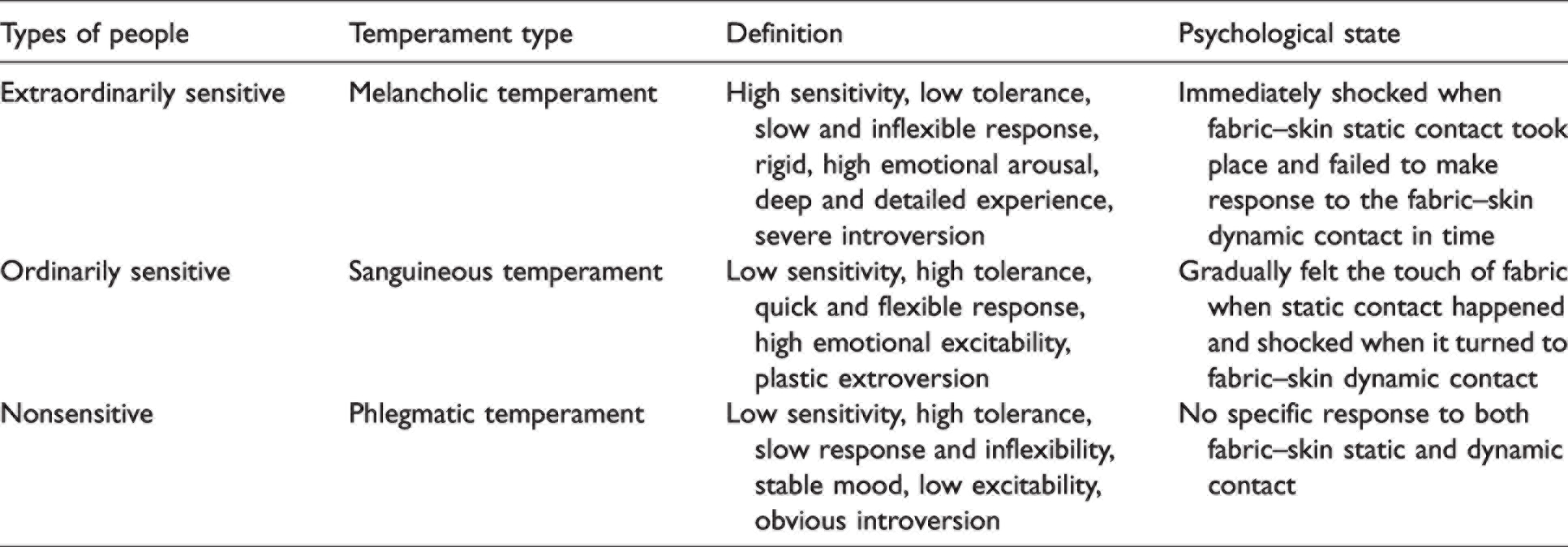
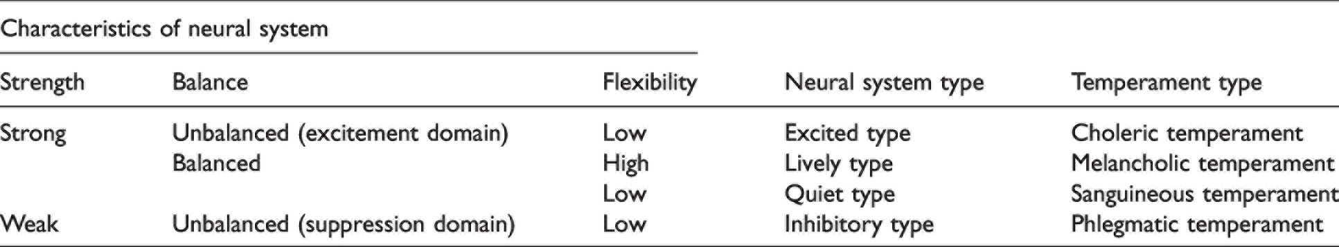
In the present study, one piece of the fabric was used to test the responses from a group of subjects in static and dynamic contact conditions. It was found that during the fabric–skin contact experiment, the electroneurophysiological responses of different subjects were totally different, and three types of subjects were identified. The differences come from neural activities of the subjects and these three types of subjects coincided well with the melancholic, sanguineous and phlegmatic temperament of Hippocrates’ temperament type theory, 34 as shown in Table 4. This theory categorizes people into four different types of temperament, namely, choleric, melancholic, sanguineous and phlegmatic temperament, which has been proved to correspond well to Pavlov’s higher neural activity type theory, 35 as shown in Table 5. The higher neural activity type theory concludes three characteristics of the neural system as strength, balance and flexibility. Based on the different features of the three characteristics, four different types of people with different neural system types are identified, namely, excited, lively, quiet and inhibitory type. Therefore, in our opinion, it is extremely important first to identify the type of subjects and separate them into different groups. These groups can be used as blocks in the experimental design and the statistical analysis to avoid systematic error.
Corresponding relationship between three different types of subjects and three different temperament types of Hippocrates’ temperament type theory
Corresponding relationship between Pavlov’s higher neural activity type theory and Hippocrates’ temperament type theory
Without doing so, this kind of error could be included as a part of the random error in the statistical analysis, and thus the results can be misleading. For example, Shimizu 8 utilized emotional curve out of EEG signals and tried to build a connection between the sentiment index and clothing comfort. The result showed large differences among different subjects and no obvious relationship could be identified. Wang et al. 9 chose six men of a similar age and height to attend the EEG experiment of clothing comfort. The energy percentages of α waves at the occipitalia were calculated as the key indicator to evaluate the human comfort perception. The authors failed to find the significant correlation between the energy percentage of α waves and the total comfort sensation. Obviously in those studies, the human subjects were treated as a sample from a uniform population, but in reality, as found in this study, three totally different types of subjects, namely, extraordinary, ordinary and nonsensitive people, may have existed in their sample, creating a systematic error in their results. In fact, we were not able to find any study that categorized subjects into different types, which could be one of the most important reasons why very little meaningful outcomes could be found in the literature of clothing comfort studies using electroneurophysiological methods such as EEG and ECG. Therefore, we conclude that different types of subjects based on physiology and psychology should be categorized before carrying out the electroneurophysiological experiment of fabric comfort study.
Based on the above findings, we propose that in studies 36 involving human subjects, the first step is to define different types of subjects so as to apply the treatments to different groups. In fact, in many population-based studies, gender, age, profession, income, educational background, ethnicity, geographic location, lifestyle and even the emotional state, as well as the hairiness of the human skin, can all be the parameters in the statistical models. For the electroneurophysiological study 37 of fabric tactile comfort, the body part touched by the fabric, the way of touching, the touching position, 38 force, speed, range and so on, can also affect the outcome. These factors could potentially influence the human electroneurophysiological responses for fabric contact. However, this study was just a preliminary study, allowing us to identify three different types of subjects, which opens a door for future electroneurophysiological studies involving these factors. Therefore, more systematic studies are certainly warranted for the influence of those parameters on human electroneurophysiological responses to clothing comfort.
Conclusions
This study analyzed the different electroneurophysiological responses of different subjects to fabric–skin contact. It was found that the subjects could be categorized into three different types, namely, extraordinarily sensitive, ordinarily sensitive and nonsensitive people, according to their electroneurophysiological responses to both static and dynamic contact with the same fabric. Their different sensitivities can be observed from the activities of α waves, the SNS and PNS. For an extraordinarily sensitive subject, when fabric–skin static contact takes place, the subject will immediately respond with a sudden intense α rhythm reaction followed by a totally suppressed α wave, as well as an increased heart rate with a decreased HRV level, enhanced activity of the SNS and a little participation of the PNS. When it comes to dynamic contact, their nervous systems were balanced instead. The α rhythm remained fluctuating normally and the PNS made a large contribution to lower the heart rate and thus increased the HRV level. For the ordinarily sensitive subjects, during the fabric–skin static contact, their α waves were gradually suppressed over time with an increased heart rate, a decreased HRV level, an enhanced activity of the SNS and near absence of the PNS. During dynamic contact, they began to respond intensely with a strong α rhythm reaction followed by a suppressed α wave, an increased heart rate, an enhanced HRV level and the joint effort of the SNS and PNS. However, the PNS failed to lower the heart rate and keep the neural system normally calm. For the nonsensitive subjects, no matter whether it comes to fabric–skin static or dynamic contact, they had no specific reaction with normally fluctuating α waves, a stable heart rate, an almost constant HRV level and a little participation of both the SNS and PNS. This preliminary study could provide an efficient way to identify different types of subjects and lay a fair ground for comparison purposes in future electroneurophysiological studies of clothing comfort.
Footnotes
Declaration of conflicting interests
The author(s) declared no potential conflicts of interest with respect to the research, authorship and/or publication of this article.
Funding
The author(s) disclosed receipt of the following financial support for the research, authorship and/or publication of this article: This work was supported by the Department of Education of Zhejiang Province (CN) under grant Y201737327, Fujian Provincial Key Laboratory of Textiles Inspection Technology (CN) under grant 2020-MXJ-02, Guiding Project of the Department of Science and Technology of Fujian Province (CN) under grant 2022H0048, External Collaboration Project of the Department of Science and Technology of Fujian Province (CN) under grant 2022I0042, and Open Competition Mechanism Project of the Department of Science and Technology of Quanzhou City (CN) under grant 2022GZ4.