
Abstract
We present a case of a female Ecuadorian patient who presented a deep facial burn injury complicated with a severe infestation of Dermatobia Hominis larvae. The burn injury was complicated by severe myiasis attributable to the poor management of the wound received at home, using tropical plants, which caused a secondary infection and severe necrosis of the tissue involving the forehead, cheeks, chin, scalp, nose, mouth and the eyes resulting in a bilateral enucleation and long inpatient hospital care.
Introduction
Myiasis is the parasitic infestation of organs and tissues by larvae of different kinds of diptera flies.1,2 It is more frequent in tropical and subtropical areas, especially Central and South America. The predisposing factors for myiasis are usually uncontrolled wound infection, poor hygiene, alcoholism or immunosuppression.3,4
This case report describes an Ecuadorian patient with a severe facial burn injury, infested by fly larvae. Although myiasis is relatively common, either incidentally 4 or as an alternative treatment for wound management, 2 uncontrolled infestation can lead to aggressive feeding on dead and infected tissues caused by the release of larval enzymes such as collagenase or protease. 5 In this case report we demonstrate that an uncontrolled wild larvae infestation can lead to aggressive ‘overfeeding’ of the tissues, which in this patient caused extensive additional damage that was resolved through aggressive surgical treatment, including a double enucleation of the eyes, complete facial eschar removal and laborious removal of individual maggots.
Case report
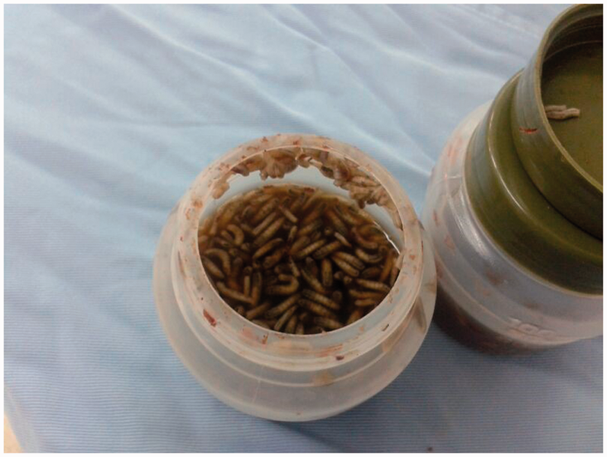
A female patient aged approximately 75 years was admitted at the San Luis de Otavalo Hospital in the province of Imbabura, Ecuador. She presented with a 2-month history of a face, scalp and left-hand burn trauma caused by boiling water. The community ‘shaman’ treated the patient with ancestral medicine based on unknown topical herbal dressings. This unconventional treatment and subsequent lack of medical attention resulted in severe secondary infestation of the wounds with copious Dermatobia Hominis larvae that forced her relatives to bring the patient to the hospital (Figure 1). On admission, a deep infected second-degree burn injury was found, with the presence of several hundred maggots covering multiple eschars on the left side of the face and additional subcutaneous fibrino-purulent secretions (Figure 2). The lesions compromised both ocular cavities and the eyes, owing to the tissue destroying effects of the larvae. The patient underwent four surgical wound debridements, and more than 300 larvae were manually extracted from the face, scalp, nose, mouth, ears and hand during the first procedure. The patient received aggressive broad-spectrum antibiotic treatment using i.v. Vancomicin + Imipenem for 10 days and oral Ivermectine 200 µg/kg/day for 2 weeks to control the larval infestation.
(a) Patient’s face at hospital arrival displaying larvae infestation. (b) Surgical debridement of the face before eyes enucleation. (c) Seventh postoperative day. (d) Patient’s condition at day 21 showing the results of the wound re-epithelialization and enucleation of the eyes. Container full of maggots.
Subsequent wound debridement was perform by otorhinolaryngology and ophthalmology specialists who subsequently removed more fly larvae from nasal, auricular, orbital and periorbital cavities (Figure 3). Finally, uniquely, she was required to suffer enucleation of the eyes due to the extension and severity of the infection.
Myiasis auricular involvement.
After reconstructive procedures were concluded, including skin grafting and wound closure, the patient returned to her home with further directions for future prosthetic replacement of the eyes.
Discussion
The patient had a long-term history of alcoholism and malnutrition. She presented late with severe burn injuries, all predisposing factors for myiasis.3,6 The lack of early adequate medical treatment and the wild maggots’ infestation resulted in severe complications.
Bacterial infections, eschars and tissue necrosis are common complications in burned patients who usually require surgical debridement. 7 Infected wounds are difficult to treat and alternatives therapies are growing in popularity, such as the use of sterile larvae; 8 however, this type of treatment requires some expertise. The type of larvae to be used needs to be properly selected; some larvae such as Dermatobia Hominis, Cordylobia Rodhaini or Chrysomya Bezziana usually require the ingestion of living tissue in order to complete their lifecycles, thus causing additional tissue damage.9–11 Thus, selecting the proper species is mandatory in order to avoid complications. The common green-bottle, Phaenicia sericata, is the most appropriate species to use for wound debridement by practitioners today. 12 The use of larvae has some complications such as pain, anxiety and bleeding. 5 As far as we know, there are no prior studies showing ‘overfeeding’ by larvae; however, there is one study where ‘wild maggots’ were removed due to the risk of affecting healthy tissue and causing more damage.4,5
Surgical debridement of the eschars and meticulous extraction of the fly larvae was performed since alternative methods of removing maggots such as the application of lateral pressure 13 and suffocation by occlusion of the punctum with mineral oils, 13 petroleum jelly, 14 mineral turpentine, ethanol spray 15 or even pork fat, 11 are often ineffective and necessitate more invasive interventions.
Conclusion
Meticulous attention to detail has achieved in the re-modelling of a face virtually destroyed by a burn, subsequent bacterial infection and gross larval infestation.
Footnotes
Declaration of conflicting interests
None declared.
Funding
This research received no specific grant from any funding agency in the public, commercial or not-for-profit sectors.