
Abstract
Ocular trauma accounts for a significant proportion of cases in any emergency eye outpatient department. There are very few cases reporting ocular injuries after leech exposure. Leeches are annelids belonging to the subclass Hirudinea; they dwell in fresh water and the sea and can be terrestrial. An oral sucker present anteriorly helps to attach to the host by releasing an anesthetic that prevents its recognition as a foreign body. Although many methods of leech removal have been proposed, its removal with forceps after instillation of local anesthetic remains a safe and effective method.
Keywords
Introduction
Leeches belong to a large phylum of annelids and the subclass Hirudinea. They live in fresh water, marine water and can be terrestrial. The haemophagic leech attaches to its host and disengages after finishing sucking. An anterior (oral) sucker helps it to connect to the host by releasing an anesthetic thereby preventing the host from sensing the leech. It also releases an anticoagulant enzyme to prevent the host blood from coagulation. As well the skin, a leech can also attach to the mucosal surfaces of the eyes and the nasal mucosa.1,2
Case report
A 24-year-old man presented to the ophthalmology outpatient department after being referred by an intern with a diagnosis of iris prolapse of the right eye. He was a farmer by occupation. Symptoms appeared 4 h after washing his face with stream water. There was no history of eye trauma. Visual acuity (VA) was 6/6 in both eyes. The left eye examination was unremarkable. In the right eye, on examination with a pen light, a brown cucumber-shaped foreign body was seen adherent to the temporal bulbar region. Slit lamp biomicroscopy revealed a black foreign body approximately 1 × 2 mm in size attached to the temporal bulbar conjunctiva at the 3 o’clock position to the limbus (Figure 1). The foreign body was observed to move, and after manipulating it with a sterile cotton eye bud, it was revealed to be a living leech sucking the bulbar conjunctiva.
Leech attached to bulbar conjunctiva of right eye.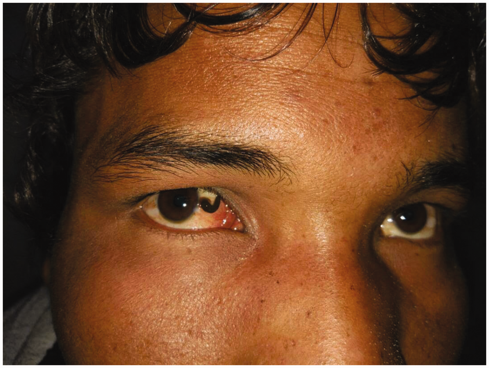
Neither cotton swab nor forceps could detach the leech but two drops of 0.4% oxybuprocaine paralysed it, whereupon it was removed easily from the eye along with sucking apparatus intact with the help of forceps (Figure 2).
Leech removed with help of forceps.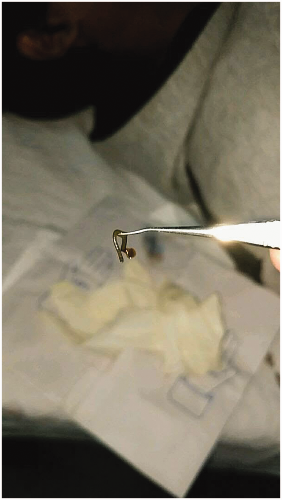
The anterior and posterior segments of the right eye were normal after mydriatic dilation. Topical chloramphenicol and flouromethonol prophylaxis were recommended q.i.d. for two weeks. The patient was followed up after one day and three days for evidence of any signs of inflammation. No subsequent evidence of inflammation, scleral perforation or uveal tissue prolapse was noted. The specimen was identified as a freshwater leech, Hellobdella stagnalis.
Discussion
In the past, ocular leeches have been documented for the treatment of acute congestive glaucoma and draining periorbital hematomas.3,4 Ocular leech infestation should be considered in patients after exposure to stream water, sea or lakes with a history of ocular complaints.
Topical local anesthetic is preferable to other methods, such as the application of cooking salt.5,6 Different methods for the removal of leeches have been proposed. Their removal with forceps after instillation of local anesthetic lidocaine should be tried first. The other proposed methods for the removal of leeches include irrigation with normal or hypertonic saline 7 and/or suxamethonium injection into the leech, 8 which have potential complications. There have been no long-term complications reported in the literature so far. In the present case, topical anesthetic was instilled and the leech body, including the sucker, was removed carefully with forceps. It is therefore recommended to remove the leech with forceps after instillation of anesthetic.
Conclusion
Ocular leech bulbar conjunctival infestation is a rare instance of conjunctival foreign body. Contact with fresh water, stream water or lakes must be enquired. Removal of the leech with forceps using local anesthetic should be attempted first as it is a safe method.
Footnotes
Declaration of conflicting interests
The author(s) declared no potential conflicts of interest with respect to the research, authorship, and/or publication of this article.
Funding
The author(s) received no financial support for the research, authorship, and/or publication of this article.