



Abstract
Scalp wounds with exposed calvarial bones continue to be a challenge especially when no local flap options are available and no microvascular flaps can be performed. Our prospective study looked at 19 patients (14 males) where customized negative pressure wound treatment was used till the complex scalp wounds, mostly from animal bites, were covered with healthy granulation and grafted. Scalp wounds ranged from 6 × 4 cm to 17 × 11 cm in size whereas the area of exposed bone ranged from 1 × 2 cm to 10 × 10 cm. No major complication was seen, and wounds were rapidly healed.
Introduction
The scalp consists of layers of skin and subcutaneous tissue covering the calvarial bones which protect the brain. Exposed bone, devoid of scalp, may lead to bone desiccation, sequestration, and infection of the bone and underlying intracranial contents. 1 Exposure of dural venous sinuses, which is life-threatening, may result.
The primary goal of reconstruction of a full thickness scalp defect is to cover the exposed calvarium, and hair restoration form the secondary goal. 2 Scalp defects may arise as a result of trauma, burns, surgery, autoimmune diseases, infection, or osteoradionecrosis. 1
Local flaps are rarely feasible in large defects with traumatized adjoining scalp, and micro-vascular tissue transfer requires special equipment and expertise and patient fitness to withstand prolonged surgery. Negative-pressure wound therapy (NPWT), initially described by Morykwas et al. in 1997, is an option to be considered. 3
Patients and methods
Our single-arm, intervention-only prospective study was conducted in the Department of Plastic and Reconstructive Surgery of our institution from July 2014 to June 2019. We included scalp wounds with exposed bone where local flap options were not feasible, in co-operative patients who consented, and could be relied upon not to disrupt the dressings. We excluded wounds with exposed dura or deeper structures, patients with GCS scores < 15 (Figure Suppl.) and uncooperative patients, and those with a cerebrospinal fluid leak. The research was conducted according to the Declaration of Helsinki after obtaining clearance from our institutional ethical committee (IEC-SKIMS).
Customized NPWT, fabricated in our hospital, was used in all patients (Figures 1–3). 4 Scalp hair about 5 cm beyond the wound margins was shaved off and the wounds debrided and haemostasis achieved before application of every NPWT dressings (Figures 1(a), 1(b), and 3(a)). Multiple unicortical holes were drilled into the exposed bone to expose the diploie to negative pressure, thus encouraging speedy granulation. For this, a manual drill with a 1.5 mm bit was used for smaller exposed bone areas and a Hudson brace with a 10 mm burr for larger areas with holes made c. 0.5–2 cm apart respectively. Careful precaution was taken to avoid breaching the inner bone cortex.

(a) Case of Bear maul with extensive craniofacial injuries with necrosed scalp over frontoparietal region. (b) Lateral view showing areas of frontoparietal bones denuded of skin, soft tissue, and periosteum. (c) Burr holes drilled into diploe to promote early granulation with NPWT. (d) Customized NPWT applied over exposed calvarial bones. (e) Exposed bones covered well with granulation following eleven NPWT dressings. (f) Wound reconstructed by split -thickness skin graft with NPWT. (g) Stable graft at follow- up.

(a) Post-traumatic compound defect biparietal region scalp with areas of graft loss and exposed calvarial bone; extensive scarring of adjoining scalp. (b) Holes drilled into diploe to promote early granulation with NPWT. (c) Customized NPWT applied over wound and connected to wall-mounted suction. (d) Granulation tissue sprouting through drilled holes to cover exposed bone. (e) Wound reconstructed successfully with a stable STSG.
The wound dressing was changed after every 48–72 h except in case of leakage or filling of the collecting bottle, it was changed immediately. 5 Our primary outcome variable was a graftable wound covered with healthy granulations. Once this desired result was achieved (Figure 1(e)), a split-thickness skin graft was placed covered by conventional dressing or NPWT as deemed suitable by the operating surgeon (Figure 1(f)). Patients were followed up till December 2020. Any complications encountered were recorded. For analysis, categorical variables were summarized as frequency and percentage. Age was categorized as 10-year age intervals.
Results
Most patients [8(42.1%)] were in the third decade of life (Table 1). The commonest mode of injury was an animal bite/maul which was seen in nine patients, burns in four, road crash in four, and blast injury in two. The scalp wounds ranged from 6 × 4 cm to 17 × 11 cm in size, whereas the areas of exposed bone ranged from 2 × 1 cm to 10 × 10 cm. Relatively larger exposed bone areas were seen in ten; scalp defects located in the parietal area in six, the occipital area in five, the frontal area in four, the temporal region in three, and the parieto-occipital region in one. The minimum number of dressing changes required to cover the wound with granulation tissue was five, whereas one patient required 16 dressing changes in 34 days to get the desired result (Table 2). All wounds were grafted (Figure 1(f)). Follow-up ranged from nine months to three years. No major complication was seen during the study and all patients had stable grafts at follow- up (Figures 1(g), 2(d), and 3(e)). Minor complications were encountered in seven patients; two patients had excoriation of surrounding scalp, while 5 patients experienced pain on dressing removal.
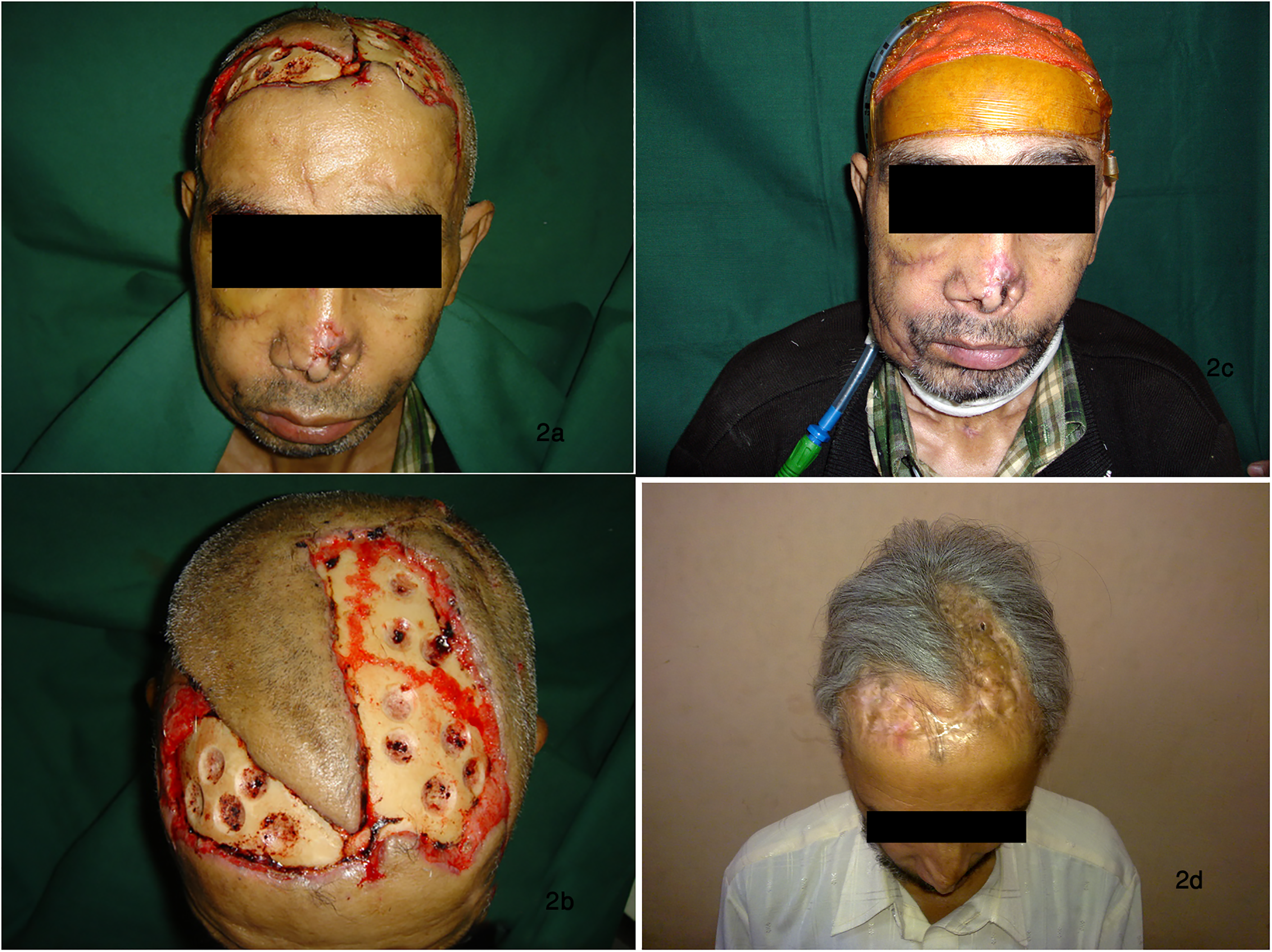
(a) Post bear maul composite soft tissue loss over bilateral frontoparietal regions with exposed underlying calvarial bones. (b) Burr holes drilled into diplooe to promote early granulation with NPWT. (c) Customized NPWT applied over complex wound. (d) Wound reconstructed by split-thickness skin graft stable at follow- up.
Age distribution of patients.

Number of NPWT dressing changes required to cover the whole wound with granulation tissue.

Discussion
The management of scalp wounds with exposed skull bone depends upon various factors, particularly the site and size of the defect, and the condition of the surrounding scalp. Smaller defects may be closed primarily or by advancing the surrounding scalp by scoring the galea aponeurotica. Medium-sized defects with exposed bone may be reconstructed by local flaps.
Large defects may require a regional flap or microvascular free tissue transfer. The former is awkward with special disadvantages in the form of donor site morbidity, long duration of surgery, complicated operative procedures, and difficult recuperation. 1 NPWT has revolutionized the management of wounds difficult to treat. 6 The latter may not be available in many low resource centres.
Wounds which would have required extensive surgical procedures have been successfully managed with NPWT. It is sometimes referred to as micro-deformational wound therapy (MDWT) and is popularly known as Vacuum assisted closure (VAC). The optimal negative pressure to be effective has been demonstrated to be between −50 to −200 mm Hg. 3 They conducted experiments on pigs and demonstrated that controlled negative pressure applied to a wound result in quicker granulation tissue formation and early closure of the wounds. The mechanism of action is by reducing wound oedema, and wound debris, thus angiogenesis. 7 NPWT also reduces the amount of fluid that must be cleared through the lymphatic system. The semi-occlusive drape acts as a thermal insulator to maintain wound warmth and also prevents evaporative water losses, thereby preventing desiccation of the wound. Bacterial contamination is reduced by the suction. 8
NPWT has been used successfully in almost all areas of the body and the scalp is no exception. 6 Open fractures and exposed bones, particularly of the lower extremity, are frequently treated successfully with NPWT, thus obviating complex wound care. 8 A retrospective study showed that NPWT used in wounds with exposed bones reduced the time before a flap or skin graft could be applied. 9
Although NPWT has revolutionized the management of wounds, it is not without its own share of complications, including bleeding, infection, pain, foam retention within the wound, and tissue adherence. It is contraindicated in the presence of necrotic tissue or eschar, untreated osteomyelitis, non-enteric and unexplored fistulas, malignancy, exposed vasculature, exposed nerves, exposed anastomotic site and exposed organs. Caution must be exercised with its use in patients on anticoagulant therapy or with a bleeding diathesis.
The commercially available VAC® dressing is more than 20 times as expensive as customized NPWT dressing, without any real advantage. 4
Conclusion
Though our study was small, and we had no control group (which was ethically difficult to justify), we have clearly shown that NPWT is a very useful tool to promote granulation of extensive wounds of the scalp, particularly where bone is exposed. NPWT can readily be manufactured locally.
Footnotes
Acknowledgements
We would like to acknowledge the guidance and support provided by the Ex. Head of our department and the help of medical and paramedical staff in conducting this study. This research did not receive any specific grant from funding agencies in the public, commercial, or not-for-profit sectors.
Declarations of conflicting interest
All the authors declare that there is no conflict of interests.
Funding
This research did not receive any specific grant from funding agencies in the public, commercial, or not-for-profit sectors.
