



Abstract
Mycosis fungoides (MF) is the most common type of cutaneous lymphoma. However, granulomatous MF remains a rare subtype. Its incidence is estimated in the literature to be 6.3%. Clinical and pathological diagnosis of this entity is difficult owing to clinical heterogenicity and various histopathological mimics. We report one such case.
Introduction
Granulomatous mycosis fungoides (MF) was first described by Ackerman and Flaxman in 1970.1–3 This mimics granulomatous dermatitis, lupus vulgaris, or sarcoidosis and thus the correct diagnosis of lymphoma may be delayed. 4 Where tuberculosis or leprosy are the most common differentials the diagnosis of MF is challenging, both clinically and on histopathological examination.

Haematoxylin and eosin-stained (H&E; 400×) showing multi-nucleated giant cells along with dense lymphocytic inflammatory infiltrate.

Haematoxylin and eosin-stained (H&E; 400×) showing intra-epidermal lymphocytes (epidermotrophism).
Case report
A 40-year-old male presented with painless indurated plaques over his back and abdomen for 10 years, with hyperpigmentation of his whole body over the last two years. Initially, these started as whitish patches on the back measuring ∼4 × 2 cm and then by similar lesions on the abdomen. Figure 1 shows hyperpigmented patches over abdomen.
Living in a rural area, on the advice of a local practitioner, he was started on multi-drug anti-tubercular therapy (ATT) for six months. No improvement was seen; indeed the lesions increased in size.
A skin biopsy diagnosed lupus vulgaris five years previously and a further two years of ATT had again proved no improvement. At his presentation six months previously, with profound mycosis and prominent plaque-like lesions at various places all over the body, another skin biopsy showed focal thinning and irregular epidermal acanthosis. Intra-epidermal lymphocytes (Fig. 2 shows intraepidermal lymphocytes) were out of proportion (epidermotropism) with spongiosis found, with a few showing pleomorphism. The upper and lower dermis showed a dense lymphocytic inflammatory infiltrate and evenly distributed epithelioid cell granulomata with multi-nucleated giant cells (Fig. 3 shows multinucleated giant cells with dense lymphocytic inflammatory infiltrate). There was no evidence of necrosis. Special stains (Fite, Ziehl-Neelsen, Periodic Acid-Schiff) all failed to show micro-organisms. Immunohistochemistry (IHC) showed lymphocytes positive for CD3 (Fig. 4 shows membranous positivity for CD3), CD4 (Fig. 5 shows membranous positivity for CD4) and CD5, and negative for CD8, CD20, CD30, TIA-1 and Granzyme.
T-cell receptor gene re-arrangement was later performed which showed monoclonal T-cells. This further supported the diagnosis of T-cell lymphoma. Blood count, complete biochemical profile, purified protein derivative, computed tomography scan of chest and abdomen, cervical spine radiograph, and biopsy of abdominal fat were all normal.
The initial treatment proposed was high-power topical steroids, but the patient failed to attend the follow-up visit.

Immunohistochemistry (IHC) showing membranous positivity for CD3.

Immunohistochemistry (IHC) showing membranous positivity for CD4.
Discussion
Granulomatous MF is characterized by dermal invasion of T-cells with associated granuloma formation comprised of epithelioid and multi-nucleated giant cells. It typically affects middle-aged adults and may present initially as a patch and then progresses to plaque and eventually evolves into a tumour in some individuals. 4 ATT treatment may alter the histology of the lesions; therefore, examination of a most recent lesion is crucial for correct early diagnosis.
Granuloma formation may also occur in Sézary syndrome, cutaneous anaplastic large-cell lymphoma, subcutaneous panniculitis-like T-cell lymphoma, and primary cutaneous B-cell lymphoma.
Epidermotropism is a crucial finding for the histopathological diagnosis of MF per se. It is however absent in ∼47% of cases. 3 Malignant cell infiltrate is also difficult to identify owing to obscuring granulomata. The exact pathogenetic mechanism of granuloma formation in cutaneous lymphomas is unknown. It has been postulated that for the histopathological diagnosis of granulomatous cutaneous lymphoma, the granulomatous component must comprise at least a quarter of the infiltrate. 3 This is important because extra-cutaneous manifestations involving lymph nodes, or rarely lungs, nasopharynx, and bones are more frequent in granulomatous MF when the overall 5-year survival rate is 66%, worse than classical MF. 5
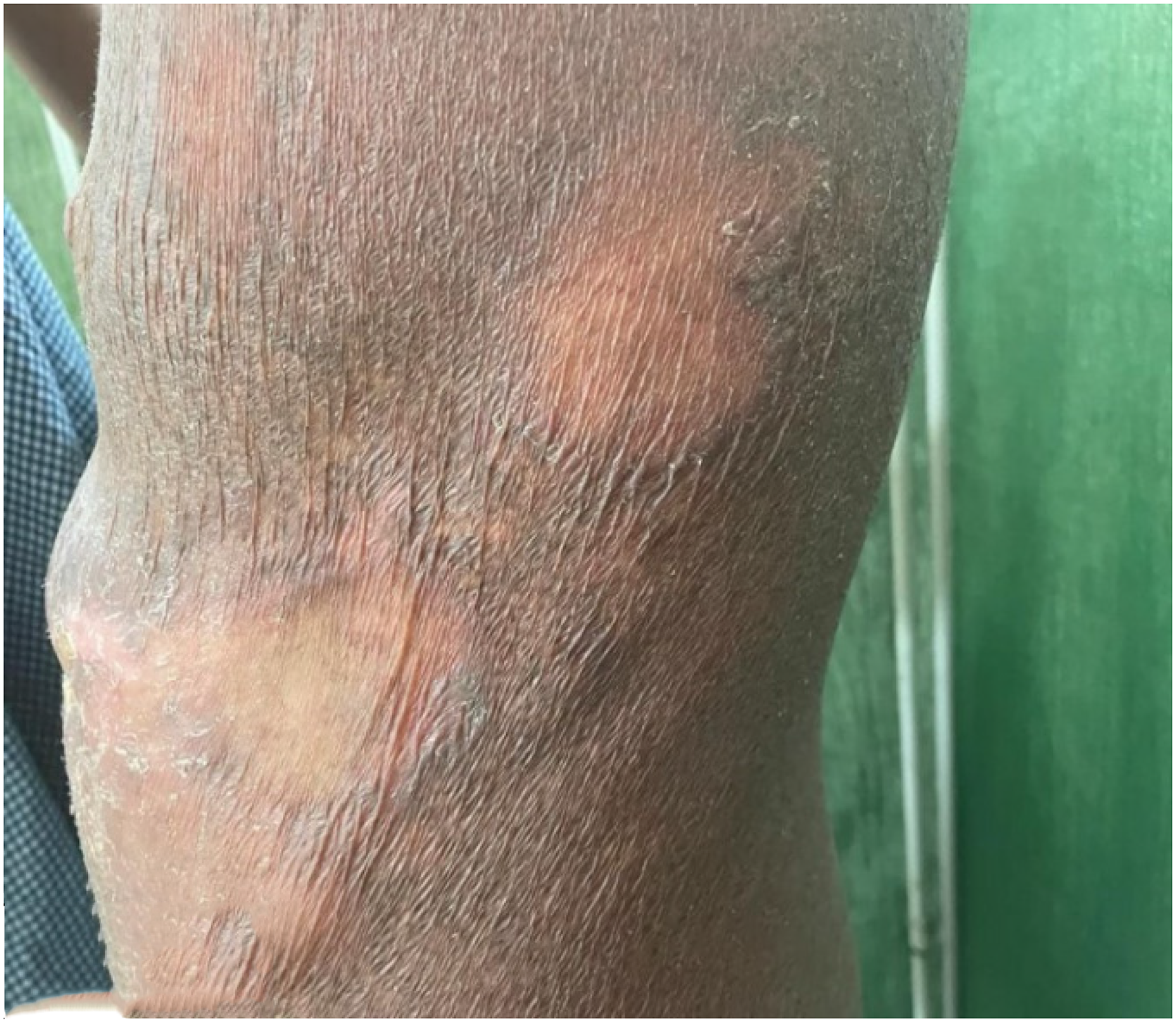
Hyperpigmented patch over abdomen.
Footnotes
Declaration of conflicting interests
The authors declared no potential conflicts of interest with respect to the research, authorship, and/or publication of this article.
Funding
The authors received no financial support for the research, authorship, and/or publication of this article.
