
Abstract
Septic cavernous sinus thrombosis (CST) is a rare but life-threatening complication of facial infections, typically arising from the midface ‘danger triangle’. Rarely, lateral facial infections may also lead to CST through spread via deep venous pathways. We report a previously healthy adolescent boy who developed fever, bilateral orbital swelling, and painful ophthalmoplegia shortly after the resolution of a furuncle over the left temporal part of the forehead. Magnetic resonance imaging confirmed CST. He was treated with intravenous ceftriaxone, vancomycin, and low molecular weight heparin, resulting in clinical recovery. This case highlights the importance of considering CST even when the primary infection lies outside the classical anatomical danger zone.
Case report
A previously healthy 15-year old boy developed a painful boil on the left forehead, accompanied by fever and purulent discharge. Oral antibiotics were prescribed, leading to the resolution of the local lesion, but the fever persisted. Over subsequent days, he developed bilateral periorbital swelling, headache, and irrelevant speech, prompting emergency evaluation (Fig. 1).

Clinical photograph showing bilateral orbital swelling and ptosis (more prominent on the right side) (a) and healed furuncle with surrounding skin discoloration over the left temporal forehead (b, arrow).
On admission, examination revealed bilateral ptosis and painful ophthalmoplegia involving cranial nerves III, IV, and VI, more pronounced on the right side. Pupillary reflexes were preserved, and there was no optic nerve involvement. Systemic examination was otherwise unremarkable.
Laboratory investigations revealed a total leucocyte count of 20 × 109/L with 92% neutrophils. Cerebrospinal fluid (CSF) analysis showed neutrophilic pleocytosis (2.85 × 10⁹ cells/L, 90% neutrophils) and hypoglycorrhachia (glucose 1.44 mmol/L), consistent with a pyogenic meningeal inflammatory response. Blood and CSF cultures were sterile, probably due to prior antibiotic therapy. Contrast-enhanced magnetic resonance imaging (MRI) of the orbits showed soft tissue swelling on both sides (more on the right), extending posteriorly toward the orbital apex with pachymeningeal thickening and cavernous sinus thrombosis (CST) (Fig. 2). HIV serology and primary immunodeficiency work-up (serum immunoglobulin levels and nitroblue tetrazolium test) were normal.
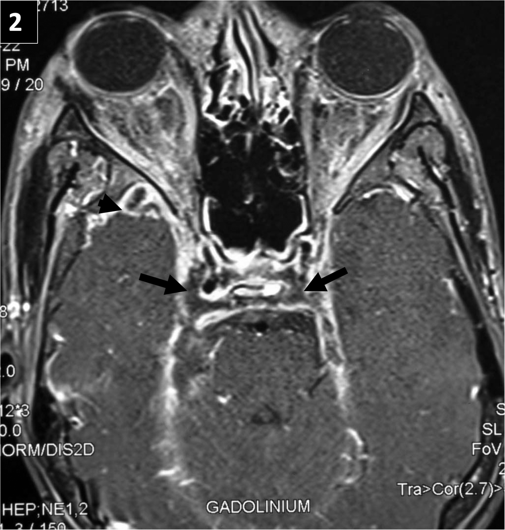
Axial post-contrast T1-weighted magnetic resonance imaging (MRI) image showing pachymeningeal thickening (arrowhead) and bulky cavernous sinuses (arrows) with non-enhancing areas suggestive of thrombosis.
He was treated with intravenous ceftriaxone, vancomycin, and low molecular weight heparin. Over four weeks, he improved significantly, with resolution of fever and gradual recovery of ocular movements.
Discussion
Septic CST typically arises from infections within the midface ‘danger triangle’, where valveless facial and ophthalmic veins permit retrograde intracranial spread.1,2 However, infections outside this region, including periorbital or temporal forehead areas, can also rarely lead to CST.2,3 As illustrated in our case, temporal forehead infections may spread via deep venous connections involving the superficial temporal vein, deep facial vein, and pterygoid venous plexus, which communicate with the cavernous sinus through emissary veins.2–4
Typical presentations include fever, headache, periorbital oedema, and ophthalmoplegia due to the involvement of cranial nerves III, IV, and VI, which traverse the cavernous sinus.1–4 Bilateral ocular involvement may occur rapidly due to intercavernous sinus communication.2,3 Contrast-enhanced MRI is the diagnostic modality of choice, characteristically showing cavernous sinus enlargement with non-enhancing thrombus, pachymeningitis, and orbital inflammation.3,4
The common causative organisms include Staphylococcus aureus and streptococci.1,3,4 Timely initiation of empirical intravenous antibiotics is critical. A combination of ceftriaxone and vancomycin is commonly used for broad-spectrum coverage and adequate CSF penetration. Surgical intervention may be required for complications such as orbital abscess, subdural empyema, or refractory sinusitis. Anticoagulation therapy is generally recommended to prevent thrombus propagation and facilitate venous recanalisation unless contraindicated.4,5
Painful ophthalmoplegia following a recent facial infection should always raise suspicion for septic CST. 1 Our case reinforces the importance of maintaining clinical suspicion even when the primary lesion is located outside the traditional danger triangle. Early empirical antibiotics and prompt imaging are essential to minimise complications and improve clinical outcomes.
Footnotes
Author contributions
AS: collected patient data, drafted the manuscript, and patient management; SS, BS, SA, VB: collected patient data and patient management; AKP: conceptualisation, collected patient data, drafted and revised the manuscript, and patient management.
Declaration of conflicting interests
The authors declared no potential conflicts of interest with respect to the research, authorship, and/or publication of this article.
Funding
The authors received no financial support for the research, authorship, and/or publication of this article.
Patient's consent
Written informed consent is obtained. The patient and father were explained about confidentiality, and the case information will be used for educational purposes only.