
Abstract
The simultaneous occurrence of pelvic organ prolapse and cervical adenocarcinoma is rare and can pose diagnostic challenges owing to overlapping symptoms. We present the case of a 60-year old multiparous, postmenopausal woman with a six month history of vaginal prolapse involving a mass. After undergoing vaginal hysterectomy with pelvic floor repair, histopathological examination revealed the presence of stage 1A2 endocervical adenocarcinoma. Post-operative imaging showed no metastasis or lymph node involvement.
Introduction
In India, pelvic organ prolapse affects a significant proportion of women, ranging from 20–65%, with the highest incidence observed in those around 60 years of age. 1 Various demographic and health factors, including age, socioeconomic status, obstetric history, and occupation, contribute to this prevalence. In contrast, cervical carcinoma is the leading genital cancer in India, but is rare in postmenopausal women, with whom no well-established management protocols. 2
Diagnosing malignancy imposes a significant emotional burden on both physician and patient, underscoring the importance of remaining alert to rare conditions, even when symptoms and signs do not readily indicate a specific diagnosis, in order to avoid belated management.
Case presentation
A 60-year old postmenopausal woman with no known co-morbidity presented with a six month history of a mass descending vaginally, accompanied by urinary disturbances. She had never undergone cervical cancer screening and had a history of several vaginal deliveries attended by untrained personnel.
On examination, there was a third-degree uterovaginal prolapse with a grade 2 cystocoele. Additionally, a healed decubitus ulcer was observed on both lips of the cervix, without any associated discharge or bleeding on palpation (Fig. 1). Manual examination revealed a normal uterus and adnexa with free parametrium, and the rectal mucosa appeared normal. A PAP smear examination showed no intra-epithelial lesions or malignancy.
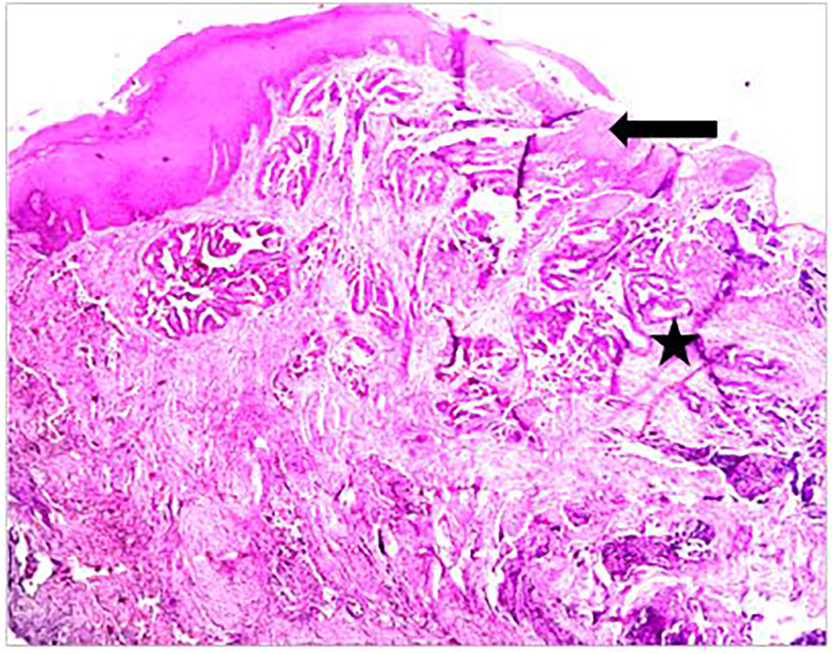
Four times magnification showing ulceration, hyperkeratosis, acanthoses (arrow); crowding (asterix).
After a comprehensive pre-operative evaluation, a vaginal hysterectomy with pelvic floor repair was performed. Intraoperatively, friability of the cervix prompted further investigation. Histopathological examination revealed a 0.7 × 0.2 cm tumour in the cervix, confined to the endocervix, with an aggressive glandular pattern and hyperchromatic nuclei, displaying a 5 mm depth of stromal invasion and free margins (Figs. 2 and 3). There was no tumour involvement in the ectocervix nor uterine fundus, and no evidence of lympho-vascular space invasion. The lesion was classified as endocervical adenocarcinoma of standard type and retained it as FIGO 2018 stage 1A2. 3 Further imaging studies revealed post-operative changes in the vaginal cuff, with no signs of distant metastasis nor lymph node involvement. Further management was to follow up the patient periodically with imaging and examination, with no need for adjuvant therapy at present.

(a) Ulceration, hyperkeratosis, acanthosis; (b) cribriform pattern; and (c) papillary pattern.

Gross image of pelvic organ prolapse with keratinisation.
Discussion
Pelvic organ prolapse and carcinoma of the cervix are common conditions, but their concurrence is rare. A nationwide data analysis on patients who underwent hysterectomy identified 17,074 cases of early-stage cervical cancer, of which only 4.9% were invasive. 4
A retrospective review to determine the incidence of concurrent gynaecological malignancy in patients with uterovaginal prolapse concluded that pre-malignant cervical conditions increased the incidence of cervical cancer from 0.3–4.2%. 5 The mean age of diagnosis is 74.4 years, with a range from 54 to 89 years. Most patients are asymptomatic; however, blood-stained vaginal discharge may be a symptom. 3 A chronic non-healing decubitus ulcer should raise a suspicion of cancer.
Local staging of squamous carcinoma by ultrasound or magnetic resonance imaging (MRI), characterised by hypo-echogenic, richly vascularised tumour, while adenocarcinoma is identified as an iso- or hyperechogenic, highly perfused lesion, contrasting with healthy residual cervical stroma. On T2-weighted MRI, cervical cancer typically appears as an iso- or hyperintense mass, regardless of histopathologic type, and when small, is surrounded by a hypo-intense normal cervical stroma. 4 Currently, there is no evidence-based management for cervical cancer associated with uterovaginal prolapse. The decision for management must consider staging, the extent of spread, and the degree of procidentia and cystocele–rectocele involvement. 5
Footnotes
Acknowledgements
We would like to express our gratitude to the patient who provided their consent to report the case. The Department of Obstetrics and Gynaecology at Aarupadai Veedu Medical College in Puducherry made this work possible.
Declaration of conflicting interests
The authors declared no potential conflicts of interest with respect to the research, authorship, and/or publication of this article.
Funding
The authors received no financial support for the research, authorship, and/or publication of this article.
Informed consent
We affirm that we obtained informed consent from the patient. This report contains no information that could identify the patient.