



Abstract
Objective
This study aimed at describing routine electroencephalogram (EEG) findings among children and adolescents with a clinical diagnosis of epilepsy and determines how interictal EEG abnormalities vary with the psychiatric comorbidities.
Methods
We conducted a cross-sectional study among children and adolescents with epilepsy aged 5–18 years receiving care from a regional referral hospital in Southwestern Uganda. Psychiatric comorbidities were assessed using an adapted parent version of Child and Adolescent Symptom Inventory-5. Thirty-minute EEG samples were taken from routine EEG recordings that were locally performed and remotely interpreted for all participants.
Results
Of the 140 participants, 71 (50.7%) had normal EEG findings and 51 (36.4%) had epileptiform abnormalities while 18 (12.9%) had non-epileptiform. Of those who had epileptiform abnormalities on EEG, 23 (45.1%) were focal, 26 (51.0%) were generalized, and 2 (3.9%) were focal with bilateral spread. There was no significant association between the different psychiatric comorbidities and the interictal EEG abnormalities.
Conclusions
Among children and adolescents with a clinical diagnosis of epilepsy in Southwestern Uganda, only 36% showed epileptiform abnormalities on their EEG recordings. There was no association between the interictal EEG abnormalities and psychiatric comorbidities.
Introduction
Epilepsy is a neurological disorder with predominant childhood onset1,2 with 80% of the affected persons living in low- and middle-income countries.3,4 In Uganda, epilepsy has a prevalence of about 10.3 per 1000 people. 1 Available records at Mbarara regional referral hospital (MRRH) show that epilepsy accounts for 4% of all admissions and 53.5% of all disorders among children and adolescents attending the mental health clinics. Epilepsy diagnosis is mainly made clinically through history-taking with witness account being the most important with EEG being only supportive.5,6 Unfortunately, the accuracy of clinical diagnosis depends on several factors such as patient history and clinician accuracy which increases the risk of misdiagnosis. 7 EEG provides information about presence or absence of epileptiform brain wave discharges and their location. 8 This is important in supporting diagnosis and classification of epilepsy syndromes, assessing prognosis, and selection of antiepileptic drugs. EEG is also useful in monitoring and determining when to discontinue the antiepileptic treatment.9-11 However, in Uganda, even though EEG is known to have a role in the diagnosis of epilepsy and its management, 12 its diagnostic effectiveness is not well known. Since epilepsy may become a chronic condition, misdiagnosis and poor management can complicate the clinical outcome including predisposition to various psychiatric disorders.13,14 Additionally, some psychiatric disorders may persist even after treatment of epilepsy15-17 and, hence, when not assessed and treated, they significantly affect the person’s quality of life and functioning. A range of EEG abnormalities, often subtle, have been described in psychiatric conditions including personality disorders. Some studies give an overall impression that there is a low EEG yield in patients presenting with pure psychiatric manifestations. This is likely to be the case in younger subjects (27). Several studies have explored the association between EEG abnormalities (frequencies) and different psychiatric disorders such as mood disorders, attention deficit hyperactivity disorder (ADHD), autism, psychotic, anxiety, trauma, and stressor-related disorder.18-20 However, there is scanty literature exploring the association between the interictal EEG abnormalities and emotional, behavioral, developmental, and psychosis-related psychiatric comorbidities. In the middle aged or elderly with new onset of psychiatric illness, the EEG is helpful in detection of organic brain syndromes with diffuse abnormalities indicating a neurodegenerative or encephalopathic process, or focal slow activities alerting to a space occupying lesion. There is also a need to determine routine EEG findings among young patients with a clinical diagnosis of epilepsy and also to assess the association between interictal EEG abnormalities and the different psychiatric disorders. In this study, we determined the proportion of children and adolescents with the clinical diagnosis of epilepsy that had corresponding epileptiform EEG findings. We also determined how interictal EEG abnormalities varied with psychiatric comorbidities, that is, emotional, behavioral, developmental, and psychosis-related disorders.
Methods
Study design and setting
This was a cross-sectional study in Southwestern Uganda at MRRH psychiatry department, and 3 other community outreach sites: St Joseph’s Rubindi Health Center III (RHC III), Mushanga Health Centre III (MHC III), and Tukore Invalids Primary School (TIPS). MRRH is the biggest public hospital in the Southwestern Uganda, located in Mbarara Municipality, about 265 km from Kampala city. It has about 350 beds and offers general, specialized, and consultant as well as emergency medical services. The psychiatry ward has about 40 beds with a total of over 1000 new patients per year, more than half of them having suspected epilepsy. The department offers both inpatient and outpatient mental health services alongside epilepsy treatment. In addition, a team from MRRH composed of a psychiatrist, psychiatry residents, a psychiatric clinical officer, undergraduate students, and a counselor hold Mental Health Clinics every first, third, and last Thursday of the month at Rubindi Health Centre III, Mushanga Health Centre III, and Tukore Invalids Primary School, respectively. These clinics offer assessment, diagnosis, and treatment of various psychiatric disorders and epilepsy.
Study population
The study included all children and adolescent aged 5–18 years with epilepsy who had at least 1 seizure in the last 12 months, in order to capture all cases of active epilepsy and had come with their parents/adult carer to provide collateral information. Those who had any emergency medical condition due to serious infections and high temperatures and those with status epilepticus or suspected severe medication side effects at the time of the study were excluded.
Sample size, sampling, and recruitment procedure
We recruited 140 participants (determined by using OpenEpi satistics software for sample size calculation for cross-sectional studies) 21 with two-sided significance level (α) = .05; power (1-β, % chance of detecting) = 80%. 22 We identified potential participants through reviewing their medical records and recruited them consecutively until the desired sample size was obtained. These were further assessed by a psychiatry resident, psychiatrist, pediatrician, or internist to confirm the clinical diagnosis of epilepsy. Only those who had a positive diagnosis of epilepsy were recruited for the study.
Data collection and management
Data were collected from March to September 2018 and participants responded to a pretested investigator-designed questionnaire which collected data about baseline characteristics and the parent version of the Child and Adolescent Symptom Inventory-5 (CASI-5). The questionnaire was administered by research assistants using tablets. EEG recordings were either carried out on the same day of the interview or the participants were booked for a later date according to their convenience. We used Stellate Harmonie EEG machine for all recordings with each taking between 20 and 60 minutes. The participants were prepared by taking them through the recording procedure so that they could know what to expect. For young children aged 5–8 years, sleep EEGs were recorded. Their parents were advised to have an overnight sleep deprivation for the child a day before the recording. This would be accompanied by administration of fast release oral melatonin 2–10 mg. For older children and adolescents, most EEGs were recorded in a wakeful state. The EEG electrodes were placed on the scalp following the 10-20 International system. 23 Standard silver chloride scalp electrodes were used. EEG recording was performed using a 24-channel stellate Duo Digital EEG and Video recording system. The EEG transcription recording settings were Sampling rate 400 Hz. sensitivity 1-1000 μV/cm, HF15-120 Hz, LF 0-10 Hz, and time scale 15-60 mm/s. The EEG was digitally recorded and referentially stored on local hard drive media. During the recording process, standard activation procedures of hyperventilation and photic stimulation were performed in order to increase the yield of epileptiform abnormalities and the possibility of getting a seizure that could be captured on EEG. The EEG recordings were interpreted locally by trained personnel with support by epileptologists from TeleEEG charity UK, the Royal London hospital, and Harvard University. Participant identity was protected by using the assigned unique numbers for each patient while transferring the EEG recordings to TeleEEG UK and the Royal London Hospital. The data would later be entered into the corresponding participant number and submitted electronically to the central server.
Study tool: Child and adolescent symptom inventory (CASI-5)
This is a rating scale for emotional, behavioral, and developmental disorders developed according to fifth edition of the Diagnostic Statistical Manual. 24 The behavioral disorders included oppositional defiant disorder and conduct disorder. Developmental disorders included ADHD, motortics, vocaltics, autism spectrum disorder, nocturnal enuresis, and encopresis. Emotional disorders included selective mutism, separation anxiety, social anxiety, generalized anxiety, specific phobia, manic episode, post-traumatic stress disorder, panic disorder, obsessions, compulsions, somatic symptoms, major depressive episode, persistent depression, prosocial emotions, binge eating, hair pulling, and skin picking. Psychosis-related disorders include schizophrenia and schizoid personality disorder. The adapted version of CASI-5 was translated to Runyankore/Rukiga prior to being used for this study in our setting. 25 The data collection questionnaire also had a section for entering EEG findings for each child.
Data analysis
Collected data were downloaded from the central server in form of an excel sheet which was cleaned and imported to STATA version 13 for analysis. The children and adolescents who had epileptiform abnormalities as per their EEG recording were expressed as a proportion/percentage of the entire study sample (those with a clinical epilepsy diagnosis). Proportions of participants having (or not having) the different emotional, behavioral, and developmental disorders were determined in relation to the interictal abnormalities (focal or generalized) as per the EEG findings. Association was determined using odds ratios and considering 5% level of significance.
Ethical considerations
Privacy of respondents
Respondents were first advised about the sensitivity of some questions and were informed that they could choose whether to answer or not if they found them difficult. Interviews were conducted in a private room having only the investigator, participant, and, where necessary, the parent/care taker.
Confidentiality
Respondents’ confidentiality and integrity were ensured. Participants were assigned unique identification numbers for the purpose of this study strictly and their names, signatures, and those of their relatives were not included in the final report. Participants were given opportunity to seek clarification on any matter that was not clear to them.
Consent to participate
We obtained written informed consent from the parents or the responsible adult of children aged 5–17 years while these children assented. Children aged 18 years provided written informed consent.
Results
Social demographic characteristics
A total of 161 participants were assessed for psychiatric disorders (Figure 1). However, only 140 had EEG recordings and those are the only ones that were included in the analysis. Flow diagram showing the recruitment of participants.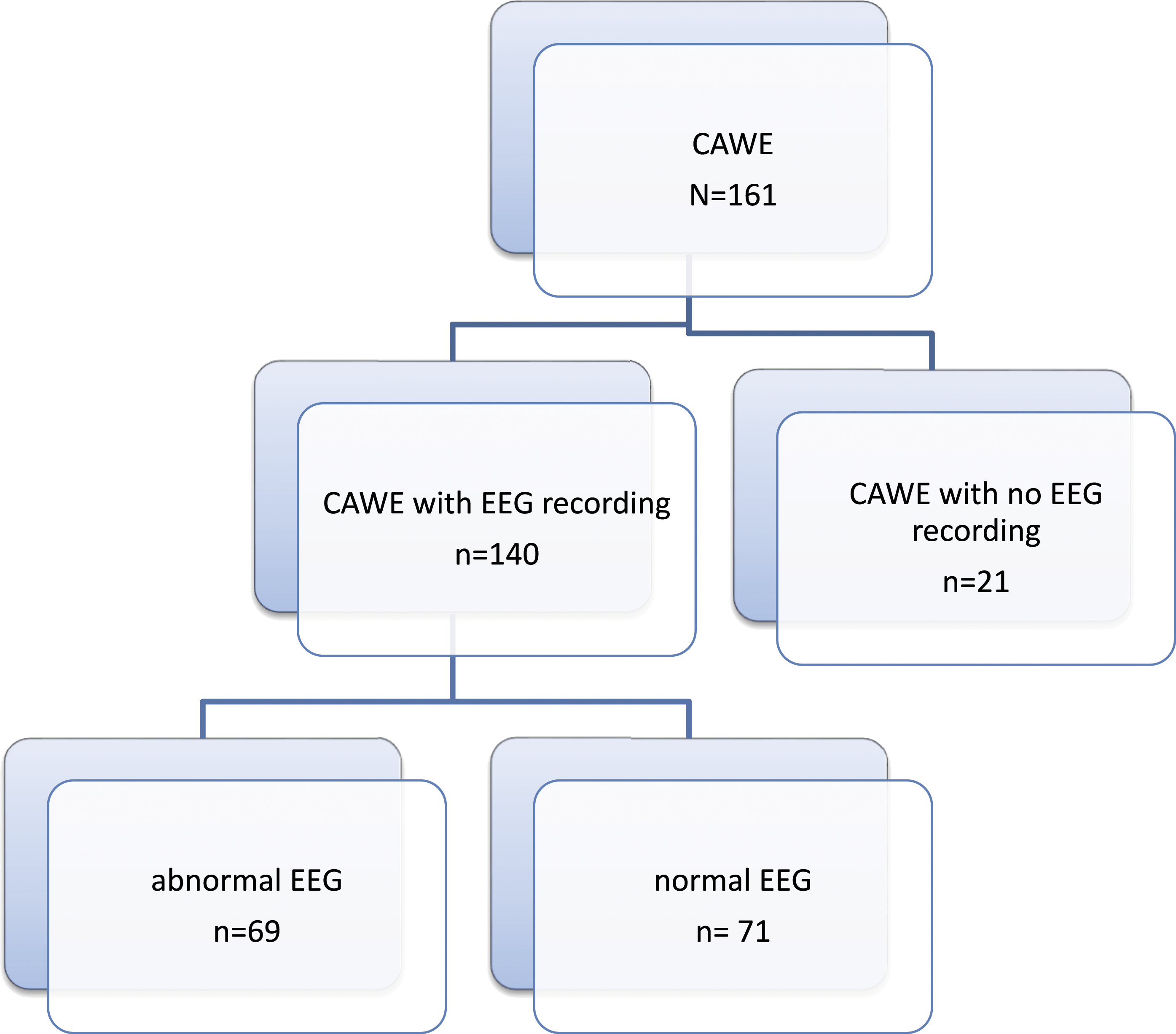
Socio-demographic characteristic of participants whose EEGs were recorded (n = 140).
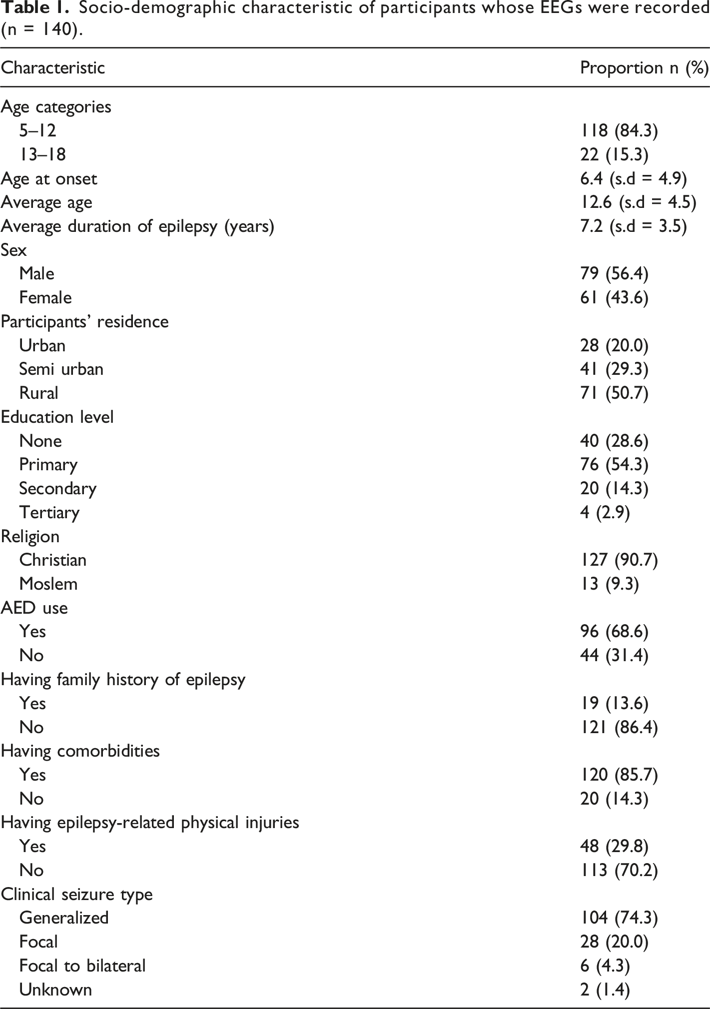
Of these, 93 (66.4%) were from Mbarara district and 18 (1.9%) came from Sheema district while 29 (20.7%) were from other different districts. The average duration of epilepsy was 7.2 (s.d = 3.5) years, whereas that of antiepileptic drug use was 4.2 ± 4.2 years. Of the 96 participants who were using antiepileptic drugs, 76 (79.2%) were using 1 drug and the rest using 2 drugs. The most commonly used antiepileptic drug was carbamazepine (70.8%).
Participant electroencephalogram findings (n = 140).
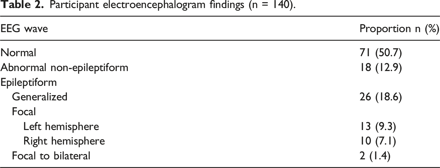
Of those who had epileptiform discharges, generalized discharges (generalized spike and wave or polyspike and wave discharges depicting the generalized seizure type) were the commonest (52%). Those with abnormal non-epileptiform EEG, findings such as rhythmic focal slow and intermixed generalized slow with multifocal sharp wave elements suggestive of a variety encephalopathies, for example, metabolic, toxic, endocrine, and neurodegenerative complications of virus infections and/or mental retardation were found.
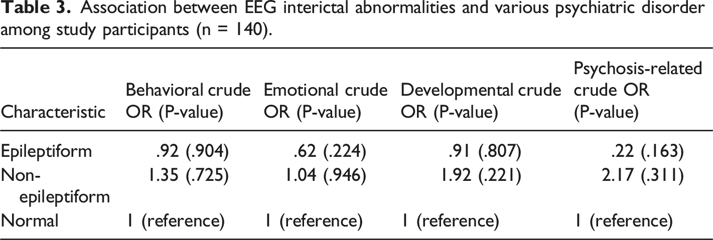
Association between EEG interictal abnormalities and various psychiatric disorder among study participants (n = 140).
Discussion
The aim of this study was determine the proportion of children and adolescents with both the clinical and the EEG confirmed diagnosis of epilepsy. It also aimed to determine the variation between EEG abnormalities (depicted seizure type) and psychiatric disorders in the same study population. We found that only 36.4% of the patients with a clinical diagnosis of epilepsy showed epileptiform discharges in their EEG. We also found that 50.7% of the patients with a clinical diagnosis of epilepsy had normal EEG findings. Of those who had epileptic activities on EEG, 23 (45.1%) were focal, 26 (51.0%) were generalized, and 2 (3.9%) were focal with bilateral spread (secondary generalization). About 13% had abnormal EEG traces that were not specific to depict the corresponding type of epilepsy. Surprisingly, there was no significant association between psychiatric comorbidities and the interictal EEG abnormalities.
Our study findings are surprising but parallel with results from other settings as reported by Smith which indicated sensitivity of EEG to range from 25 to 56%. 27 The study acknowledges the wide range and multiple factors such as diversity in case selection and varying requirements in making diagnosis of epilepsy among others. Our study also indicates that EEG abnormalities may not have a primary psychiatric diagnostic specificity. This may be especially so in young patients. However, our finding has a lower percentage than that found in other studies.28-30 In these studies which were mainly conducted among adults from high-income countries, the prevalence of epileptiform abnormalities on EEG range from 41% to 68% depending on the type of epilepsy. These studies employed various EEG recording modalities such as longer sleep recordings, drug-induced sleep EEGs, 6–24-h ambulatory and prolonged recordings, and long-term video telemetry recordings in addition to thorough assessment of the cases using investigations of neuroimaging such as brain magnetic resonance imaging. This setting is not available in Uganda and hence the need for the clinicians in our setting to consider that a majority of children and adolescents with a clinical diagnosis of epilepsy may have normal EEGs. The low proportion found in our study could also be explained by the fact that the routine EEG recordings were for a short duration and so they did not increase the diagnostic yield of the epileptiform abnormalities and many did not capture seizures. There is a need to improve and enhance our EEG technology to perform longer EEG recordings to increase the diagnostic yield. In our study, most participants recruited were already on epilepsy drug treatment and this possibly led to the elimination of the epileptic abnormalities on the EEG, particularly in patients with genetic epilepsies like childhood absence, juvenile absence, and juvenile myoclonic epilepsies if they were on the right treatment and probably this also influenced the possibility of no lack of association between interictal EEG abnormalities and the psychiatric comobilties in these patients. The other possible explanations for the differences would be that the etiology of the epilepsies and psychiatric comorbidities of the participants in our study were different compared to the ones in some of the studies in the more developed world and also the fact that patients that were able to reach our center for epilepsy treatment did not include patients with severe epileptic syndromes and psychiatric comorbidities that often are associated with specific EEG characteristics as seen in symptomatic generalized epilepsies and epileptic encephalopathies like in cases of Lennox-Gastaut syndrome and progressive myoclonic epilepsies. Nonetheless, our study emphasizes the importance of the EEG as a tool to assess epilepsy and to refine its diagnosis. The EEG is expensive and not easily accessible in resource-limited developing countries like Uganda. Our study highlights the important point that selective EEG investigations and protocols may be the way forward for accurate diagnosis and classification of epilepsy as noted in various studies.28,29,31,32 The study findings highlight the importance of thorough and accurate history taking which forms the basis of diagnosing epilepsy supported by EEG and the low EEG yield in patients presenting with psychiatric comorbidities On the other hand, this study also highlights the possibility that several children and adolescents may be misdiagnosed with epilepsy clinically which may be purely psychiatric manifestations. This may consequently expose them to unnecessary emotional and psychological distress which comes with the notion of having a chronic and highly stigmatized condition in the low- and mid-income countries. The misdiagnosis and hence treatment with AEDs also increases the health cost of affected families living in an already financially constrained environment.
We also found several abnormal EEG patterns which were not necessarily epileptiform in nature. Several of these abnormalities required further investigation and assessment to establish the exact cause. Our study also proves and emphasizes the fact that there are several other organic and non-organic brain disorders that may present with epileptic seizure-like manifestations mimicking real epileptic convulsions like in cases of psychogenic or dissociative non-epipletic disorders usually with no associated EEG abnormalities. It also highlights the need for a multidisciplinary team of clinicians dealing with epilepsy diagnosis and management which is a very complex neurological disorder. Our study highlights the vital role of EEG in the clinical context of the diagnosis and management of epilepsy in the resource-limited developing world.
The study found no significant association between any of the psychiatric disorder categories and the EEG interictal abnormalities (depicting specific seizure types). This is the first such study in our setting and this finding depicts that CAWE experiencing any type of seizure are at risk of having emotional behavioral developmental or psychotic disorder with no specific predilection or preference. This is in line with findings from several other studies that also documented no specific association between EEG findings and specific psychiatric disorders.18-20 However, some studies have found association between specific psychiatric disorders such as ADHD and seizure types such as frontal lobe and absence epilepsy based on EEG.33,34 Therefore, it is vital for clinicians to routinely screen for all psychiatric comorbidities among people with epilepsy in order to promptly manage the disorders.
Our findings are interesting but the study was conducted from 1 institution and may not reflect findings from large and broader community and hence the need to conduct a large community study all over the country with longer EEG recordings for further assessment of the association between interictal abnormalities and psychiatric comorbidities.
Conclusions
Among children and adolescents with clinical diagnosis of epilepsy in this Southwestern Uganda, only 36% showed epileptiform EEG abnormalities. There was no association between the interictal EEG abnormalities and psychiatric disorders. This finding raises questions and highlights the need for larger community studies with higher power to verify our findings.
Footnotes
Acknowledgments
We appreciate Mbarara University Research Training Initiative for funding this study. We are also thankful to Mbarara Regional Referral Hospital administration especially psychiatry department and other study site administrators for all the technical and logistic support rendered during the study. In a special way, I would like to thank Dr. Moses Kibuuka of Royal London and his entire team for all the logistical and technical support offered to our psychiatry department in order to improve the diagnosis and management of epilepsy. Lastly, we thank all the study participants who volunteered useful information for the study.
Research reported in this publication was supported by the Fogarty International Center and co-funding partners (NIH Common Fund, Office of Strategic Coordination, Office of the Director (OD/OSC/CF/NIH); Office of AIDS Research, Office of the Director (OAR/NIH); National Institute of Mental Health (NIMH/NIH); and National Institute of Neurological Disorders and Stroke (NINDS/NIH)) of the National Institutes of Health under Award Number D43TW010128. The content is solely the responsibility of the authors and does not necessarily represent the official views of the National Institutes of Health.
Author Contributions
K.J. participated in the conception of the idea, designing the study, proposal writing, data collection, management, analysis and interpretation as well as manuscript writing. G.Z.R. and M.K. contributed and provided technical assistance on idea conception, designing the study, proposal writing, data collection, analysis and interpretation. They also actively participated in manuscript writing. All authors have read and approved the manuscript.
Declaration of Conflicting Interests
The author(s) declared no potential conflicts of interest with respect to the research, authorship, and/or publication of this article.
Funding
The author(s) disclosed receipt of the following financial support for the research, authorship, and/or publication of this article: This work was supported by the Fogarty International Center grant number D43TW010128.
Ethics Approval
Before starting the data collection process, we obtained ethical approval from Mbarara University of Science and Technology Research Ethical Committee (MUST-REC number 15/09-17) and Uganda National council of Science and Technology (UNCST number SS 4522). This was presented to all the study site administrators for permission to collect data from their centers.
