
Abstract

A 5-year-old girl presented with a gradually growing tumor on her right nostril and associated nasal blockage of several months’ duration. Anterior rhinoscopy detected a smooth, well-demarcated, protruding, 0.5 × 0.5-cm tumor of the right nasal vestibule (figure 1). Multiple small, ectatic blood vessels were also identified on the surface.
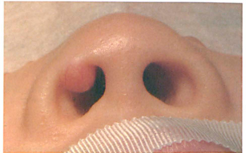
At presentation, the pinkish hypervascular tumor is seen at the right nasal vestibule.
The patient was administered general anesthesia, and the tumor was resected en bloc along with the vestibular skin without damaging the underlying structure. Histopathologically, the dermal lesion contained a mixture of histiocytes, foamy macrophages, lymphocytes, and scattered Touton giant cells (figure 2). Based on the histopathology, a diagnosis of juvenile xanthogranuloma (JXG) was made. At 4 years postoperatively, no sign of recurrence was evident.
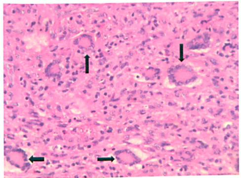
Specimen contains multiple Touton giant cells (arrows). Their nuclei display a wreath-like peripheral arrangement (H&E, original magnification x40).
JXG was first reported in 1905 by Adamson, who described a 2-year-old boy with multiple yellow cutaneous papules that eventually regressed spontaneously. 1 It is a rare, benign, non-Langerhans histiocytic lesion that typically arises in the head and neck, trunk, upper extremities, and lower extremities. 2 The development of such a lesion in the nasal cavity has seldom been reported in the literature.
The etiology and pathogenesis of JXG are still unclear. A clonal proliferation of histiocytic/dendritic cells has been assessed. 3 Furthermore, an association between JXG and both neurofibromatosis type 1 and chronic myelogenous leukemia is well known. 4
The histopathologic hallmark of JXG is the Touton giant cell, a multinucleated giant histiocyte whose rings of nuclei separate a central homogeneous core from a foamy periphery. The differential diagnosis of JXG should include other histiocytic lesions, Rosai-Dorfman disease, and infectious causes. 5 Because JXG spontaneously regresses, it usually does not require any treatment. However, excision may be carried out for diagnostic, cosmetic, or symptomatic reasons.