



Abstract
Significance statement
Vocal fold cysts are common, and conservative treatment with voice therapy is tried before surgical interventions are recommended. However, superimposed vocal fold scar can complicate clinical outcomes, as vocal fold scar can impair mucosal waves. For this reason, it is important to identify vocal fold scar preoperatively in patients undergoing surgical treatment of vocal fold cysts.
A 47-year-old female, alto singer (classical, pop, and jazz) and classroom preschool teacher with an undergraduate degree in voice performance presented with a 4-year history of dysphonia. Her symptoms included breathiness, voice fatigue, weak projection, and loss of high notes. She had stopped regular singing lessons 8 years prior to the presentation. She had a history of reflux but was not taking reflux medications. She had mild seasonal allergies but no other relevant medical history. She had a 15-pack-year history of cigarette smoking but had stopped smoking 10 years prior to presentation in our office.
Strobovideolaryngoscopy (SVL) revealed a left vocal fold cyst and right reactive mass with associated right hypervascularity and a vessel running perpendicular to the vibratory margin at the base of the reactive mass (Figure 1). At the base of the left cyst, the vocal fold was indented, and a thin line of white fibrotic scar was present. She also had compensatory muscle tension dysphonia and fluctuating paresis. Laryngeal electromyography was normal in the distribution of the left recurrent laryngeal nerve. She had moderate paresis noted in the distributions of the right recurrent laryngeal nerve, with decreased muscle recruitment in the thyroarytenoid and cricothyroid muscles.
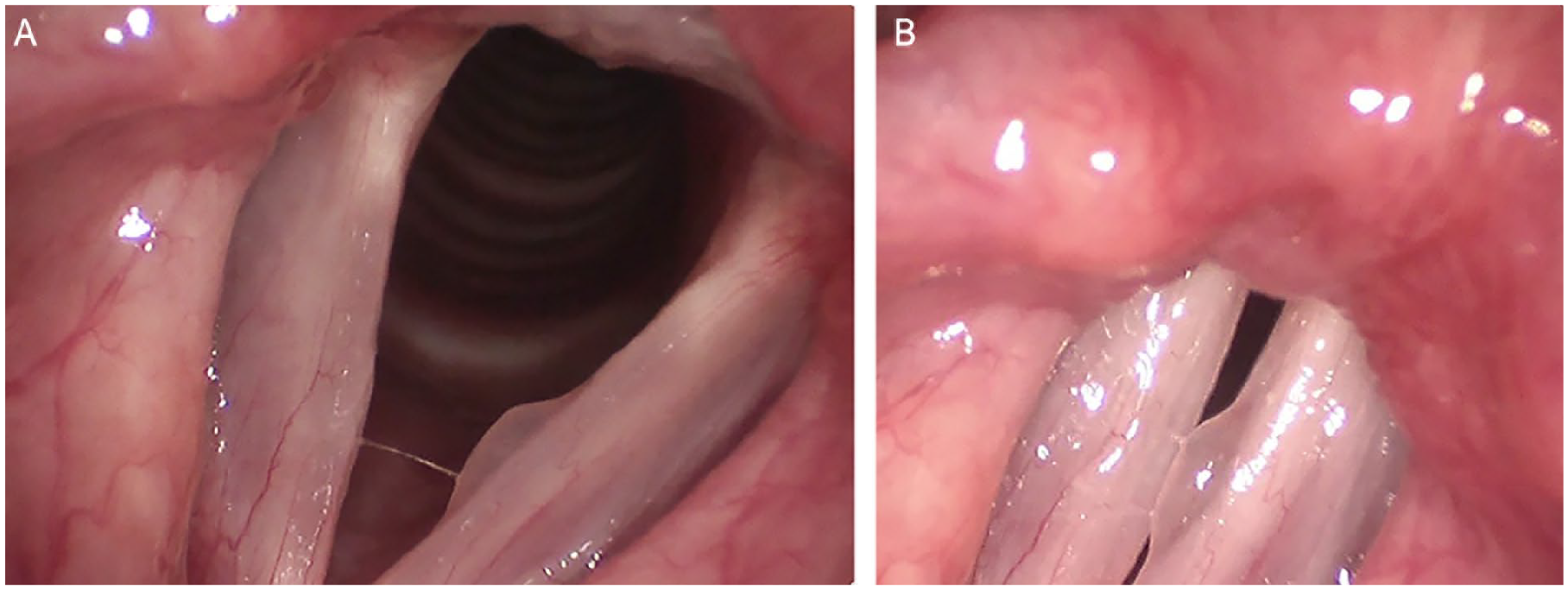
Left vocal fold cyst and right reactive mass in abduction (A) and adduction (B). Fibrotic indentation can be seen at the base of the left cyst, and the right reactive mass shows avascular fibrosis. There is a draining vessel on the right perpendicular to the vibratory margin and associated with the base of the reactive mass.
After a course of voice therapy, she underwent excision of the cyst and corresponding reactive mass using a mini-microflap technique.1,2 The left vocal fold scar and base of the right reactive mass were also injected with dexamethasone. Contact endoscopy revealed that blood flow was away from the right reactive lesion (draining vessel rather than feeding vessel); thus, the vessels were not treated.
The patient recovered well from surgery. Minimal residual stiffness at the site from which the left cyst had been excised could be seen with SVL under high magnification, but the patient’s voice returned to normal. While the slight residual scar (which appeared improved from its preoperative condition) was asymptomatic in alto, it might have been symptomatic in a high soprano. Since our patient had been aware of the scar prior to surgery, and even though the minimal stiffness was more obvious to her after the mass had been removed, she was completely satisfied with the result. That was due partly to her excellent voice outcome but also to the fact that she expected at least some scar in the area that had been identified to her as scarred preoperatively.
Footnotes
Author Contributions
K.M. and R.T.S. contributed to writing the final manuscript. M.J.H. identified necessary case information and strobovideolaryngoscopy photos.
Declaration of Conflicting Interests
The author(s) declared no potential conflicts of interest with respect to the research, authorship, and/or publication of this article.
Funding
The author(s) received no financial support for the research, authorship, and/or publication of this article.
Informed Consent
Written informed consent was obtained from the patient.
