
Abstract
Esophageal innominate artery fistula (EIF) is a rare cause of life-threatening gastrointestinal hemorrhage. This condition occurs most often secondary to an aneurysm, tumor, or esophageal foreign body. After stabilization of airway and circulation, the management options vary from open to endoscopic and endovascular repair. The initial presentation, diagnosis and management of EIF in the literature focuses on adults primarily. In this case report, the clinical presentation of EIF in a pediatric patient, secondary to delayed recognition of a foreign body, highlights management considerations in this population.
Summary
Esophageal innominate artery fistula (EIF) is a rare cause of life-threatening gastrointestinal hemorrhage. This condition occurs most often secondary to an aneurysm, tumor, or esophageal foreign body. 1 Frequently, it presents as a triad including chest pain, sentinel bleed and massive hemorrhage. 2 After stabilization of airway and circulation, the management options vary from open to endoscopic and endovascular repair. The initial presentation, diagnosis and management of EIF in the literature focuses on adults primarily. In this case report, the clinical presentation of EIF in a pediatric patient, secondary to delayed recognition of a foreign body, highlights management considerations in this population.
Background
EIF is a rare but life-threatening cause of gastrointestinal hemorrhage. It is most common with foreign bodies, malignancies, and aneurysms that erode through the esophagus, creating a fistula tract from the esophagus to the innominate artery. 3 In children, esophageal foreign bodies are relatively common; however, progression to an EIF is rare and scarcely described in the literature. It frequently presents as chest pain and life-threatening gastrointestinal hemorrhage, often preceded by a sentinel hemorrhage, but these symptoms can be difficult to pinpoint in the pediatric population. CTA is the first diagnostic test, and management of an EIF is aggressive fluid resuscitation, measures to control bleeding, and endovascular aortic and esophageal repair. 1 In the literature, EIF is scarcely reported and often has a fatal outcome, so early detection and intervention are crucial. 4 In our case report, we add to the literature of innominate esophageal fistula secondary foreign body.
Case Presentation
A 25-month-old male presented to the Emergency Department for episodes of hematemesis. He had a sore throat, neck pain, decreased oral intake, and a cough for 2 weeks. Of note, he had been taken to a sick clinic by his caregiver twice preceding this event and had been diagnosed with strep pharyngitis.
In the Emergency Department, vital signs revealed tachycardia without oxygen desaturation. The physical exam revealed lethargy, dark stools, and mild stridor. Notable values from initial blood tests included WBC 24.9 and hemoglobin 7.9. X-ray of the soft tissues of the head and neck was concerning for tracheal deviation. A preliminary CT of the neck revealed an ill-defined and prominent upper thoracic esophagus with food residue and inflammatory changes in the superior mediastinum, with possible extraluminal air Figure 1. Flexible laryngoscopy was normal, except for blood emerging from the esophagus in the postcricoid region. The patient was then admitted to the medical floors for further evaluation. CT scan demonstrating esophageal foreign body, possible extraluminal air and erosion of innominate artery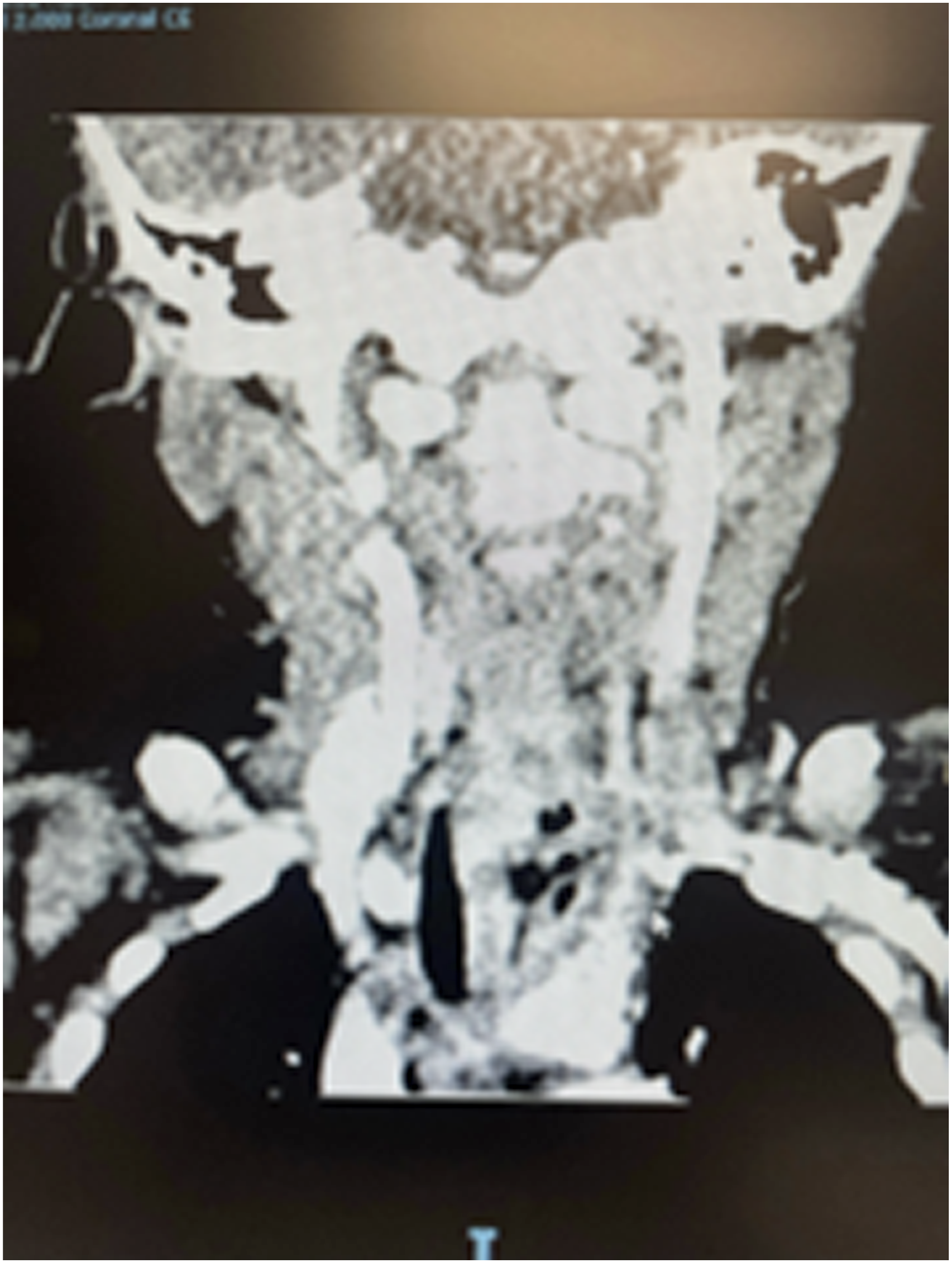
Later that night, a rapid response was called due to low blood pressure and large, melanotic stools. The patient was given a blood transfusion and fluids. The patient was examined a few hours later and he appeared diaphoretic and was unresponsive to name and sternal rub, so another rapid response was called. The patient was further transfused, and a massive transfusion protocol (MTP) was ordered. A decision was made to transfer the patient to the PICU, and the suspected cause of the bleeding was still unknown.
After the transfer to the PICU, copious amounts of blood were coming from the mouth and nose, and the patient was persistently hypotensive. Pressors were started, and he was intubated. The patient was persistently hypotensive even after MTP, so emergent exploration in the OR with pediatric surgery, otolaryngology, and CT surgery was planned. During that time official CT scan read became available, which showed a possible foreign body in the esophagus, with tracheal deviation and abutment against the innominate artery. There was no active extravasation of contrast.
Treatment
The patient had a left neck exploration, median sternotomy with repair of the innominate artery and esophageal fistula, as well as a flexible esophagoscopy with removal of the foreign body with pediatric and cardiothoracic surgery. In the operating room, the patient was hemodynamically unstable and required multiple pressors as well as methylene blue. The patient had one minute of intraoperative cardiac arrest treated with compressions, and ROSC was achieved. The procedure was successful, and the patient was returned to the PICU.
Management of the EIF is broken down into two main steps: 1. Control of the innominate artery bleeding and 2. repair of the esophagus and artery. To repair the artery or aorta, endovascular repair is suggested due to its speed and safety. 5 Another repair method discussed in the literature is primary closure with adjuncts, such as an aortic shunt or a pleural flap. The current literature describes many methods to close an esophageal defect, ranging from primary esophageal closure 6 to interposition of pleura or muscle to cover the defect. Other methodologies for closing the esophagus include patching the defect with bovine pericardium, esophagostomy, esophagectomy, grafts, and endovascular stents, depending on the severity of the case. 4 Commonly, there are two methods of entry: median versus lateral thoracotomy. The literature supports the use of a left posterolateral thoracotomy for descending thoracic aorta repair 7 and a median thoracotomy for repair of the innominate artery and proximal aortic arch.
In the case above, the fistula was found in the posterior aspect of the innominate artery and the anterior esophagus. Therefore, entry into the mediastinum was performed via a median sternotomy. The innominate artery defect was repaired primarily with autologous pericardial pledgets, and the esophageal defect was closed primarily with a pedicled sternocleidomastoid flap.
Outcome and Follow-Up
After surgical intervention, the patient was brought to the PICU in critical condition. There was concern about transfusion-associated circulatory overload (TACO) and transfusion-related acute lung injury (TRALI) in the setting of massive transfusion. In the week following these events, the patient continued to have evidence of multiple organ dysfunction, likely from the persistent hypotension that occurred with the bleeding from the innominate artery fistula. EEG revealed diffuse neurologic dysfunction concerning for hypoxic ischemic brain injury, and MRI/MRA showed multiple acute cerebral infarctions involving the cerebellum and cerebral hemispheres bilaterally. Barium esophagram was negative for a leak, and the patient was started on a modified diet. He was discharged to inpatient rehabilitation due to decreased gross/fine motor skills, functional mobility, and dysphagia.
Discussion
EIF or aortic artery fistula is a rare but life-threatening clinical entity. The initial management includes patient stabilization with aggressive fluid resuscitation, an esophageal tube with a balloon to tamponade any bleeding, after which operative management is required.
3
One study reported 32 cases of aortic-esophageal fistula secondary to a foreign body. They reported that all 13 non-operative cases died, while 3 of 19 surgically treated patients survived.
8
This highlights the importance of early intervention to increase the likelihood of survival. Alreheli examined cases of EIF survival and described the causes, presenting symptoms, and management. In our case, as well as in all 17 cases reported by Alreheli et al, hematemesis was the presenting symptom. The etiology of EIF in these 17 patients ranged from a needle, pin, or button battery to vascular malformations. Our case is unique in that it was a blue plastic toy piece Figure 2. All of these cases, including ours, were managed surgically.
4
Esophageal foreign body (plastic toy) after removal via flexible endoscopy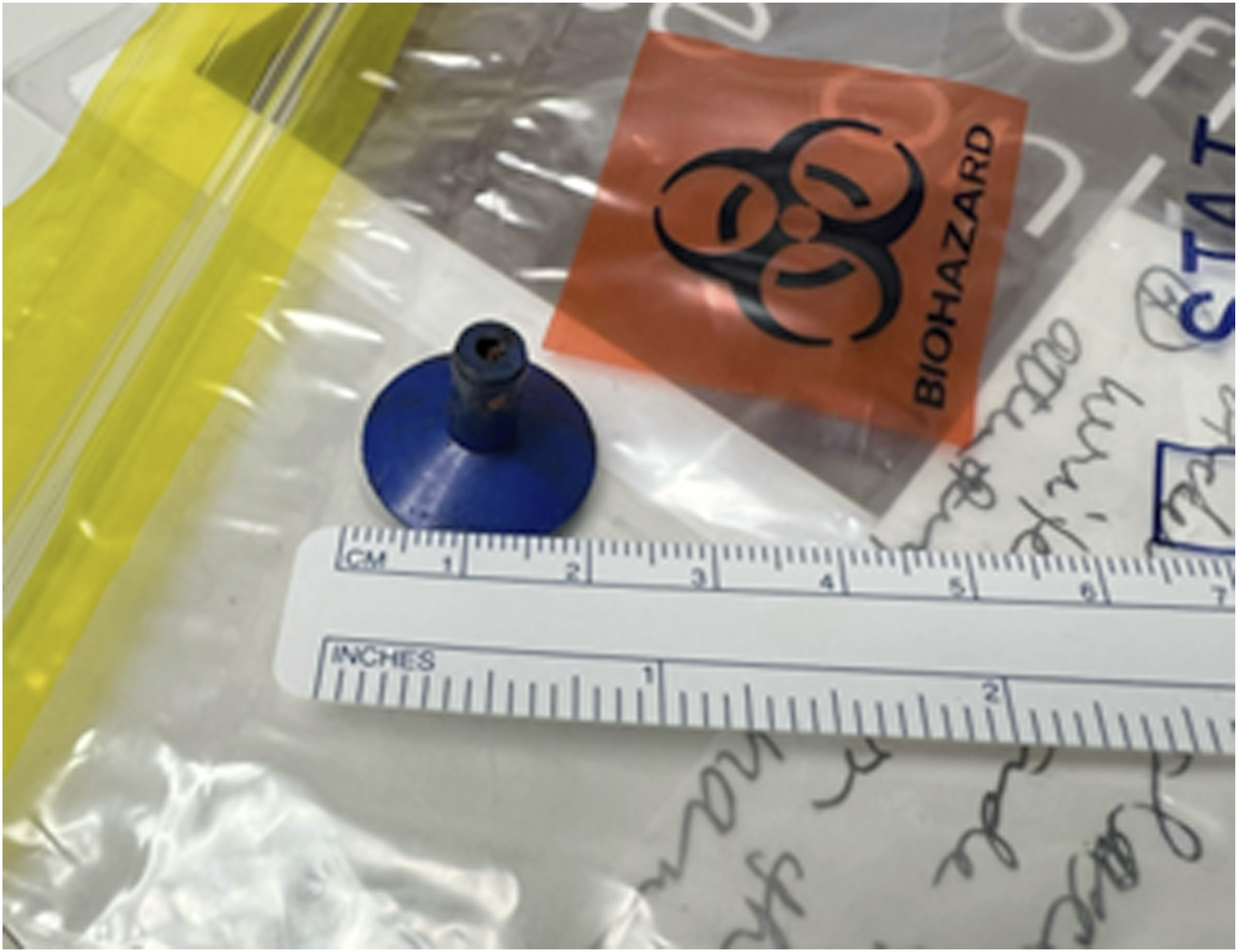
The reason EIF can be so difficult to detect: early symptoms are non-specific, a foreign body is not witnessed, and these patients present with a self-sentinel bleed before true exsanguination. Dysphagia, a child less than 4 years old, and recurrent ED visits, without resolution of symptoms, should be early warning signs for EIF. 4 In our case presentation, the patient initially presented with a sore throat and decreased PO intake, which was attributed to strep pharyngitis at a prior sick visit. There was no witness of foreign body aspiration, and the lack of overt hemorrhage at the time lowered clinical suspicion for an esophageal foreign body. These factors combined likely contributed to the delayed diagnosis and intervention for our patient. If earlier intervention had been taken, it could have decreased the risk of vascular erosion and subsequent hemorrhage and post-op complications.
Given the rarity of this complication and rapid progression, diagnosis may still be delayed even when CT imaging is obtained early. Preliminary read may not show clear vascular erosion, and by the time the final radiographic read is present, the patient may have already clinically deteriorated. In the setting of massive hemorrhage and hemodynamic instability, immediate fluid resuscitation and operative planning take priority, potentially delaying recognition of subtle radiographic findings. In addition, it may be difficult to identify foreign bodies on imaging, especially if they are radiolucent, small, or if there is significant inflammation in the surrounding area. Understanding early warning signs and reasons for the delayed diagnosis, as described above, will help clinicians facilitate early intervention before a life-threatening hemorrhage occurs.
Conclusion
This case report presents a rare case of an innominate esophageal fistula secondary to a retained foreign body. We highlight early clinical signs, pitfalls, and management of this life-threatening condition. Increased awareness can help improve early diagnosis and intervention, likely improving overall survival. Further studies are needed to optimize and standardize diagnostic and operative management.
Footnotes
Ethical Consideration
IRB was not required for this study as it was a case study of individual patient. The materials of this case study were not previously distributed or published.
Funding
The authors received no financial support for the research, authorship, and/or publication of this article.
Declaration of conflicting interests
The authors declared no potential conflicts of interest with respect to the research, authorship, and/or publication of this article.
Data Availability Statement
Data can be accessed upon request to the corresponding author.