
Abstract
Decline in executive functioning (EF) is a hallmark of cognitive aging. We have previously reported that faster vagal recovery from cognitive challenge is associated with better EF. This study examined the association between vagal recovery from cognitive challenge and age-related differences in EF among 817 participants in the Midlife in the U.S. study (aged 35–86). Cardiac vagal control was measured as high-frequency heart rate variability. Vagal recovery moderated the association between age and EF (β = .811, p = .004). Secondary analyses revealed that older participants (aged 65–86) with faster vagal recovery had superior EF compared to their peers who had slower vagal recovery. In contrast, among younger (aged 35–54) and middle-aged (aged 55–64) participants, vagal recovery was not associated with EF. We conclude that faster vagal recovery from cognitive challenge is associated with reduced deficits in EF among older, but not younger individuals.
Keywords
Decline in executive functioning (EF) is a hallmark of cognitive aging (Jurado & Rosselli, 2007; Lachman, Agrigoroaei, Murphy, & Tun, 2010; Royall et al., 2002; Tun & Lachman, 2006, 2010). EF is a higher order cognitive ability that is essential for planning, executing, and monitoring complex goal-directed behaviors in novel situations. The formal definition and components of EF are still disputed (Jurado & Rosselli, 2007; Packwood, Hodgetts, & Tremblay, 2011; Royall et al., 2002), and the concept has been criticized for its lack of clarity and excess of terms (e.g., Jurado & Rosselli, 2007; Miyake et al., 2000). Four cognitive structures, partially controlled by the prefrontal cortex (PFC), are essential for executive function: attention, working memory, preparatory task set, and response monitoring (Barkley, 1997). Studies also emphasized the importance of shifting mental sets, updating/monitoring information, and response inhibition (Miyake et al., 2000) among a wide spectrum of other cognitive abilities (see Jurado & Rosselli, 2007; Packwood et al., 2011; Royall et al., 2002 for the review). Although these three cognitive abilities are distinct, they are also interrelated, so EF may be viewed as both unitary and nonunitary construct (Miyake, Emerson & Friedman, 2000). The exact nature of EF as a unitary (Duncan, Emslie, Williams, Johnson, & Freer, 1996; de Frias, Dixon, & Strauss, 2006) or nonunitary construct (Godefroy, Cabaret, Petit-Chenal, Pruvo, & Rousseaux, 1999) is, however, still disputed. In the analysis of over 60 studies of executive function, Packwood, Hodgetts, and Tremblay (2011) proposed a functional EF definition in such a way that EF could be viewed as a system responsible for executing a task, determining rules, and guiding behavior; this definition is similar to the g factor definition of intelligence.
According to the inhibitory deficit theory of cognitive aging, a deficit in the inhibitory control system may be the main underlying reason for age-related deficits in EF (Hasher, Lustig, & Zacks, 2007; Hasher & Zacks, 1988). Set shifting is negatively influenced by advancing age (Wecker et al., 2005), although the evidence has been contradictory (Salthouse et al., 2000). Although age-related deficits in EF are extensively documented, evidence suggests that age-related decline in EF is not universal. Importantly, studies find greater individual differences in EF among older adults than among their younger counterparts (Ardila, 2007; Jurado & Rosselli, 2007). Therefore, the effort to identify contributors to the heterogeneity in EF among older individuals is an important research focus.
Cardiac vagal control (CVC), a measure of parasympathetic nervous system function, appears to be one factor contributing to the differences in EF in older cohorts. CVC has been viewed as an index of behavioral flexibility and ability to adapt to environmental challenges (Porges, 2007; Thayer & Lane, 2009; Thayer, Åhs, Fredrikson, Sollers Iii, & Wager, 2012). CVC is measured as heart rate variability (HRV; Task Force of the European Society of Cardiology and the North American Society of Pacing and Electrophysiology, 1996). High-frequency (0.15–0.4 Hz) power of HRV is considered to be one of the most accurate measures of CVC (Task Force of the European Society of Cardiology and the North American Society of Pacing and Electrophysiology, 1996). Cross-sectional research has shown a positive association between CVC and EF (Hansen, Johnsen, & Thayer, 2003); while studies that used aerobic exercise training interventions demonstrated an increase in CVC and improved EF in sedentary older individuals (Albinet, Boucard, Bouquet, & Audiffren, 2010). The rationale for linking CVC and EF is provided by the neurovisceral integration model that holds that both CVC and EF are governed by the same network of brain regions controlled by PFC (Thayer & Lane, 2009). Indeed, emerging evidence has linked activation of prefrontal cortical structures to CVC (Gianaros, Van Der Veen, & Jennings, 2004; Shapiro et al., 2000). A number of methodological issues, however, limit the interpretation of these previous findings, including small sample size, restriction in age range within the study cohort (Albinet et al., 2010; Hansen et al., 2003), inclusion of only male participants (Hansen et al., 2003) and absence of dynamic CVC assessments, such as the size and duration of vagal response to stress (Albinet et al., 2010). According to polyvagal theory (Porges, 2007), dynamic changes in CVC in response to psychological stress reflect can be used as an index of attentional control and effort (Tattersall & Hockey, 1995; Croizet, Despres, & Gauzins, 2004). There is preliminary evidence that cardiovascular recovery from psychological stress may be a stronger predictor of cardiovascular morbidity than reactivity (Heponiemi et al., 2007; Stewart, Janicki, & Kamarck, 2006). Moreover, dynamic assessments of CVC may improve the ecological validity of the findings. Since HRV is viewed as an index of adaptability to the constantly changing environment, HRV dynamics may better reflect behavioral flexibility and adjustment than resting HRV. The aim of this study was therefore to investigate CVC as one contributor to the observed heterogeneity in EF among older individuals, using a more representative sample and including dynamic CVC assessments, such as vagal recovery from psychological stress.
Using a sample of 817 (aged 34–86 years) participants from the second wave of the Midlife in the U.S. study (MIDUS II), we have recently reported a significant positive association between vagal recovery from cognitive challenge and EF (evaluated as task switching) that was not seen in either global EF factor or in any of the four other cognitive tests comprising this factor (i.e., speed of processing, working memory, verbal ability and speed, fluid intelligence; Kimhy et al., 2013). The present investigation extends and expands upon this finding by focusing on age differences and examining whether vagal recovery moderates age-related deficits in EF (evaluated as task switching). Because Kimhy et al. (2013) did not find any association between CVC dynamics and EF evaluated as a global factor, we do not address age-related differences in the links between CVC dynamics and EF factor (or any of its other four components), but focus exclusively on task switching instead. Specifically, we hypothesized that while older individuals would overall perform worse on the task-switching test compared to their younger counterparts, those older individuals who demonstrated faster vagal recovery from cognitive challenge would perform better on the task-switching test compared to their peers who demonstrated slower vagal recovery from cognitive challenge. We expected to observe this effect before and after controlling for respiratory rate.
Methods
Participants
The data for the current study are from the second wave of MIDUS II, a 9-year follow-up of the MIDUS I cohort. Our sample is based on those MIDUS respondents for whom we had assessments of EF and who also participated in the psychophysiology protocol. Of note, our report uses the same sample as utilized in Kimhy et al. (2013; N = 817).
Procedures and Measures
Assessments of executive function and psychophysiology protocol
EF was evaluated using the Stop and Go Switch Task (SGST; Tun & Lachman, 2006, 2010). Briefly, the SGST is an executive-function test that taps key abilities of attention switching and inhibitory control. The test includes two single-task blocks and a mixed-task block that requires switching between two sets of response rules. A minimum of 75% accuracy on each of the SGST conditions was required for inclusion in analyses, in order to ensure that the participants were performing the task correctly. Following Kimhy et al. (2013) and the approach used in the previously published MIDUS reports (Agrigoroaei & Lachman, 2011), we used the average reaction time to the switch and nonswitch trials of the mixed-task block as our measure of EF.
The psychophysiology protocol was administered in the morning after a light breakfast with no caffeinated beverages. ECG electrodes were placed on the left and right shoulders, and in the left lower quadrant. Respiration bands were put on chest and abdomen. The participant was seated, and a keypad for responding to the stress tasks was secured in a comfortable position relative to the dominant hand. The stressors used included a Mental Arithmetic task (Turner et al., 1986) and the Stroop color-word conflict task. Both tasks were computer-administered (see Figure 1). Task order was counterbalanced. Responses were entered on a keypad, and participants were instructed to remain silent throughout the procedure. At the start of the experimental period, including recovery, the participants provided verbal stress ratings on a scale of 1–10 (just one number was given to the experimenter) and then they were reminded to remain silent. At the end of the each stress task and immediately prior to the start of the recovery period, the participants were instructed to “please sit quietly and try to relax.” The recovery period consisted of sitting in the same position with no distractions present. The experimenter was present in the room during the entire protocol.
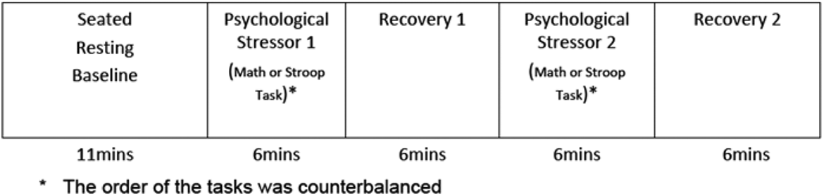
Psychophysiology protocol.
Assessments of EF were performed 1–61 months (average 24.18 ± 14.09 months) prior to the psychophysiology protocol. Table 2 describes age-related differences in this time lag. These differences were significant, F(2, 814) = 3.36, p = .04. Therefore, we controlled for the time lag in all analyses.
The Sample’s Demographic and Clinical Characteristics.

Note. COPD = chronic obstructive pulmonary disease; CVC = cardiac vagal control; GED = general equivalency diploma; TIA = transient ischemic attack.
Age-Related Differences in Time Lag Between the Cognitive Assessments and the Psychophysiology Protocol.

CVC was evaluated using high-frequency (HF) HRV (Berntson et al., 1997). Following previously reported procedures (Crowley et al., 2011; Kimhy et al., 2013; Shcheslavskaya et al., 2010), analog ECG signals were digitized at 500 Hz by a National Instruments A/D board and passed to a microcomputer for collection. The ECG waveform was submitted to an R-wave detection routine implemented by proprietary event detection software, resulting in an RR interval series. Errors in marking R-waves were corrected interactively (Dykes et al., 1986). Spectral power in the high-frequency (0.15–0.50 Hz [HF]) band was computed. Spectra were calculated on 60-s epochs using an interval method for computing Fourier transforms similar to that described by DeBoer, Karamaker, and Strackee (1984). Prior to computing Fourier transforms, the mean of the RR interval series was subtracted from each value in the series and the series was then filtered using a Hanning window (Harris, 1978) and the power, that is, variance (in ms2), over the low frequency and HF bands was summed. Estimates of spectral power were adjusted to account for attenuation produced by this filter (Harris, 1978). Respiratory rate was also calculated based on 1-min epochs. Because HF data were skewed, natural log transformation was performed prior to the statistical analysis.
Assessment of vagal recovery
Following previously reported procedures (Crowley et al., 2011), we averaged ln HF data for both challenges, associated recovery periods, and 5 min to 10 min of the baseline period (Kamarck, 1992). Vagal recovery was computed by subtracting aggregated ln HF during the two challenges from the aggregated ln HF during the two associated recovery periods. Thus, greater number represented larger post-stress increases in ln HF.
Statistical Analysis
Data were analyzed using SPSS PASW (Predictive Analytics Software, version 18) and SAS (Statistical Analysis Software, version 9.2). To facilitate interpretation of our results, we reversed the reaction time to the task-switching test, so greater value represented faster reaction time (i.e., superior EF). Using multiple linear regression, we tested a model that included main effects for age, vagal recovery, and the Age × Vagal recovery interaction (the latter term to capture the hypothesized moderating effect of age) as predictors of EF. To adjust for the effects of vagal reactivity and age-related differences in vagal reactivity (as greater age was significantly negatively associated with vagal response to cognitive challenges), we also entered vagal reactivity (computed by subtracting averaged ln HF during the challenges from the baseline ln HF), and the Age × Vagal reactivity interaction in the model.
All analyses were adjusted for the time (in months) between the EF assessments and the Psychophysiology Protocol, and for the demographic, lifestyle, and medical factors influencing CVC and EF. Following a classification scheme we used previously with MIDUS data (Crowley et al., 2011; Shcheslavskaya et al., 2010), we classified menopausal status as pre-, peri- and postmenopausal, with premenopausal status serving as a reference in the model. To control for 10 diseases and medications that can alter CVC and EF (detailed in Table 1), we created a dummy variable that categorized the participants as either (1) having at least one of these diseases or taking at least one of these medications or (2) disease and medication-free. We created three continuous exercise/physical activity variables (vigorous, moderate, and light; in hours per week) to adjust for the three respective types of exercise/physical activity evaluated separately in MIDUS II. For smoking status, we created three dummy variables; two of them (current smoker and ex-smoker) were entered in the model, while the third (never smoked) served as a reference category. Table 1 provides description of these covariates.
Analyses were conducted in four steps. We entered (1) age, vagal recovery, and Age × Vagal recovery as predictors of EF (adjusting for vagal reactivity, Age × Vagal reactivity, and the time lag between the Cognitive and the Biomarker Projects); and then further adjusted our model, first for the effects of (2) demographic covariates, then sequentially (3) biological covariates (diseases and medications affecting CVC are described in Table 1), and (4) health behaviors.
As HRV is known to be influenced by respiration (Grossman, Wilhelm, & Spoerle, 2004), we followed the standard approach used in the literature (Crowley et al., 2011; Cyranowski, Hofkens, Swartz, Salomon, & Gianaros, 2011; Sloan et al., 2001) and conducted the analyses before and after adjusting for respiratory rate. To adjust for respiration, we conducted within-subject regression analyses with respiratory rate as a predictor of ln HF on a minute-by-minute basis (Crowley et al., 2011; Kimhy et al., 2013; Sloan et al., 2001). We used the resulting unstandardized residual scores as an estimate of the variance in ln HF that cannot be explained by the effect of respiratory rate, and replicated all analyses using this estimate.
Results
Sample and Measures
The demographic, biological, and lifestyle characteristics of the sample are described in Table 1. As illustrated by Figure 2, CVC declined from baseline to the task and then increased during the recovery period; these changes were observed before and after adjusting for respiratory rate (4.93 ± 1.21, 4.59 ± 11.12, 4.99 ± 1.14 Hz; and 5.77 ± 1.79, 4.39 ± 1.46, 6.00 ± 1.74 Hz, respectively). Figure 2 also shows CVC changes for the each age-group.
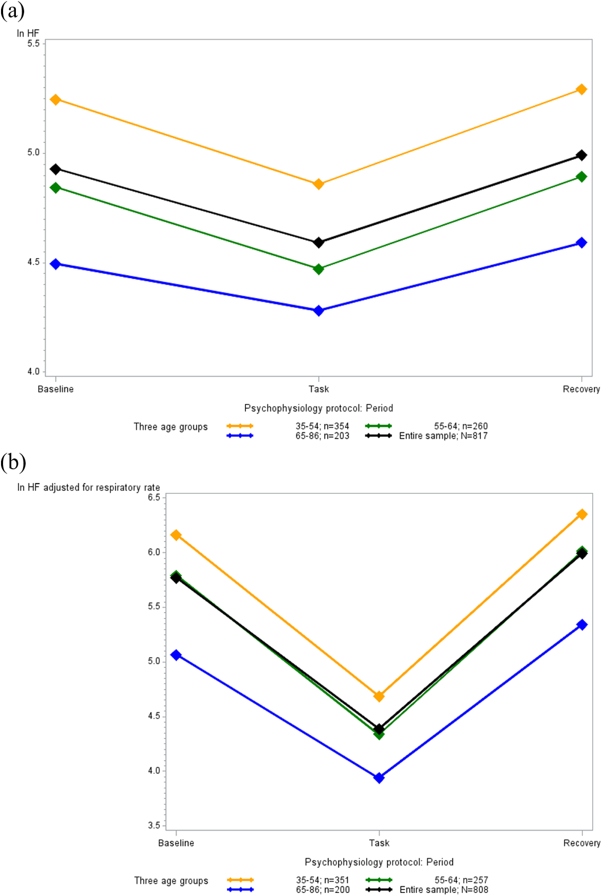
Age-related differences in cardiac vagal control (CVC) during the psychophysiology protocol.
We observed substantial age-related deficits in EF as evidenced by the significant negative correlation between age and average reaction time to the switch and nonswitch trials of the mixed-task block of the SGST test (r = −.274, p = .000). Table 3 describes age-related differences in EF. Age also correlated negatively with vagal reactivity and vagal recovery before (r = −.098, p = .006; r = −.086, p = .014, respectively) and after (r = −.142, p = .000; r = −.121, p = .001, respectively) adjusting for respiratory rate. Table 4 describes the correlations between CVC and baseline, task, and recovery periods before and after adjusting for respiratory rate, respectively.
Age-Related Differences in EF.

Note. EF = executive functioning.
Correlations between ln HF at Baseline, Task, and Recovery Values: Before Adjusting for Respiration and After Adjusting for Respiration.

Note. HF = high frequency.
The Moderating Effect of Vagal Recovery on the Association Between Age and EF
Regression analyses controlling for the time lag between the EF assessments and administration of the psychophysiology protocol, vagal reactivity, and age-related differences in vagal reactivity demonstrated that age, vagal recovery, and the Age × Vagal recovery interaction were significantly associated with EF (see Table 5, Step 1). All effects remained significant in models controlling for demographic, biological, and health behavior covariates (Table 5, Steps 2–4). Therefore, vagal recovery moderated the association between age and EF.
The Impact of Age, Vagal Recovery, and their Interaction on EF: Before Adjusting for Respiratory Rate and After Adjusting for Respiratory Rate.

Note. Unstd. = unstandard.
To understand the nature of this moderating effect, we next sought to examine the relationship of vagal recovery to EF among three age-groups: younger (35–54 years; n = 354), middle-aged (55–64 years; n = 260), and older (65–86 years; n = 203) adults. We next reran the moderation models using age-group as a categorical variable to estimate the slopes and the intercepts for the younger, middle-aged, and older groups, with the three intercepts centered on the grand mean. In this model, the interaction term of Age-group × Vagal recovery was significantly associated with EF (p = .016). The three slopes resulting from this effort visually portrayed the differential strength with which vagal recovery was associated with EF for younger, middle-aged, and older individuals. As illustrated by Figure 3, vagal recovery related significantly to EF among older participants (p = .002), but not among their younger and middle-aged counterparts (p = .740, p = .115, respectively). After adjusting for respiratory rate, the findings remained significant (Table 5). As before, the term of Age-group × Vagal recovery significantly predicted EF (p = .012). Specifically, Figure 3 demonstrates that, greater post-challenge increases in CVC were associated with faster reaction time to the EF task among older participants (p = .0002), but not among their younger and middle-aged counterparts (p = .172, p = .240, respectively).

(a) Age-related differences in the relationship of vagal recovery to task switching: Before adjusting for respiratory rate. (b) After adjusting for respiratory rate.
Discussion
The primary finding of the present report highlights the role of vagal recovery in moderating age-related EF deficits, particularly among older individuals. Although these associations have been documented previously, previous investigations were limited by samples that were small in size (Albinet et al., 2010; Hansen et al., 2003), consisted of only male participants (Hansen et al., 2003), restricted the age within the study cohort (Albinet et al., 2010; Hansen et al., 2003), and relied exclusively on the assessments of the resting CVC levels (Albinet et al., 2010). Our results extend these findings by demonstrating this association in a large, demographically heterogeneous sample and for using dynamic CVC assessments, such as recovery from psychological stress. Although greater age was associated with significantly longer reaction time to the task-switching test, those older adults who had faster vagal recovery from cognitive challenge had faster reaction time compared to their peers who had slower vagal recovery. Adjusting for respiratory rate did not change this finding. Task switching, a task that encompasses attention switching and inhibitory control, the key components of EF, may be the purest measure of EF available in the MIDUS data set (Tun & Lachman, 2010). Indeed, the correlation between the task-switching test and the Trail Makings A and B measures (r = .32, r = .43, respectively) and Digit Symbol Substitution (r = −.47) measures, the established standards for EF assessment, was stronger than the respective correlations between these measures and other tests in the MIDUS II cognitive battery that tap EF, such as speed of processing, working memory, verbal ability and speed, and fluid intelligence (Tun & Lachman, 2010).
As we observed the association between EF and vagal recovery only among older individuals, our results offer limited support to the neurovisceral integration theory. If CVC and EF are associated, because both are governed by the same network of brain regions controlled by PFC, it is not clear why the association between vagal recovery and task switching was restricted to just one age-group. Interestingly, previous studies found significant age-related differences in PFC activation during performance on executive function tasks. For example, Smith et al. (2001) reported that older adults (aged 65–72) and younger adults (aged 18–29) who performed poorly on a task-switching test recruited left PFC during their performance, but younger adults who performed well did not show this prefrontal activation. If the younger MIDUS II participants had little or no left PFC activation during the psychophysiology protocol and EF assessments, while older participants had greater left PFC activation, this may have contributed to the fact that we only saw the association between vagal recovery and EF in older participants. However, PFC activation was not evaluated in the psychophysiology protocol. Thus, we do not have sufficient evidence to conclude whether our results are consistent with the neurovisceral integration theory. Of note, faster vagal recovery may be considered not a mechanism, but rather a marker of better EF among older individuals. Specifically, vagal recovery may reflect another moderator that influences EF among older individuals. In other words, age may serve as a proxy for another moderating variable, particularly given the unique size and representativeness of our sample. Age-related differences in the association between CVC and EF may also be explained by the greater variability in EF among older participants. Indeed, previous studies showed that older individuals have greater heterogeneity in cognitive functioning compared to their younger counterparts (Ardila, 2007).
Our results may be interpreted within the context of the previous evidence linking cardiovascular functioning in older adults to their performance on cognitive tasks. Thus, Pearman and Lachman (2009) found that faster heart rate recovery from a challenge that evaluated working memory was associated with better performance on that challenge among older adults (aged 60–85 years), but not among their younger counterparts (aged 18–23 years). Keary et al. (2012) found that slower heart rate recovery was associated with poor performance on EF tests that assessed speed of processing among older adults (aged 53–83 years). Our study adds to this evidence by reporting the link between vagal recovery and executive function among older individuals.
Our results have implications for future investigations. Future studies should address whether older MIDUS participants with faster vagal recovery and better EF had overall superior level of functioning within their age-group. Of note, a recent analysis of the impact of social, mental, and physical activities on the association between risk factors for cardiovascular disease and cognitive and neuroendocrine functioning reported that older MIDUS participants (aged 60–84 years) who engaged in physical activities (defined as frequency of engaging in leisurely sports, such as light tennis, slow or light swimming, low-impact aerobics, golfing without a power cart, brisk walking, and mowing the lawn with a walking lawnmower during summer and winter time) more frequently had better episodic memory compared to their less physically active peers (Lin, Friedman, Quinn, Chen, & Mapstone, 2012). Thus, physical activity may have protective effect on certain aspects of cognitive functioning. Recent reports indicate such benefits also extend to clinical populations, even those with severe psychopathology (Kimhy et al., 2014, 2015).
The limitations of this study should be considered. An important conceptual limitation of our study is that the PFC control of HR is attenuated in older individuals (Thayer et al., 2009), which questions the relevance of the neurovisceral integration model as a conceptual framework for our study. We do not, however, know about either age-related differences in the PFC control of HF power of HRV, which reflects the vagal contribution to HR (Task Force of the European Society of Cardiology and the North American Society of Pacing and Electrophysiology, 1996), or age-related differences in the PFC control of the CVC dynamics, such as recovery from psychological stress. Another limitation of our study is methodological in nature. Assessments of EF and CVC recovery were separated in time, and while we adjusted for this time lag in all our analyses, it still may have influenced our findings. There is a possibility of the potential confound of longer intervals for those participants who experienced greater EF decline. Indeed, EF might have declined at a different rate among older participants in the MIDUS study. Also, older participants who report better health had higher retention rates in the MIDUS study compared to their peers who reported better health (Radler & Ryff, 2010), thereby limiting the interpretation of our findings. We did not have data on the intensity of fitness training or aerobic fitness among MIDUS participants and relied on self-reported amount of time spent performing tasks that required different physical activity levels. The absence of information about the menstrual cycle, an important determinant of CVC (McKinley et al., 2009), further limits our results.
Conclusion
In summary, we found that faster vagal recovery from cognitive challenge is associated with attenuation of age-related deficits in EF, as reflected in reaction time to a task that tapped switching and inhibitory control. Therefore, vagal recovery may be one contributor to heterogeneity in EF in older individuals, a prominent feature of age-related EF decline.
Footnotes
Acknowledgment
We thank the staff of the Clinical Research Centers at the University of Wisconsin–Madison (UW), UCLA, and Georgetown University for their support in conducting this study.
Declaration of Conflicting Interests
The author(s) declared no potential conflicts of interest with respect to the research, authorship, and/or publication of this article.
Funding
The author(s) disclosed receipt of the following financial support for the research, authorship, and/or publication of this article: This work was supported by a grant from the National Institute on Aging (P01-AG020166; to C.D.R) to conduct a longitudinal follow-up of the MIDUS (Midlife in the U.S.) investigation as well as by a grant from the National Institute of Mental Health (K23-MH077653; to D.K.). The original study was supported by a grant from the John D. and Catherine T. MacArthur Foundation Research Network on Successful Midlife Development (to C.D.R). The study also received support from M01-RR023942 (Georgetown), M01-RR00865 (UCLA) from the General Clinical Research Centers Program and 1UL1RR025011 (UW) from the Clinical and Translational Science Award (CTSA) program of the National Center for Research Resources, National Institutes of Health and from the Nathaniel Wharton Fund.