



Abstract
Porous hydrogel dressings show breathability and possibility to carry and release basic fibroblast growth factor (bFGF) to promote wound healing. However, the difficult replacement may lead to the secondary damage. Thus, there is an urgent need to develop a method platform to control the degradation rate of hydrogel, so as to realize the on-demand replacement. The present study fabricated a porous hydrogel from co-polymized N,N′-bis(acryloyl) cystamine (BAC), allyl polyethylene glycol 500 (APEG500) and acrylic acid (AA) with the presence of polycaprolactone (PCL). BAC contains disulfide bond, which crosslinked the hydrogel. The pore size of the porous hydrogel was 400–600 μm. Higher content of BAC indicated higher crosslinking density, which reduced swelling ratio of hydrogel, while promoted hydrogel storage modulus. At the same time, the presence of PCL reduced swelling ratio of hydrogel, while promoted hydrogel mechanical properties, endowing hydrogel with tough feature. Porous hydrogels that crosslinked by disulfide bonds immersed in glutathione solution were found to degrade spontaneously and quickly due to the response to glutathione. Both crosslinking density and PCL content affected the degradation rate. The porous hydrogel carrying bFGF was applied to wound, promoting angiogenesis, thus accelerating wound healing within 12 d. Due to the spontaneous and rapid degradation of optimized porous hydrogel on wound within 3 days, there was no operation of removing dressing during treatment, avoiding damage during dressing replacement.
Introduction
Traditional dressings, such as gauze, bandage and cotton with the advantages of low cost are the most widely used wound dressings in clinical practice. 1 However, these dressings show no significant positive effect for the cells in the wound site to keep high activity, especially show no bioactivity towards chronic and refractory wound. 2 Basic fibroblast growth factor (bFGF) plays an important role in accelerating wound repair, and has been applied in clinic.3,4 The dosage forms are sprays and freeze-dried powders, which tend to lose their activity and cannot release controllably. 4
Hydrogel dressing is a kind of new wound dressings and has received extensive research focus. Hydrogel dressings create optimal environment for wound healing, effectively promoting epithelial cells migration and angiogenesis, so as to promote wound healing.5,6 Specially, the crosslinked networks provide the possibility for functionalizing the hydrogel dressings, such as carry and transport of bioactive molecules.5,6 Various hydrogels, such as chitosan hydrogel and collagen hydrogel have been reported to carry growth factors for sustained release to promote wound healing.4,7,8 Of note, the degradation of hydrogel is important. Because the dressings are easy to adhere closely to the wound and difficult to replace, leading to the re-destruction of the new granulation tissue and secondary damage.9,10 Thus, there is an urgent need to develop a method platform to control the degradation rate of hydrogel, so as to realize the on-demand replacement.
Disulfide bonds are widely present in proteins and are important mechanisms for ensuring the normal function of proteins.11–13 The most prominent feature of disulfide bonds is that they can exchange with free sulfhydryl groups and can be broken under the action of glutathione.14–16 Of note, disulfide bond used in materials has been reported to be cell responsive.17,18 Disulfide bond can be cleaved by various molecules containing sulfhydryl groups that produced by cells and tissues.17,18 Disulfide bond has been used widly in the field of drug release.19,20 However, the application of disulfide bond to control the degradation rate of biomaterials is rare.
In the present study, N,N′-bis(acryloyl) cystamine (BAC) that contained a disulfide bond was synthesized, and co-polymized with allyl polyethylene glycol 500 (APEG500) and acrylic acid (AA) with the presence of PCL to fabricate a porous hydrogel with the assistance of NaCl particles. The porous structure, swelling behavior, mechanical properties, degradation rate and protein release of the porous hydrogel were evaluated. Then the porous hydrogel carried bFGF was placed on wound to evaluate its effect on wound healing (Figure 1). Schematic diagram of preparation and application of porous hydrogels.
Materials and methods
Synthesis of BAC
Cystine dihydrochloride (5.8 g, 0.025 mol) was dissolved in 30 mL of deionized water, then placed in an ice water bath and cooled to 0°C. Acryloyl chloride (4.7 g, 0.05 mol) was dissolved in 5 mL of dichloromethane. Sodium hydroxide (4.0 g, 0.1 mol) was dissolved in 10 mL of water, which was then dropped slowly and alternately drop by drop into the two solutions. The mixture was stirred at room temperature for 6 h. The suspension after the reaction was filtered by suction, and the white solid obtained was washed with deionized water for three to four times. After the obtained substance was fully dissolved with ethyl acetate at 55°C, it was cooled and recrystallized naturally.
Sample (10 mg) was dissolved in deuterated dimethyl sulfoxide. Tetramethylsilicon (TMS) was employed as the internal standard. Qualitative and quantitative analysis were carried out by nuclear magnetic resonance spectrometer (avance 500 MHz, Bruce, Switzerland). The scanning times were 64.
Fabrication of porous hydrogel
Composition of PEG-SS-AA/PCL porous hydrogels with different component ratios.

Scanning electron microscopy (SU-1500, HITACHI, Japan) was used to investigate the pore structure of porous hydrogel. To investigate swelling behaviors, the dry hydrogels were immersed in deionized water and weighed at different time until they reached swelling equilibrium. The swelling ratio was calculated according to the following formula: Qt = (Wx−W0)/W0, where Wx was the wet weight of the hydrogel, and W0 was the dry weight of the hydrogel. DHR-1 rheometer (TA Instruments, USA) with a 12 mm diameter parallel geometry was used to measure the storage modulus (G′) of hydrogels with the frequency scanning range of 0.1–100 Hz and the strain of 0.01%. The Q800 (TA Instruments-Waters LLC, USA) was used to compress the PEG-SS-AA/PCL porous hydrogel. The samples were placed in the center of the fixture. The maximum compression rate was 90%, and the compression rate was 5% strain/min at room temperature.
In vitro degradation of porous hydrogel triggered by glutathione
PEG-SS-AA/PCL hydrogels (swelling and equilibration in pH = 7.4 PBS buffer) were cut into pieces with equal size and shape, and weighted. These wet porous hydrogels were immersed into glutathione PBS buffer solution (5 mM, pH = 7.4). The hydrogels were weighted at different time points. The weight loss rate of hydrogel was calculated by using the following formula: R = (Wt−W0)/W0, where Wt was the wet weight of the hydrogel at different time points, and W0 was the initial wet weight of the hydrogel.
Cell adhesion test
Fibroblasts, L929, were pre-labeled with fluorescent 3,3′-dioctadecyloxacarbocyanine perchlorate (Dio) dye (Molecular Probes, USA), and seeded into the porous hydrogels. After 12 h, hydrogels carrying cells were observed with confocal laser scanning microscopy (CLSM).
Wound treatment on nude mice
All of the animal care, housing, and study procedures for mice were performed in accordance with the Guide for the Care and Use of Laboratory Animals and approved by Shanghai Tenth People’s Hospital Ethics Committee. 10 nude mice were divided into two groups: experimental group (EXP., treated with porous hydrogel, 30-1) and control group (CON., untreated). Mice were anesthetized by 4 wt% chloral hydrate. Each mouse carried two circular full-layer wounds with diameter of 5 mm on the dorsum. The wounds were covered and fixed by silicone rings. After surgery, porous hydrogel was placed onto the dorsal wounds, followed by being wrapped with film to avoid damage. Wounds were observed on day 3, 6 and 12. A new porous hydrogel dressing was used to cover wound after the degradation of previous dressing. Total four dressings were used during 12 d treatment. The proportion of wound area was obtained via
At 12 d, the skin samples were collected and fixed with 4 wt% paraformaldehyde and embedded in paraffin, followed by being sliced into pieces with 4 μm thickness. The sections were histologically stained with hematoxylin-eosin (H&E) and masson. At the same time, samples at 6 d were collected for immunohistochemical staining of CD31.
Statistical analysis
All bar graph data were expressed as the mean ± standard deviations (SD). Significance was assessed as indicated for each experiment with one-way ANOVA followed by the posthoc Tukey’s test applied for multiple comparisons. p < 0.05 was considered statistically significant.
Results and discussion
Synthesis of BAC
BAC was synthesized from cystine dihydrochloride and acryloyl chloride (Figure 2(a)). The successful synthesis of BAC was confirmed by the corresponding assignment of characteristic peaks in 1H-NMR spectra (Figure 2(b)). Due to the electron pushing ability of disulfide bond, the chemical shift was low (2.82 ppm). The peak at 3.42 ppm belonged to site b. Due to the strong electron absorption ability of carbonyl and N atoms, the chemical shift at site c was high (8.30 ppm). The peaks at 5.61 ppm and 6.11 ppm belonged to d and e on the double bond in BAC molecule, respectively. The integral area ratio of the five peaks (a: b: c: d: e) was 2:2:1:1:2. Synthesis of BAC and fabrication of DMF gel. (a) The synthetic equation of BAC. (b) 1H-NMR spectra. (c) Co-polymerization to form DMF gel.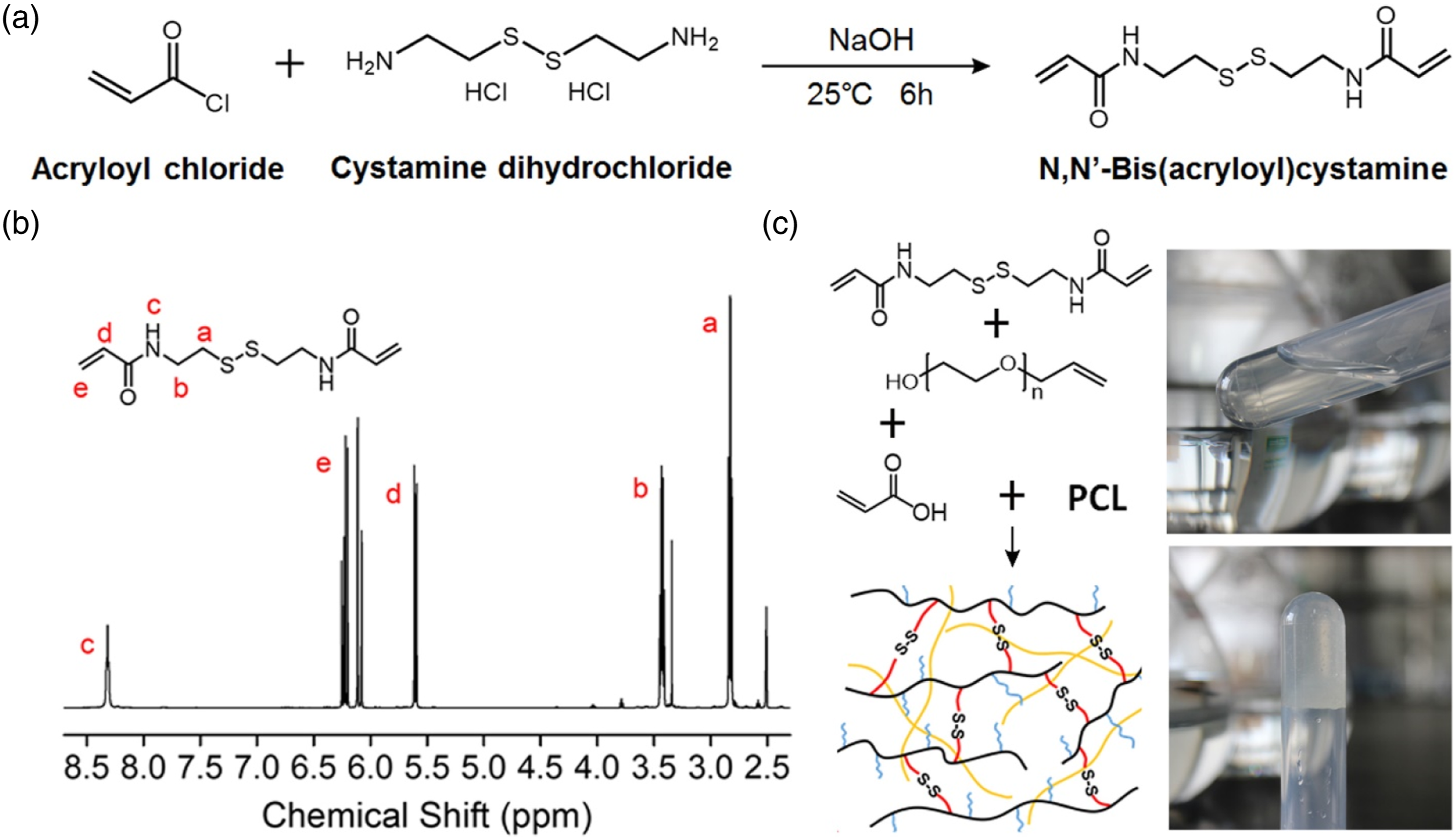
Fabrication of porous hydrogel
PEG-SS-AA/PCL porous hydrogels were prepared by UV initiated radical polymerization. As shown in Figure 2(c), a solid DMF gel was formed after 15 min UV irradiation. After immersing in H2O to replace DMF and remove NaCl particles, a porous hydrogel was achieved (Figure 3(a)). The dry porous hydrogel could adsorb plenty of water to form a water containing hydrogel that still possessed porous structure (Figure 3(b) and (c)). This feature was important, which indicated that porous hydrogel could absorb wound exudate while maintain breathability at the same time. General and micro morphology of porous hydrogel. (a) General observation of dry porous hydrogel. (b) Porous hydrogel adsorbing water. (c) Porous structure of wet porous hydrogel observed by stereomicroscope (Bar scale: 400 μm). (d, e, f) SEM images of PEG-SS-AA porous hydrogel without PCL. (g, h, i) SEM images of PEG-SS-AA porous hydrogel with PCL.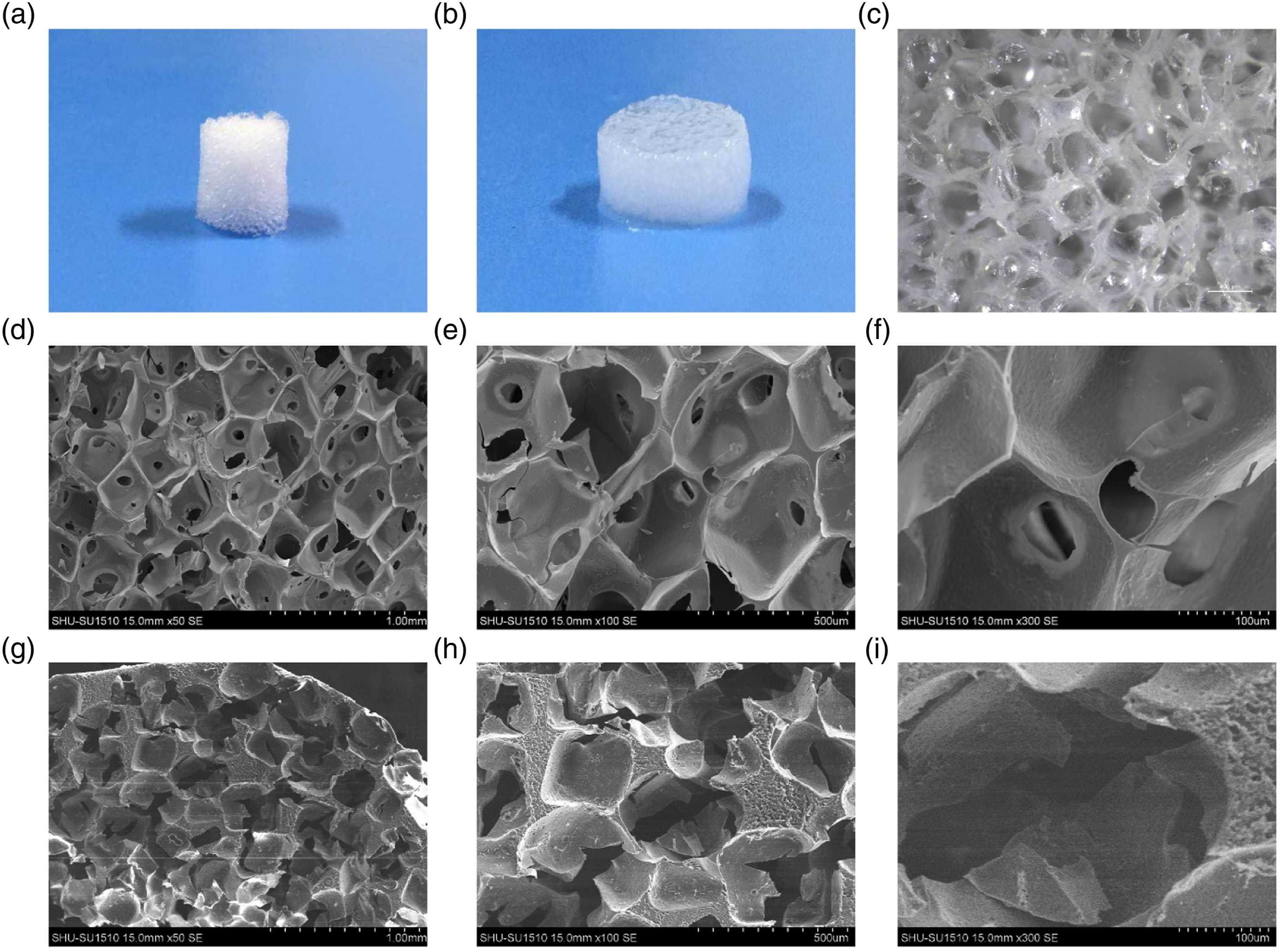
At the same time, the present study fabricated a PEG-SS-AA porous hydrogel without PCL. According to SEM images of porous hydrogels, it was found that the dry hydrogels possessed porous structure. However, the inner surface of PEG-SS-AA hydrogel without PCL was smooth (Figure 3(d), (e), (f)), while the inner surface of PEG-SS-AA hydrogel with PCL was rough (Figure 3(g), (h), (i)).
Properties of hydrogels
The present study fabricated six hydrogels with different BAC (disulfide bond) content and PCL content. As shown in Figure 4(a), different PEG-SS-AA/PCL porous hydrogels showed different swelling degrees. For one thing, the crosslinking density greatly affected the swelling property of porous hydrogel. More BAC content meant higher crosslinking density. Increasing crosslinking density increased the density of crosslinking network, leading to smaller swelling degree. Thus, hydrogel with higher BAC possessed lower swelling ratio (lower than 40%), while hydrogel with lower BAC possessed higher swelling ratio (higher than 110%). For another, with the increase of PCL content, the swelling degree of hydrogel decreased. For example, the swelling degree of hydrogel containing 0% PCL (10-0) was 143%, while the swelling degree of hydrogel containing 1% PCL (10-1) was 126%. Therefore, the introduction of PCL reduced the swelling degree of PEG-SS-AA hydrogels. Properties of hydrogel. (a). Swelling ratio of porous hydrogels. (b). G′ of hydrogels. (c) Compressive stress-strain curves of hydrogels. (d) General observation of mechanical performance of hydrogel with/without PCL. *p < 0.05.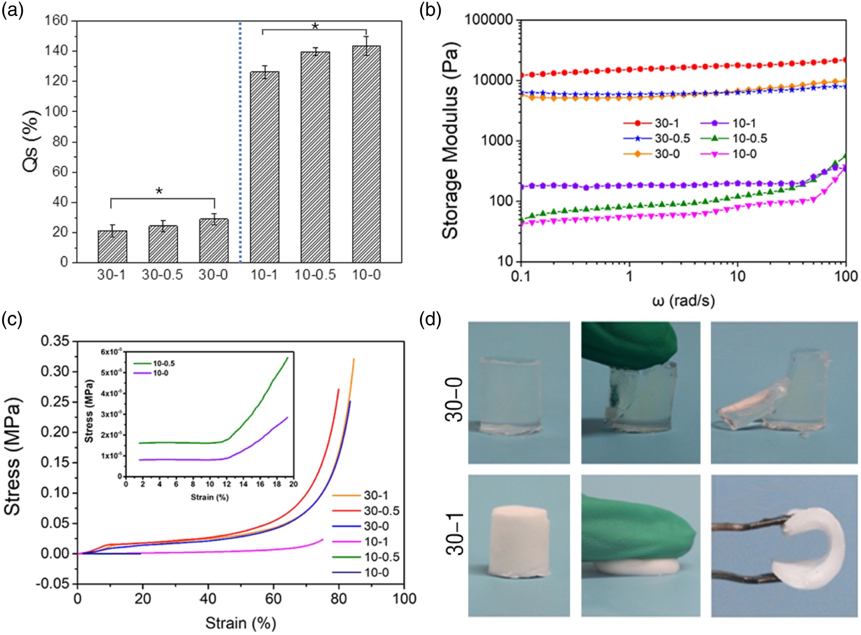
Increasing crosslinking density also increased the storage modulus (G′) of hydrogel. As shown in Figure 4(b), hydrogels (30-0, 30-0.5, 30-1) with higher crosslinking density showed G′ values near 1 × 10⁴ Pa. While hydrogels (10-0, 10-0.5, 10-1) with lower crosslinking density showed G′ values near 1 × 101 Pa, which was significantly lower than that of hydrogels with higher crosslinking density. In addition, the presence of PCL also improved the G′ of hydrogels significantly. For example, G′ of hydrogel 30-1 with 1% PCL was 12,179.1 Pa, significantly higher than that of hydrogel 30-0 without PCL (5791.52 Pa). This might be related to that the hydrophobic PCL provided rigidity to the hydrogel.
At the same time, the compression test showed similar results with rheological test. The increase of crosslinking density made the hydrogel network more stable, so that the hydrogel could bear higher load under the same strain. PCL in hydrogel networks also promoted compressive stress (Figure 4(c)). Of note, the introduction of PCL endowed the PEG-SS-AA hydrogel with toughness. As shown in Figure 4(d), PEG-SS-AA hydrogel without PCL was brittle, and was easy to be broken. However, PEG-SS-AA hydrogel with PCL was tough, showing larger deformation. Accordingly, hydrophobic interaction is a method to improve the mechanical properties of hydrogels. PCL is a hydrophobic polymer. The introduction of PCL into hydrogel restrained excessive swelling of network, increased the degree of freedom of molecular chain motion, thus promoting toughness of hydrogel.
Degradation of hydrogels
Glutathione was used for simulating in vivo degradation experiment. As shown in Figure 5(a) and (b), porous hydrogels with lower crosslinking density (10-0, 10-0.5, 10-1) in glutathione solution showed significant degradation within 12 h. While the degradation of porous hydrogels with higher crosslinking density (30-0, 30-0.5, 30-1) in glutathione solution was slower. Significant swelling of porous hydrogels with higher crosslinking density was observed at 16 h, which indicated significant destroy of disulfide bonds. The significant degradation of porous hydrogels with higher crosslinking density was delayed to over 24 h. Moreover, presence of PCL seemed to delay hydrogel degradation. At the same time, the present study fabricated a hydrogel from PEGDA without the presence of disulfide bonds. This porous hydrogel immersed in glutathione solution showed no significant degradation during in vitro test (Figure 5(c)). Quantitative detection further confirmed the general observation (Figure 5(d)). Significant weigh loss of hydrogel with lower crosslinking density occurred earlier than that of hydrogel with higher crosslinking density. Because there was no disulfide bonds in hydrogel prepared form PEGDA, the PEG hydrogel showed no weight loss during in vitro evaluation. It has been reported that organisms contain a large number of sulfhydryl groups.21,22 The concentration of glutathione in blood is about 2–20 μM. The concentration in the cell membrane is about 1–10 mM. Thus, in vitro quick degradation of PEG-SS-AA hydrogels revealed the similar degradation behavior in vivo. In vitro degradation of porous hydrogels in glutathione solution. (a) General observation of porous hydrogels with lower crosslinking density immersed in glutathione solution. (b) General observation of porous hydrogels with higher crosslinking density immersed in glutathione solution. (c) General observation of porous PEG hydrogel immersed in glutathione solution. (d) Quantitative detection of porous hydrogels degradation in vitro (R = weight remaining).
Cell adhesion in porous hydrogels
According to Figure 6, PEG-SS-AA hydrogel with the presence of disulfide bonds (30-0 and 10-0 hydrogels without PCL) could support fibroblast adhesion. In addition, hydrogels with higher content of disulfide bonds showed more adhered cells. This result was interesting because PEG is a commonly used non-fouling polymer, which cannot support cell adhesion.23,24 While the present PEG-SS-AA hydrogel could support cell adhesion, which might be related to the presence of disulfide bonds. According to Hong et al.,
16
the presence of disulfide bonds in hydrogels could significantly promote cell-hydrogel interaction, thus promote cell adhesion. At the same time, it was found that PCL in hydrogel could promote cell adhesion significantly. Hydrogels with higher content of PCL exhibited higher number of adhered cell. This might be related to the binding of adhesion protein molecules through hydrophobic interaction of PCL. Hydrogel with hydrophilic network usually repels protein adsorption, thus prevents cell adhesion. However, PCL in hydrogel might bind the hydrophobic domain of protein molecules through hydrophobic interaction, thus promoting cell adhesion. Fibroblast adhesion in different hydrogels. (a) Cells adhered in PEG-SS-AA porous hydrogels with higher crosslinking density. (b) Cells adhered in PEG-SS-AA porous hydrogels with lower crosslinking density. Cells were pre-labeled with Dio, and observed by CLSM. (Bar scale: 400 μm).
Porous hydrogel carrying bFGF as dressing for promoting wound healing
PEG-SS-AA/PCL porous hydrogel (30-1) was used to carry bFGF to evaluate its effect on wound healing. A new porous hydrogel dressing was used to cover wound after the degradation of the previous dressing. Thus, total four dressings were used during 12 d treatment (Figure 7(a)). Wound with no treatment was employed as control group (CON.). Wound treated with dressing was used as experimental group (EXP.). As shown in Figure 7(b) and (c), the healing rate of wound that treated with dressing (EXP.) was significantly higher than the control group at 6 and 12 d, showing higher rate of wound closure. At the same time, the wound color in control group and experimental group was significantly different. The wound treated with dressing showed lighter red color, with more whitish appearance, revealing more efficient epidermal migration. Wound healing. (a) Treatment schedule. (b) General observation images of wounds with/without porous hydrogel dressings from 0 to 12 days. (c) Wound residual area rate from 0 to 12 d. *p < 0.05. (d) (h and e), masson staining of skin samples at 12 days (Bar scale: 100 μm). (e) Immunohistochemical staining of CD31 at 6 days (Bar scale: 30 μm).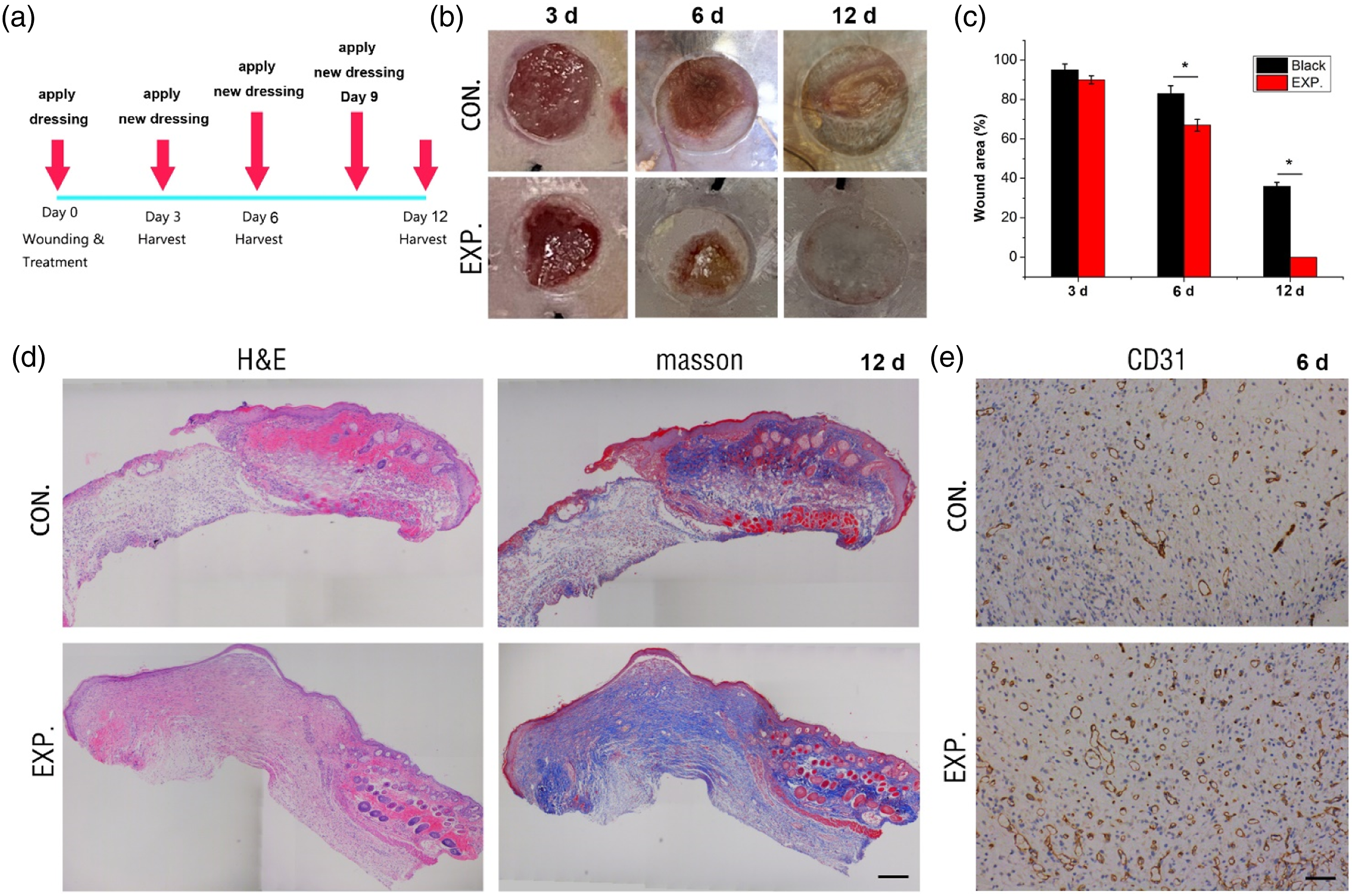
Further histological evaluation was carried out to show the structure and matrix deposition of the repaired wound at 12 d. As shown in Figure 7(d), compared with the control group, the experimental group showed denser matrix deposition and higher tissue thickness at 12 d. The epidermis of experimental group was continuous, showing dense epidermis. Masson trichrome staining showed that the collagen fibers in experimental group were thicker and denser, possessing more mature phenotype and more regular orientation, indicating that the porous hydrogel dressing exhibited positive effect on wound healing. Moreover, CD31 immunohistochemical staining of skin samples at 6 d was performed to characterize vascular endothelial cells. 25 As shown in Figure 7(e), at 6 d, the positive expression of CD31 in the wound site of experimental group was significantly higher than that of control group, illustrating the angiogenesis effect of bFGF. bFGF can significantly promote angiogenesis, which provides oxygen and nutrients to the wound site, and is critical to wound healing. The present hydrogel carried bFGF thus showed significantly positive effect on wound healing. Together with its autonomous, controllable and rapid degradation feature, the PEG-SS-AA/PCL porous hydrogel might show great potential in clinical practice to avoiding damage from dressing replacement while promoting wound healing.
Conclusion
In summary, the present study fabricated a porous PEG-based hydrogel that crosslinked by disulfide bonds with controllable degradation rate. The PEG-SS-AA/PCL porous hydrogel that crosslinked with disulfide bonds showed significant response to glutathione. The disulfide bonds content significant affected the degradation rate. The presence of PCL improved the mechanical performance and cell adhesion of the hydrogel. PEG-SS-AA/PCL porous hydrogel carrying bFGF was applied to wound to promote angiogenesis and wound healing. Due to the spontaneous and rapid degradation of PEG-SS-AA/PCL porous hydrogel on wound within 3 days, there was no dressing replacement operation during treatment, avoiding damage during dressing replacement.
Footnotes
Author Contributions
Conceptualization, writing—review and editing, supervision, S.Z. and H.C.; methodology, investigation, writing—original draft preparation, Q.W. and H.G. All authors have read and agreed to the published version of the manuscript.
Declaration of conflicting interests
The author(s) declared no potential conflicts of interest with respect to the research, authorship, and/or publication of this article.
Funding
The author(s) disclosed receipt of the following financial support for the research, authorship, and/or publication of this article: This research received funding from Shanghai Municipal Health Commission (No. 202140294), and Natural Science Foundation of Shanghai (No. 22ZR1454900).
Data availability statement
The data used to support the findings of this study are available from the corresponding author upon reasonable request.
