



Abstract
Objectives
Slough in chronic venous leg ulcers may be associated with delayed healing. The purpose of this study was to assess larval debridement in chronic venous leg ulcers and to assess subsequent effect on healing.
Methods
All patients with chronic leg ulcers presenting to the leg ulcer service were evaluated for the study. Exclusion criteria were: ankle brachial pressure indices <0.85 or >1.25, no venous reflux on duplex and <20% of ulcer surface covered with slough. Participants were randomly allocated to either 4-layer compression bandaging alone or 4-layer compression bandaging + larvae. Surface areas of ulcer and slough were assessed on day 4; 4-layer compression bandaging was then continued and ulcer size was measured every 2 weeks for up to 12 weeks.
Results
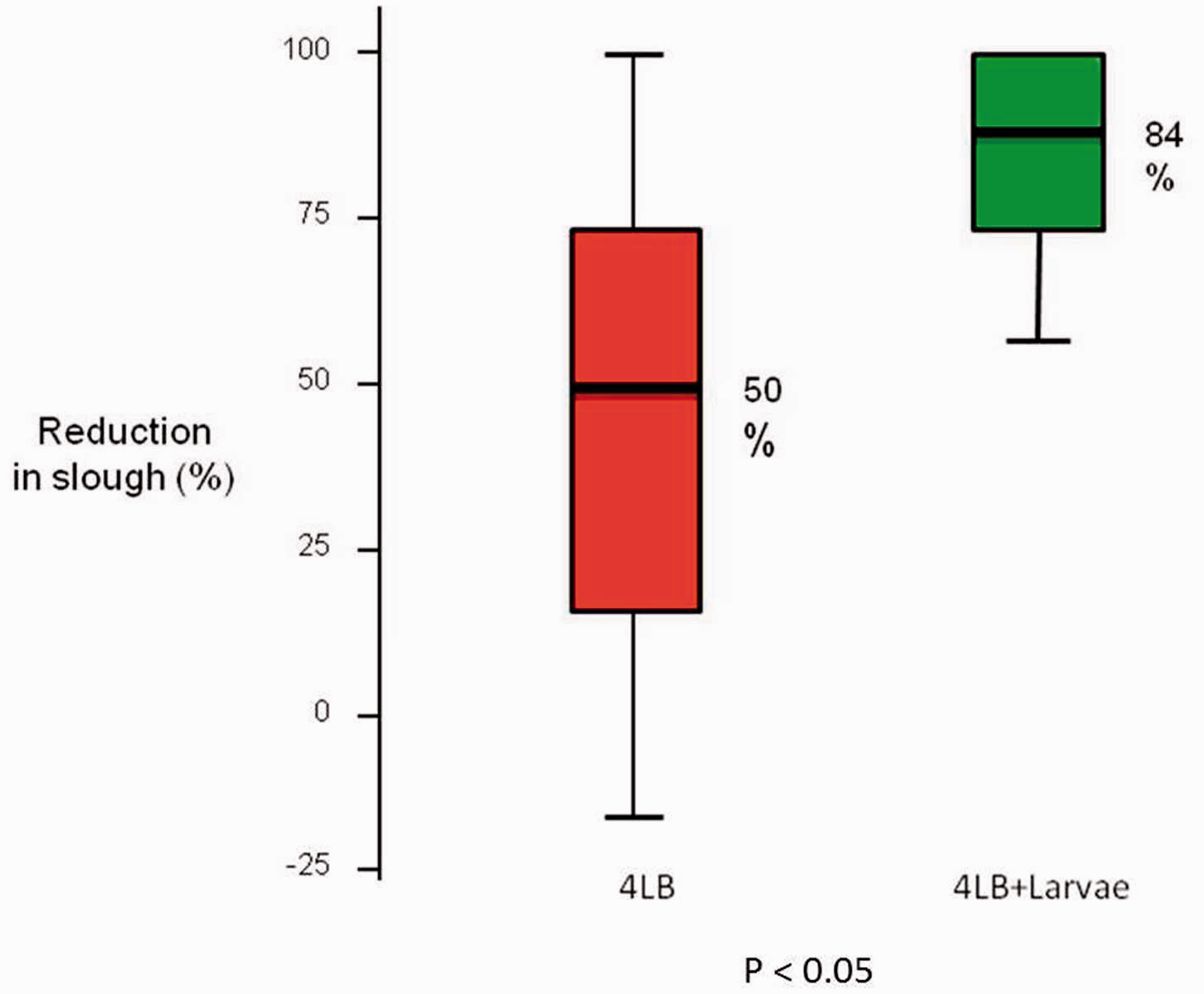
A total of 601 patients with chronic leg ulcers were screened between November 2008 and July 2012. Of these, 20 were randomised to 4-layer compression bandaging and 20 to 4-layer compression bandaging + larvae. Median (range) ulcer size was 10.8 (3–21.3) cm2 and 8.1 (4.3–13.5) cm2 in the 4-layer compression bandaging and 4-layer compression bandaging + larvae groups, respectively (Mann–Whitney U test, P = 0.184). On day 4, median reduction in slough area was 3.7 cm2 in the 4-layer compression bandaging group (P < 0.05) and 4.2 cm2 (P < 0.001) in the 4-layer compression bandaging + larvae group. Median percentage area reduction of slough was 50% in the 4-layer compression bandaging group and 84% in the 4-layer compression bandaging + larvae group (Mann–Whitney U test, P < 0.05). The 12-week healing rate was 73% and 68% in the 4-layer compression bandaging and 4-layer compression bandaging + larvae groups, respectively (Kaplan–Meier analysis, P = 0.664).
Conclusions
Larval debridement therapy improves wound debridement in chronic venous leg ulcers treated with multilayer compression bandages. However, no subsequent improvement in ulcer healing was demonstrated.
Introduction
Chronic ulceration of the leg is a common condition and has a major impact on quality of life for sufferers. 1 Furthermore, as it is an expensive drain on resources, it is justifiable to study any treatment strategy that may improve outcomes.
The use of graduated multilayer compression bandages with simple non-adherent dressings has been recognised as the gold standard in the management of chronic venous ulcers.2–4 However, many of these chronic wounds will often present with slough, or devitalised tissue, adhered to the wound bed. As devitalised tissue may provide a suitable medium for bacterial growth and potentially impede healing; prompt debridement of slough may expedite the healing process.5–7
Despite the availability of a vast array of debridement techniques and materials, little evidence exists comparing debridement with no debridement in the management of chronic venous leg ulcers. Of the techniques available, larval debridement therapy (LDT) or maggot therapy is considered an aggressive and rapid method of removing slough, with some laboratory studies demonstrating effectiveness in eradicating bacteria.8,9 In recent years, although there has been a growing interest in the use of LDT, only two randomised controlled trials have been published evaluating its efficacy with sample sizes of 12 and 267, respectively.10,11 Both the trials had two drawbacks: first, the studies compared LDT with a hydrogel dressing and second, in each study, compression was interrupted during the debridement phase as larval viability underneath compression bandages was questioned.
Aims
The aim of this study was to assess the efficacy of LDT under multilayer compression bandages compared with multilayer compression alone, evaluating early debridement and subsequent time to healing.
Methods
A two-arm parallel-group randomised controlled trial was designed with recruitment of participants sourced from an established specialist community-based leg ulcer service 12 from November 2008 to July 2012. All participants had venous leg ulcers confirmed by colour-venous duplex ultrasound imaging (Phillips IU22, Phillips, Guilford, UK) and ankle brachial pressure indices (ABPI) between 0.85 and 1.25, calculated using handheld Doppler ultrasound (Huntleigh Diagnostics, Cardiff, UK).
Patients were eligible if their ulcer had a wound surface area between 4 and 100 cm2, as it was felt that this would cover the majority of ulcers presenting to our unit and if covered by 20% or more of slough (in our unit, we would not normally consider LDT for ulcers with less than 20% slough). All patients presented with venous leg ulcers that were either new episodes or had recurred but were not previously treated with compression. In the case of multiple ulceration or bilateral leg ulceration, the largest ulcer was included in the trial. Ulcers with exposed blood vessels, tendon, muscle or bone or patients with a history of bleeding disorders were excluded from the trial.
Ethical approval was obtained from North Somerset & South Bristol Research Ethics Committee. Following written consent, participants were randomly allocated to either the control group, where they received the standard treatment of N-A dressing™ (Johnson & Johnson, New Jersey, USA) and multilayer graduated compression bandages (Profore™, Smith & Nephew, London, UK), or the larvae group, where they received the standard treatment plus a Larve BioFOAM™ dressing (Biomonde®, Bridgend, Wales) – a sterile sealed net pouch containing small pieces of hydrophilic polyurethane foam and sterile larvae of the common greenbottle fly, Lucillia sericata.
Randomisation was achieved using computer-generated random numbers prepared and sealed in envelopes by a non-clinical member of staff not allied to the research team.
Interventions
All patients attending the leg ulcer clinics were screened for suitability for the trial. Eligibility was assessed by one of the leg ulcer nurse specialists and written information was given to potential participants following verbal discussions. Patients who showed interest in the study were invited to attend for further assessment and to obtain written informed consent. Patients were randomly allocated to 4-layer compression bandaging group (4LB) or 4LB + larvae group by sequentially numbered sealed randomisation envelopes.
All participants were commenced in multilayer graduated compression bandages on day 0 and those in the 4LB + larvae group also received an application of a sterile Larve BioFOAM™ dressing at this time (Figure 1). Those in the 4LB group received an N-A dressing™.
BioFOAM larvae dressing ready for removal.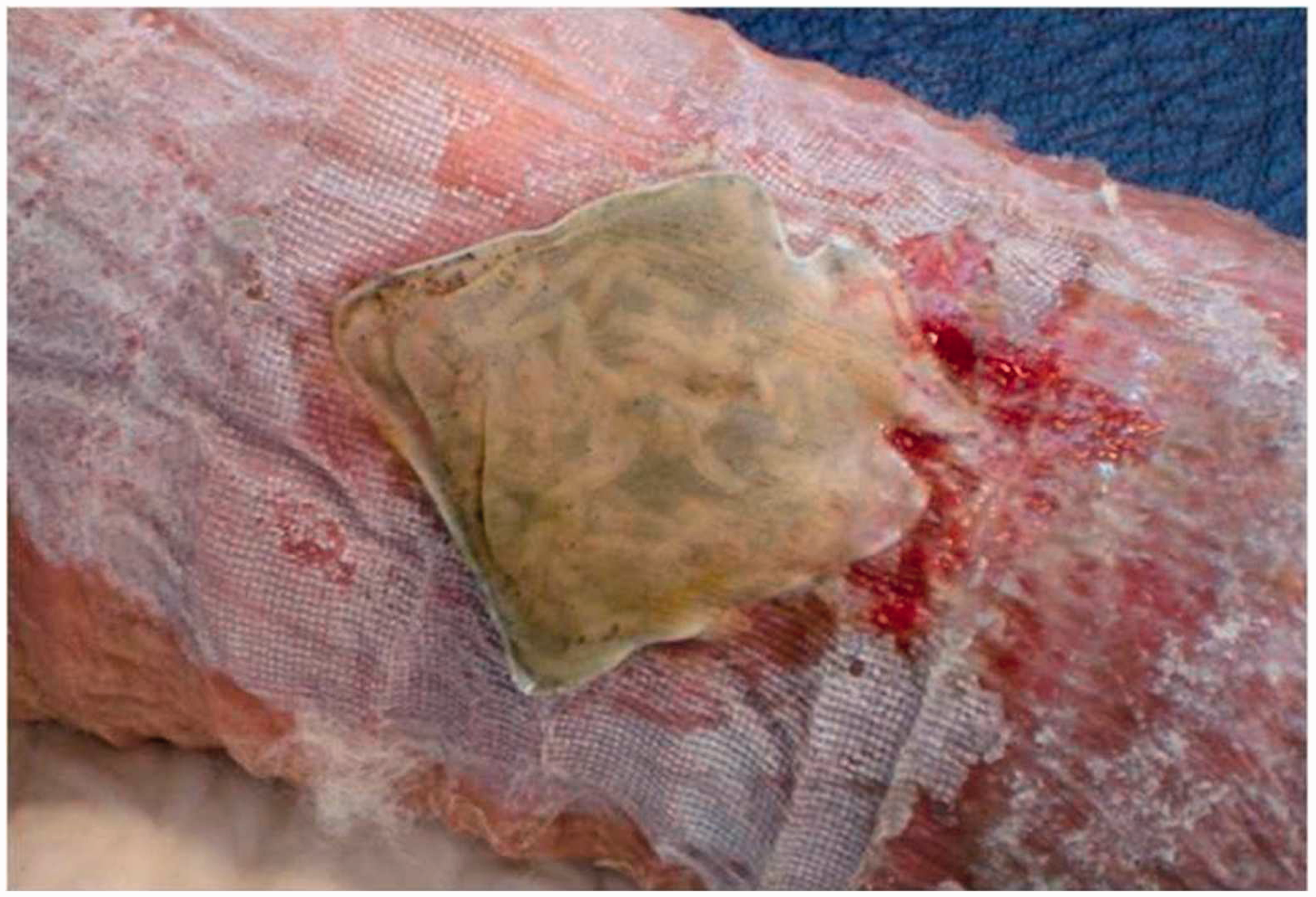
Measurements of wound surface area and area of slough were made using a portable digital planimetry system (Visitrak™, Smith & Nephew®, London, UK). All patients returned to the clinic on day 4 and dressings were removed for evaluation. Repeat planimetry measurements were made calculating ulcer area, slough area and change in slough area as a percentage; all data were recorded onto a prospective database.
All participants subsequently continued with multilayer compression bandages and were followed up every 2 weeks in the leg ulcer clinic. Shared care with a community nurse was arranged for dressing changes on alternate weeks. The total length of follow-up was 12 weeks or until the ulcer had healed, if sooner.
Outcome measurements
The primary outcome for this trial was percentage area debridement in slough at day 4. This calculation was achieved using the digital planimetry measurements made at day 0 and day 4. These measurements were made by one of the leg ulcer nurse specialists and debridement was defined as the reduction in surface area of slough. The secondary outcome was time to complete healing of the ulcer being studied. Ulcer healing was defined as complete epithelial cover in the absence of scab. Again, the leg ulcer nurse specialist assessed this and digital photographs were taken at each clinic visit to assist visual tracking of progress.
Any adverse events were logged as they occurred. Events were classified as serious (e.g. life threatening, significant disability or hospitalisation) or non-serious (pain, wound infection, wound deterioration) and a leg ulcer nurse specialist assessed the severity.
Sample size
Unpublished data from our unit involving patients selected for LDT showed that the mean percentage removal of slough after 3 days treatment was 60%. Incorporating this into the power analysis enabled the number of subjects required for each study arm to be calculated. From this it was determined that for a 90% power, 20 participants were needed in each group to detect a difference in percentage area debridement on day 4.
Statistical analysis
All analyses were performed using SPSS version 18.2 (IBM SPSS, New York, USA). Non-parametric analysis with Mann–Whitney U test, using a 5% significance level, was used to compare the two study groups with respect to percentage reduction in slough and wound surface area. To evaluate 12 week healing, Kaplan–Meier survival analysis was used with a log rank test to compare the survival distributions of the two samples.
Results
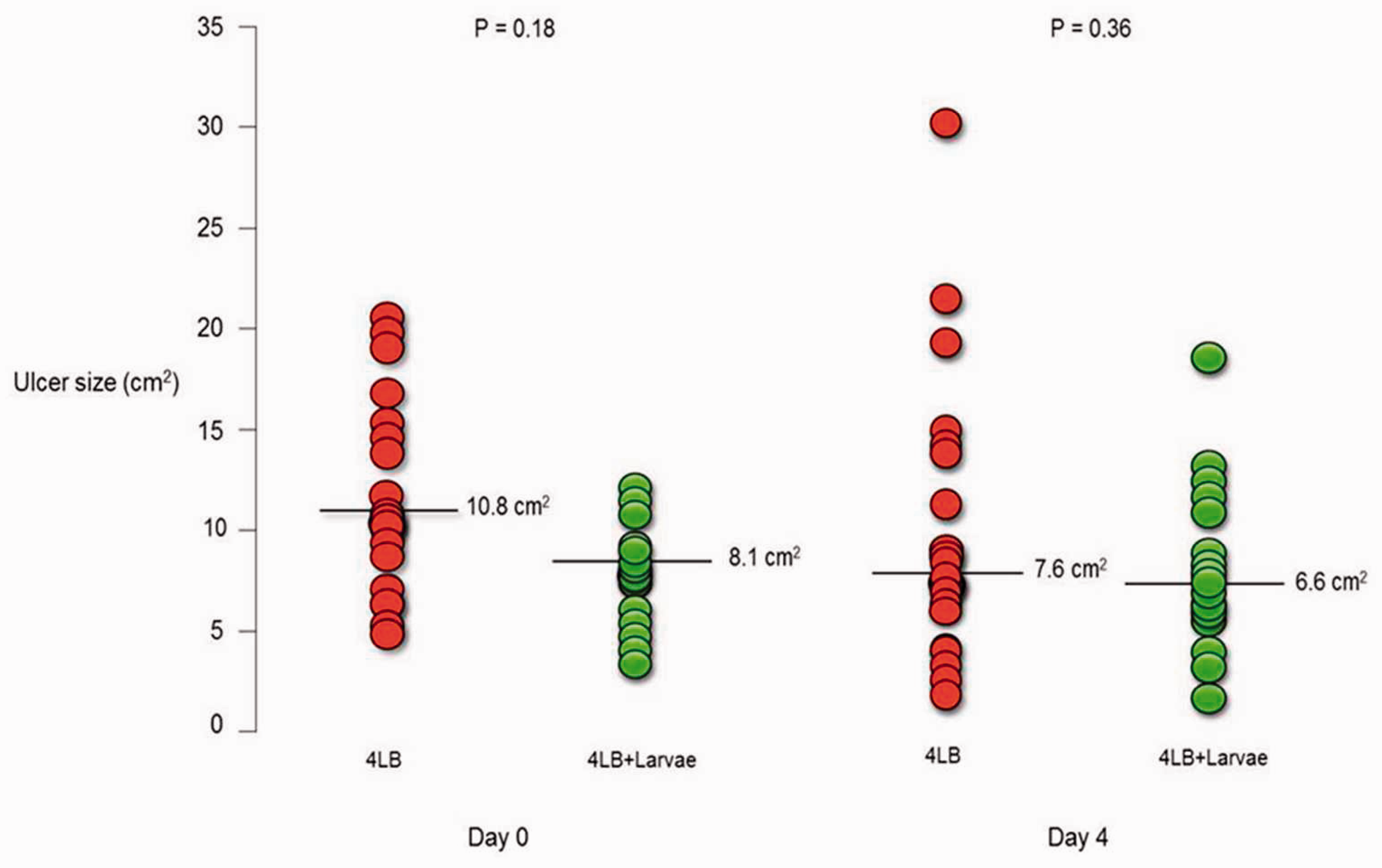
A total of 601 patients with chronic leg ulcers were screened between November 2008 and July 2012, with 40 randomised to the study: 20 to the 4LB group and 20 to the 4LB + larvae group. The flow diagram in Figure 2 shows the flow of participants, and baseline characteristics are summarised in Table 1. The median (range) ulcer size at day 0 was 10.8 (4.2–21.3) cm2 and 8.1 (4.3–13.5) cm2 in the 4LB and 4LB + larvae groups, respectively (Mann–Whitney U test, P = 0.184). On day 4, the median (range) ulcer size was 7.6 (1.9–22.4) cm2 in the 4LB group and 6.6 (1.6–18.9) cm2 in the 4LB + larvae group (Mann–Whitney U test, P = 0.366; Figure 3).
Consort diagram. Baseline characteristics of trial participants. 4LB: 4-layer compression bandaging; 4LB + larvae: 4-layer compression bandaging + larvae. Median ulcer size and range at start of trial and on day 4 (Mann–Whitney U test).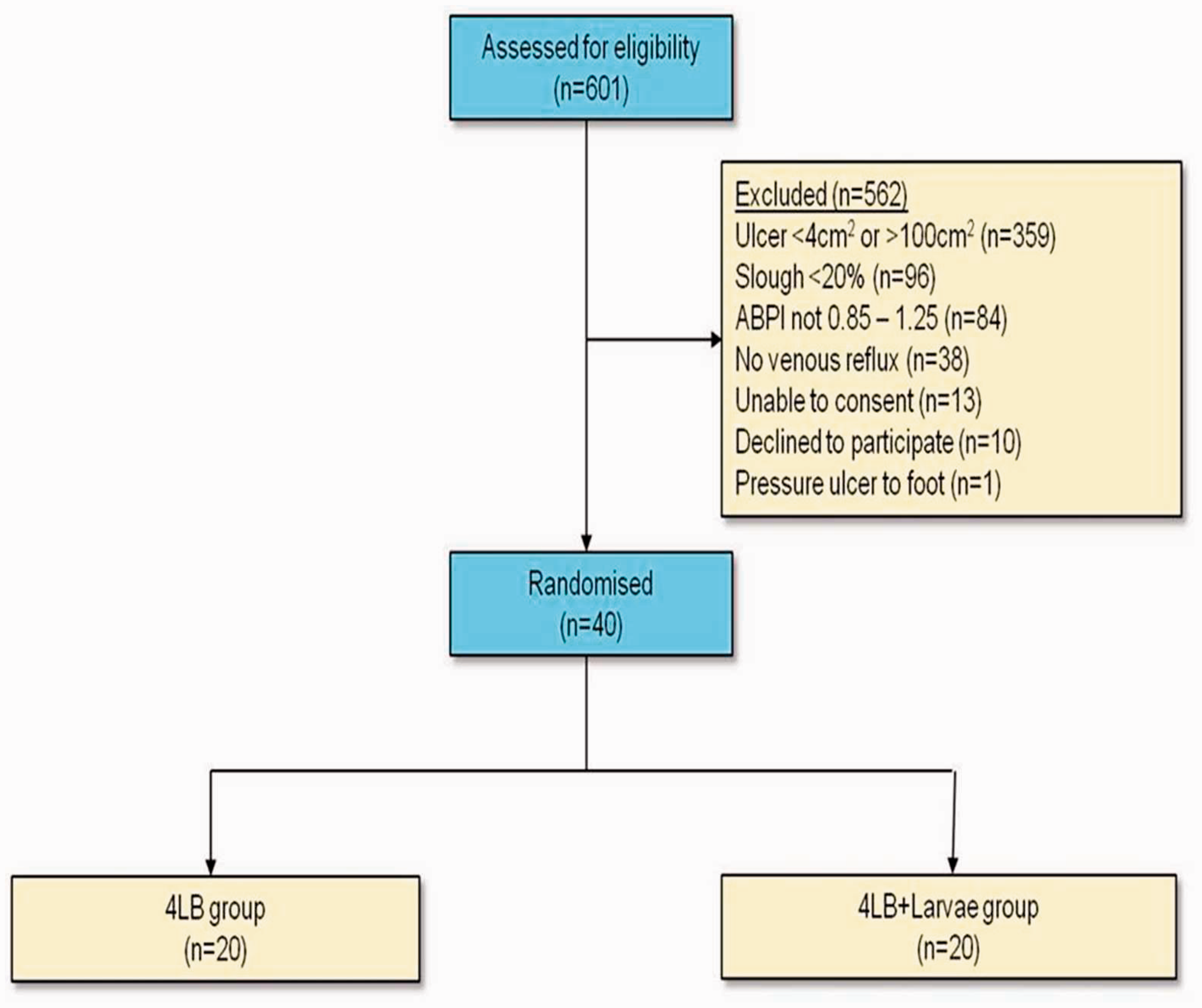

On day 4 the median reduction in slough area was 3.7 cm2 in the 4LB group (P < 0.05) and 4.2 cm2 (P < 0.001) in the 4LB + larvae group (Figure 4) This raw data translates into a median percentage debridement of slough of 50% in the 4LB group and 84% in the 4LB + larvae group (P < 0.05, Figure 5).
Median reduction in slough after 4 days (Mann–Whitney U test). Percentage area removal of slough after 4 days (Mann–Whitney U test).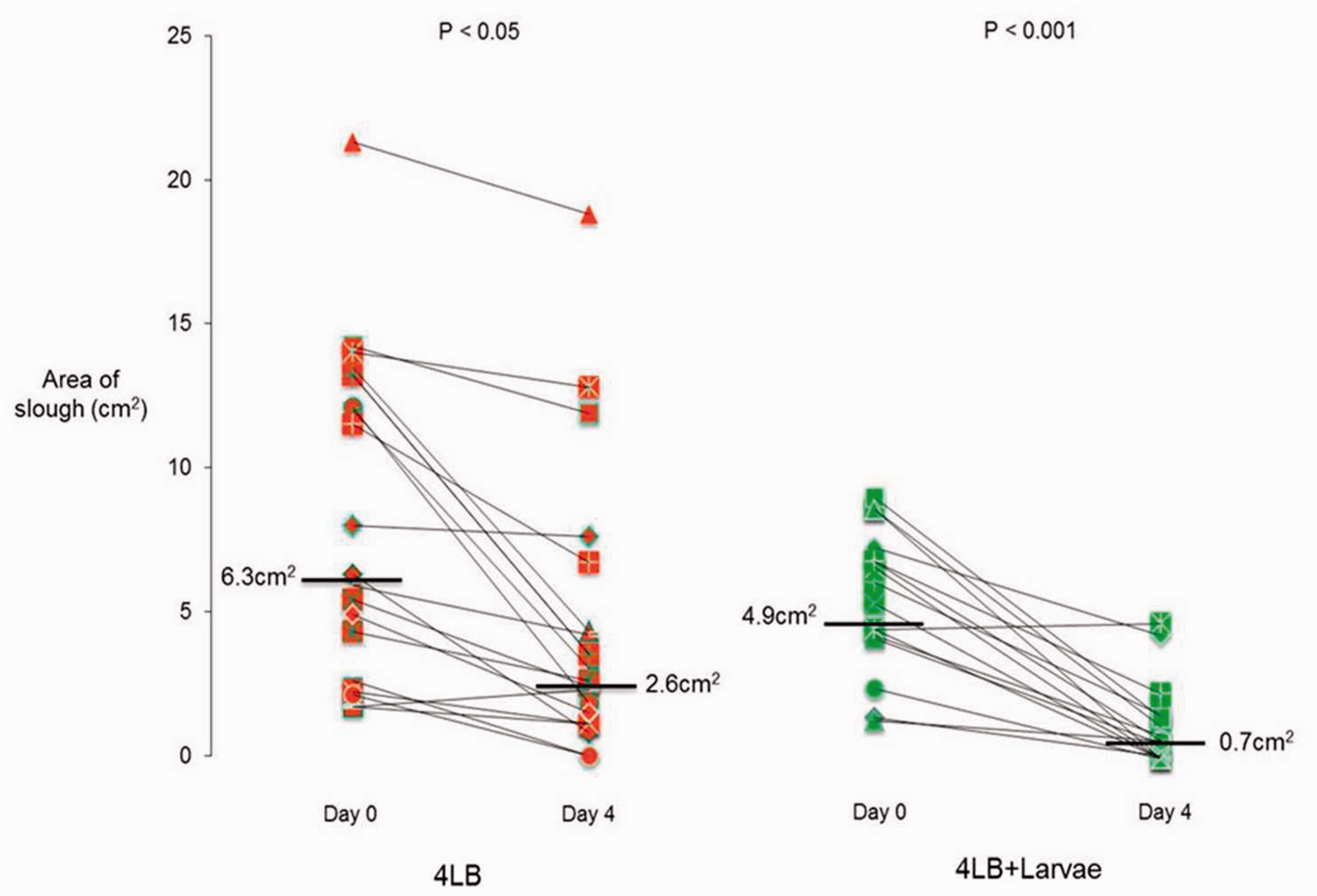
Kaplan–Meier survival analysis showed 12 week healing rates to be 73% and 68% in the control and larvae groups, respectively (Figure 6). The log rank test confirms this: χ2 = 0.20, df = 1, P = 0.664 on an intention-to-treat basis.
Kaplan–Meier analysis of ulcer healing for all legs.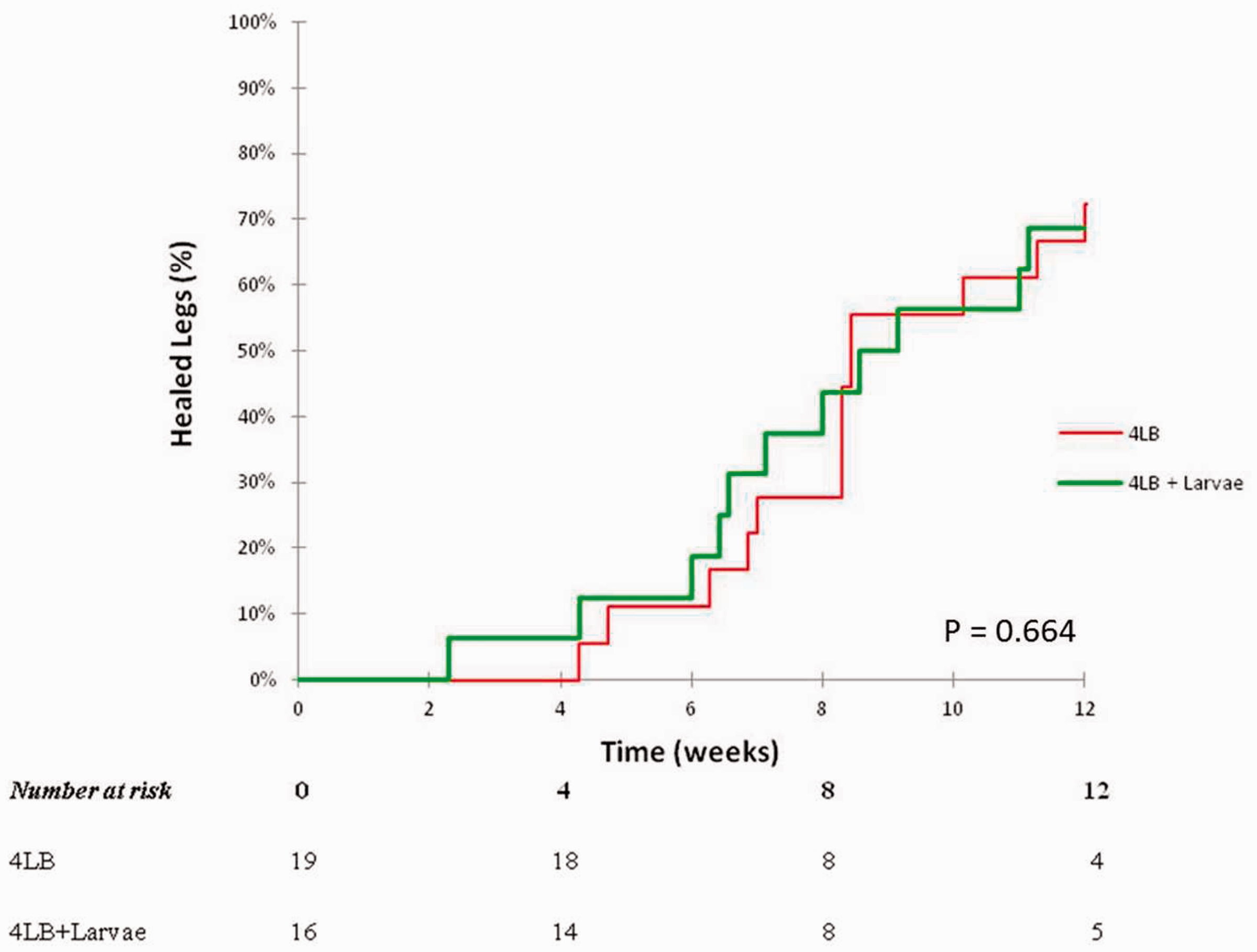
No serious adverse events were reported. However, non-serious adverse events were reported in nine participants. Of the nine participants experiencing adverse events, six were in the 4LB group and three in the 4LB + larvae group. Of these, five were due to wound infections, one due to venous stasis eczema and another three related to participants’ inability to tolerate compression.
Discussion
This study demonstrates that LDT and multilayer compression was more effective than multilayer compression alone in debriding sloughy venous leg ulcers. This finding would corroborate with the findings of the two published RCTs, which both concluded that LDT significantly reduced the time for debridement.10,11 Although there are no randomised controlled studies evaluating the viability of larvae underneath multilayer compression bandages, this study has shown that LDT was effective and not impeded, suggesting that the larvae were not harmed.
Interestingly, the study showed a significant median reduction in slough in the 4LB group. This may imply that successful correction of chronic venous hypertension with multilayer compression allows natural autolysis of slough by improving venous haemodynamics.
The secondary outcome measure showed that treatment with LDT and compression did not improve the 12 week healing rates for venous leg ulcers compared with the standard treatment of compression alone, despite the improved debridement in the first 4 days. This may suggest that in spite of the presence of devitalised tissue, venous leg ulcers may still progress towards healing if the underlying chronic venous insufficiency is adequately treated. Clearly, if the disease processes are controlled using effective graduated compression, and symptoms managed or reduced, most sloughy ulcers will debride autolytically through the action of moisture, leukocytes and matrix metalloproteinases. 13 The results on healing closely resemble those from the larger multicentre VenUS II trial 11 and the ESCHAR randomised controlled trial.14,15
The shortfall of this study was that it did not look at time to debridement nor did it seek to examine the rate of redevelopment of slough following debridement. These were outside the scope of this trial but, in view of the paucity of published data, the latter remains an unanswered question that would benefit from further research. Although the sample size was small, recruitment of participants was very slow. The study’s inclusion criteria asked for ulcers with a minimum surface area of 4 cm2 and with a minimum of 20% slough. However, from a prospective recruitment profile, the most frequently encountered reasons for non-recruitment were ulcers that were less than 4 cm2 (n = 355) or greater than 100 cm2 (n = 4) or had less than 20% slough (n = 96). This may well be due to improved community management of leg ulcers. Following the establishment of a dedicated leg ulcer service in this community for more than 19 years, more prompt assessments enable compression to be started before a referral to specialist care is made.2,12 Widening the inclusion criteria to include all ulcers above 2 cm2 would have improved recruitment rates.
Those ulcers that had a mixed aetiology were also excluded, i.e. ABPI outside the normal range 0.85–1.25 (n = 84).
The study only evaluated healing at 12 weeks and did not look at longer-term results such as 24 weeks healing.
This study is the first randomised controlled trial to compare LDT with compression against compression alone for venous leg ulcers and brings into question the role of larval debridement in the treatment of venous leg ulcers. Clearly, from our results, we have confirmed LDT is an effective method of wound debridement. However, it did not result in improved healing.
The results of the study do not support the routine use of LDT in any protocol-driven management plan. However, the treatment may be beneficial in order to expedite wound bed preparation prior to further adjunctive procedures such as pinch skin grafting, which in turn may facilitate healing of venous leg ulcers. 16 Further work is needed to confirm this.
Footnotes
Conflict of interest
None declared.
Funding
This research received no specific grant from any funding agency in the public, commercial, or not-for-profit sectors.