



Abstract
Objective
Right atrial thrombus in the setting of a large pulmonary embolus is rare and is associated with serious adverse events. This case report presents the role played by EKOS EkoSonic ultrasound system in successfully treating right atrial thrombus and massive pulmonary embolism.
Case report
A 69-year-old female presented with a massive pulmonary embolus and a large mobile right atrial thrombus. She was treated with catheter-directed lysis using the EKOS EkoSonic ultrasound system and tissue plasminogen activator, with complete resolution of her right atrial thrombus and a marked improvement in her pulmonary embolus and hemodynamics.
Conclusion
This case report provides a new and an effective option to treat right atrial thrombus associated with a large pulmonary embolus leading to a good outcome.
Keywords
Introduction
The presence of a right atrial (RA) mobile thrombus in the setting of a large pulmonary embolus is associated with adverse outcomes. RA thrombus has been treated with surgical removal, intravenous lytic therapy, or pharmacomechanical treatment (PMT). We present an alternative method to successfully treat RA thrombus and pulmonary embolus simultaneously using ultrasound-facilitated lysis using the EKOS EkoSonic system (EKOS Corporation, Bothell, WA).
Case report
We report a case of an RA thrombus and massive bilateral pulmonary emboli treated successfully with ultrasound-accelerated tissue plasminogen activator (tPA) using the EKOS EkoSonic ultrasound system (EKOS Corporation, Bothell, WA).
The patient was a 69-year-old female, who was admitted with sudden shortness of breath. Evaluation in the emergency room revealed an elevated D-dimer (13 mcg/ml). Ventilation–perfusion scan was performed and was positive for bilateral pulmonary emboli. Her main risk factor is history of familial spastic paraparesis with very limited activity, which could have been the etiology of her pulmonary embolus from a deep vein thrombosis (DVT). Her venous duplex ultrasound to the lower extremity, however, did not show the presence of thrombi that could have already migrated to her lungs. Upon her initial presentation, her systolic blood pressure (SBP) was 80 mmHg; heart rate, 120 beats/min; respiratory rate, 26/min; and oxygen saturation, 98% on 5–6 l O2 per nasal cannula. Other positive, pertinent findings were diminished lung breath sounds, jugular venous distention to 14 cm, and mild edema around the ankles.
She was placed on intravenous inotropes and unfractionated heparin. Her electrocardiogram showed evidence of a right bundle branch block, sinus tachycardia, and nonspecific ST-T abnormality. Lactic acid was 8.2 mg/dl; ProBNP, 17710 ng/l; and white blood cell, 14.03 × 10³/mm3.
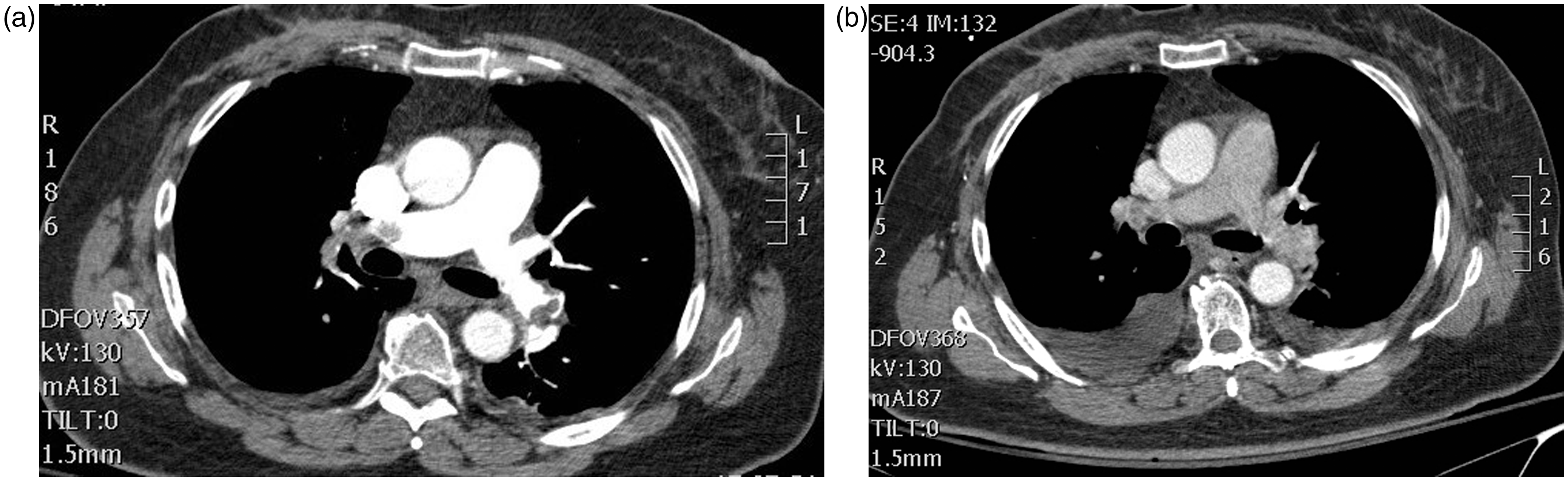
Echocardiography showed a large mobile RA thrombus (2.81 × 2.36 cm) attached to the interatrial septum, right ventricular (RV) enlargement (3.6 cm), normal left ventricular ejection fraction, pulmonary artery (PA) pressures of 48 mmHg, and flattened interventricular septum (Figure 1a). Bilateral lower extremity venous duplex showed no DVT. Chest X-ray showed no active disease. CT angiogram to PA showed extensive bilateral pulmonary emboli, more on the right than the left, with near occlusion of the distal main right PA (Figure 2a).
(a) RA thrombus seen on echocardiography (arrow) and (b) RA thrombus resolved after EKOS-facilitated lysis (white arrow points to EKOS catheter within the RV). (a) CT angiography showing pretreatment of thrombus in large bilateral main pulmonary arteries and (b) CT angiography 48 h post-treatment showing a significant resolution of thrombus from main right and left pulmonary arteries.
After informed consent was obtained, the patient was brought to the cardiac catheterization laboratory. A right heart catheterization was performed using an 8 Fr sheath placed in the common femoral vein (CFV). PA pressure was measured at 59 mmHg. Using a pigtail catheter, a pulmonary angiogram was performed and it showed a near occlusive right main PA thrombus and nonocclusive thrombi in the left PA branches. The EKOS infusion catheter (24 cm) was then placed at the junction of the RA and inferior vena cava into the right main PA extending into the thrombus. A 12 cm EKOS catheter was then deployed from a second venous sheath placed in the right CFV into the left PA. tPA was then started at 1 mg/h for each lung for a total of 12 h (total of 24 mg tPA for both lungs). A total of 250 units of heparin/h was infused in each venous sheath for the duration of tPA infusion (total of 500 units/h, no dose adjustment). A limited echo after cardiac catheterization procedure showed the persistence of the RA thrombus that was not dislodged with catheter manipulation.
Following 12 h of tPA infusion, the patient was feeling better with her heart rate 100 beats/min and SBP 110 mmHg. Her respiratory rate was 20/min, and her oxygen saturation was 96% on 3 l O2 per nasal cannula. A limited echocardiogram was performed and showed her PA pressures to be 49 mmHg. RA thrombus had completely disappeared (Figure 1b). No evidence of RV or main PA thrombi was seen. Twenty-four hours after the procedure, the PA pressure was 44 mmHg, RV size was 3.1 cm, and interventricular septal motion significantly improved. Forty-eight hours after the procedure, she had a repeat CT angiogram of the PA that showed a marked reduction in her right main PA thrombus and the left PA branches (Figure 2b). Patient was initially placed on intravenous heparin for 24 h immediately at the end of her tPA infusion (started at 1000 units/h, then adjusted for PTT of 50–60 s), which was later changed to rivaroxaban 15 mg orally twice a day for 21 days followed by 20 mg daily planned for a total of 6 months. She was discharged 1 week after her procedure to a skilled nursing facility on room air with oxygen saturation of 93%. One month later, the patient was doing well on room air with no symptoms of dyspnea.
Discussion
Pulmonary embolism (PE) carries a high rate of early mortality. 1 Patients with large pulmonary emboli and RV strain, even if normotensive, are at high risk of in-hospital and latent mortality.2–4 Aggressive intervention including thrombolysis has been recommended in patients with massive pulmonary embolus and hemodynamic compromise, but this approach remains controversial in hemodynamically stable patients.5–7 The current American Heart Association guidelines has a Class IIb recommendation (benefit more than risk) for treating patients with submassive PE (biological markers positive, enlarged RV on echocardiography, and SBP of more than 90 mmHg) and Class IIa recommendation (benefit significantly more than risk) for those with massive PE (SBP less than 90 mmHg, enlarged RV, and shock).8,9 Also, lysis has been recommended for massive pulmonary embolus by the American College of Chest Physicians 2012 guidelines (Grade 2C). 10
Thrombolysis may lead to a faster recovery of RV function and pulmonary pressures and restoration of normal interventricular septum and cardiac output. 8 Also, in the study by Kline et al., 11 patients who received lytic treatment for sub-massive pulmonary embolus did not develop a higher RV SBP than baseline at 6-month follow-up when compared with 27% of patients that received heparin alone. tPA is most commonly used either as a full intravenous dose of 100 mg over 2 h or via catheter-directed lysis (CDT) (dose of 0.5 to 2 mg/h up to 24 h). In randomized trials, however, systemic lysis was associated with a high rate of major bleeding (13%) and intracranial hemorrhage (1.8%). 12 Alternative approaches using a combined PMT to fragment, aspirate, or make the thrombus more accessible to lysis has also been adopted successfully. These techniques reduce the lytic dose and may enhance the speed of thrombus dissolution. PMT techniques include pigtail catheter fragmentation of thrombus followed by CDT, rheolytic thrombectomy with power pulse spray of a lytic agent, suction or rotational thrombectomy followed by CDT, and ultrasound-facilitated PA lytic infusion. Effective lysis is generally translated into a smaller RV size, reduction in pulmonary pressures, improvement in interventricular septal motion, increase in cardiac output, and hemodynamic stability. 13 Recently, Wang et al. 14 showed that a reduced intravenous infusion of 50 mg tPA over 2 h resulted in a reduction in RV size and pulmonary pressure similar to a 100 mg tPA dose with the advantage of reduced bleeding. The effectiveness of this regimen compared with CDT with similarly reduced dosing is unknown.
Ultrasound-facilitated lysis has been recently introduced as an effective tool to reduce thrombus burden in the pulmonary arteries of patients with submassive or massive PE and those patients with DVT. The system consists of a side-hole drug delivery catheter through which a multielement ultrasound core wire is inserted. Ultrasound pulses promote fibrin separation, facilitating active drug delivery into the thrombus. In the recent Ultrasound Accelerated Thrombolysis of Pulmonary Embolism trial (ULTIMA), a phase II, multicenter, open-label randomized trial of unfractionated heparin versus unfractionated heparin plus the EkoSonic ultrasound-assisted tPA lysis over 15 h (maximum 20 mg), the primary endpoint of RV/LV ratio was significantly reduced from baseline with ultrasound-assisted lysis versus heparin alone (at 24 h: 0.3 versus 0.03, p < 0.0001; at 90 days: 0.38 versus 0.22, p = 0.03). The secondary endpoints of death and recurrent venous thromboembolism were statistically not different between the two groups. 15 Recent data from the prospective, single arm, multicenter SEATTLE II trial 16 evaluating the EKOS system in 150 patients with massive (n = 32) and submassive (n = 119) PE showed that a total of 24 mg tPA infused over 24 h reduced RV/LV ratio by 0.42 (p < 0.0001) at 48 h after the procedure. There were no intracranial hemorrhage and no fatal bleed reported.
Our patient was also complicated by the presence of large, mobile RA thrombus arising from the interatrial septum and prolapsing into the RV. The presence of RA thrombus in the setting of PE is rare and is associated with worse outcome. 17 Embolization of a large RA thrombus could lead to significant hemodynamic compromise. 17 RA thrombus originates generally from DVT or forms in situ. Highly mobile thrombus is the result of embolization, whereas in situ thrombus tends to be nonmobile and adherent to the walls of the RA or the tricuspid valve or foreign objects such as pacemaker wires. Typically, in situ RA thrombi are more common (68.4%) than thrombi that have embolized from remote DVT. 18 In a retrospective study of RA thrombi, treatments ranged from none (9%) to anticoagulation (35.0%), surgical embolectomy (35.6%), or intravenous lysis (19.8%). Mortality was 27.1% and was highest in patients with no treatment (100%) and lowest in patients with lysis (11.3%). Patients receiving anticoagulation and surgical treatment had mortality of 28.6% and 23.8%, respectively. 19
In our patient, we elected to use the EKOS EkoSonic ultrasound system to accelerate PE and RA thrombus lysis simultaneously. The 24 cm EKOS catheter is capable of delivering ultrasound-facilitated lysis from the right atrium to the distal end of the right pulmonary vasculature, where a large thrombotic burden was seen. The catheter needs to be delivered carefully from the RA into the RV and PA. We avoided a right internal jugular vein approach as we thought the catheter and wires will have a higher chance of dislodging the thrombus as they move into the center of the RA. The CFV approach was felt to be a safer approach to advance the catheter that could be gently passed from the IVC into the base of the right atrium to the tricuspid valve avoiding the mid-RA and interatrial septum, where most of the mobile mass was seen. The presence of the catheter spanning from the RA into the RV and PA also provided a reassurance that if embolization would occur, lysis is ongoing into the RV and main PA and would help dissolving the thrombus further. After 12 h, the thrombus disappeared and was not seen in the RV or the main PA on echocardiography.
This case report presents an alternative strategy to treating RA thrombus in the setting of large PE and demonstrates the effectiveness of ultrasound-facilitated lysis, a recently approved technique by the FDA to treat PE.
Footnotes
Acknowledgement
The authors thank Debbie Litchfield and Roy Hosek for providing their expertise in the use of the EKOS EkoSonic ultrasound system at our institution.
Conflict of interest

Funding
Dr Shammas received educational grants from EKOS Corporation to the Midwest Cardiovascular Research Foundation.