



Abstract
Objectives
The present review represents a translational boundary between basic research and surgery, particularly focusing on the promising application of adipose-derived stem cells harvested intra-operatively during debridement of venous leg ulcers.
Methods
We reviewed 830 out of 5578 articles on MEDLINE starting from 1997 and sorted by the relevance option.
Results
The technique currently used for adipose-derived stem cells intra-operative harvesting is presented, including a safety evaluation on a cohort of 5089 revised patients who underwent plastic surgery and maxillo-facial surgical procedures. Complications were reported in 169 cases (3.3%). One hundred and forty-one (2.77%) patients were classified as having minor complications, specifically: nodularity/induration 93 (1.83%), dysesthesia 14 (0.26%), hematoma 12 (0.23%), superficial infection 11 (0.21%), pain 7 (0.13%), poor cosmesis 3 (0.06%), and abnormal breast secretion 1 (0.02%), while 28 patients (0.55%) were classified as having major complications, specifically: deep infection 22 (0.43%), sepsis 3 (0.06%), abdominal hematoma 2 (0.04%), and pneumothorax 1 (0.02%). Application of cell therapy in venous leg ulcer is currently used only for patients not responding to the standard treatment. The review shows the lack of randomized clinical trials for application of adipose-derived stem cells among treatments for venous leg ulcer. Finally, adipose-derived stem cells implantation at the wound site promotes a new tissue formation rich in vascular structures and remodeling collagen.
Conclusion
Adipose-derived stem cells strategy represents a great opportunity for the treatment of chronic wounds, due to the simplicity of the technique and the application of cell treatment in the operating room immediately following debridement. However, clinical studies and data from randomized trials are currently lacking.
Introduction
This review is a systematic review which aims to address the procedures and indications, which are required for the application of the adipose-derived stem cells (ADSCs) treatment strategy in the healing process of venous and mixed leg ulcers (VLUs). This method has rarely been used in this particular field; conversely, experience from other clinical fields and basic research seems to recommend the suitability of this scope of application.
With this objective, in the preliminary phase, we consulted the scientific literature which appeared after the introduction of Coleman’s procedure into clinical practice in 1995. We took three key terms into consideration: Coleman’s procedure, adipose-derived stem cells, and venous leg ulcers. We considered 5578 bibliographic references, from which we eliminated all those which were not in English, all case reports, all observational studies, and all the basic research studies which repeated findings already published with the aim of producing a review which is as up-to-date as possible and which succinctly sets out an overview of the rationale and the technical methods used alongside the results obtained from this treatment strategy.
Venous and mixed leg ulcers: a prominent problem of social medicine
Chronic venous disease (CVD) is a common condition that afflicts the general population and certainly it represents the most widespread vascular pathology. In the majority of cases, it is a minimally disabling disease, but a significant proportion may progress towards chronic venous leg ulceration (CVU). 1 In the “Edinburgh vein study” Robertson et al. 2 stated that the CVD ratio in a peer-investigated population is higher and higher with 1% year-to-year. The deep venous system was affected in one-third and the superficial venous system was affected in two-thirds of this population.
Despite superficial venous insufficiency being the pattern most frequently associated with VLUs, the prognosis is worsened when co-morbidities (rheumatoid arthritis, malnutrition, diabetes, chronic anemia, etc.), second stage peripheral arterial disease, or any other vascular pathology is involved. For this reason, we usually speak of mixed VLUs.1,3–5
Impairment of venous hemodynamic is an essential but insufficient factor in explaining the progression of the disease to the point of skin lesion or even to explain the frequent presence of non healing CVU.6,7
Venous surgery can be helpful in achieving healing and more than all the other options in preventing recurrences.8−13
However, not all CVU patients are candidates for surgery due to comorbidity conditions; compression, wound-care and debridement still remain the first-line treatments of such disabling diseases. Finally, it has been reported that CVU healing rate is absolutely variable in literature (35%–85%), and the different controlled trials report a significant proportion of recalcitrant ulceration, which respond to treatment but do not heal.14−16 In such situations, skin grafts and advanced dressings are currently used to achieve healing, and new regenerative approaches have been proposed. One of the more interesting among the latter is ADSCs: the present review topic.
ADSCs properties
In recent years, stem cell application has been suggested as a possible novel therapy for regenerative medicine. This is due to their unlimited capacity for self-renewal as well as the ability to differentiate into multiple cell types under appropriate stimuli. 17
In particular, ADSCs have attracted a lot of attention in recent years as an alternative to the use of cells that are derived from bone marrow. This is due to the ease with which they can be isolated and expanded and their large number within the adipose tissue: the frequency ranges from 1:100 to 1:1500 cells, which far exceeds the frequency of mesenchymal stem cells in bone marrow. 18 There is only 1 per 100,000 nucleated cells of mesenchymal stem cells in bone marrow and their quantity declines with age. 19
ADSCs are multi-potent stem cells and have characteristics similar to bone marrow-mesenchymal stem cells, as demonstrated by their expression of the same cell surface markers, their similar gene expression profiles and their similar differentiation potentials. 20 For this reason, ADSCs are able to form bone, cartilage, muscle and fat, under appropriate stimuli. They are multi-potent and secrete a variety of growth factors 21 such as basic fibroblast growth factor (bFGF), keratinocyte growth factor (KGF), transforming growth factor beta (TGF-β), hepatocyte growth factor (HGF), and vascular endothelial growth factor (VEGF). 22
The use of autologous stem cells in human clinical trials has several limitations, including the use of animal products for cell expansion. Animal serums could generate problems of viral transmission and immunological reactions. For this reason methods that use human-derived products in the manipulation of stem cells have been developed.
Coleman’s procedure
The most widely used technique for tissue regeneration was introduced by Coleman in 1995 23 and it was first applied for facial remodeling and for breast reconstruction and augmentation.24,25 This procedure provides lip aspiration, centrifugation, and the subsequent re-injection of autologous fat. The fat donor sites are the abdominal area, waist adipose deposit (WAD), inner sides of the thigh and knees, and flanks.
First, a tumescent solution (Kleine’s solution) of 250 ml normal saline, 20 ml of 1% carbocaine, 1 ml adrenaline, and 2 ml bicarbonate is injected into the fat harvest area. It is important to wait for approximately 10–20 min to allow for dispersion of the tumescent solution. Using a two-hole blunt Coleman harvesting cannula (Byron Medical, a division of Mentor Corporation, Tucson, AZ) and a 10-ml Luer-Lock syringe (BD Syringe Luer-Lock tip; Becton Dickinson, Franklin Lakes, NJ) fitted directly on the cannula, fat harvesting is performed and then transferred to a standard centrifuge. Coleman and Saboeiro 26 recommended the use of a 10-ml syringe for fat harvesting to reduce the pressure generated during the harvesting procedure and thereby potentially preserve the fat parcels. 27
Consequently, the samples are sealed and centrifuged at 1300 r/min (rotor size: 16 cm; g force: 580) for 5 min as this allows us to obtain a higher adipocyte concentration. After centrifugation, the samples are separated into: an upper yellow layer of oil derived from destroyed fat fragments, a middle layer composed of the adipose tissue graft with high concentration of stem cells,
28
and a bottom layer of blood. In Figure 1, we show the steps described above to harvest the adipose tissue and the stem cells using Coleman’s technique.
(a) Coleman’s technique for harvesting adipose tissue-derived stem cells. In this particular case, the harvesting is performed in the periumbilical area with tumescent local anaesthesia; (b) Adipose tissue aspirated. The areas with more fat are not always the best to harvest tissue for regenerative purposes; (c) The adipose tissue is then centrifuged in order to separate the different components; (d) The lower part of the adipose layers is particularly rich in stem cells, containing more than 90% ADSCs.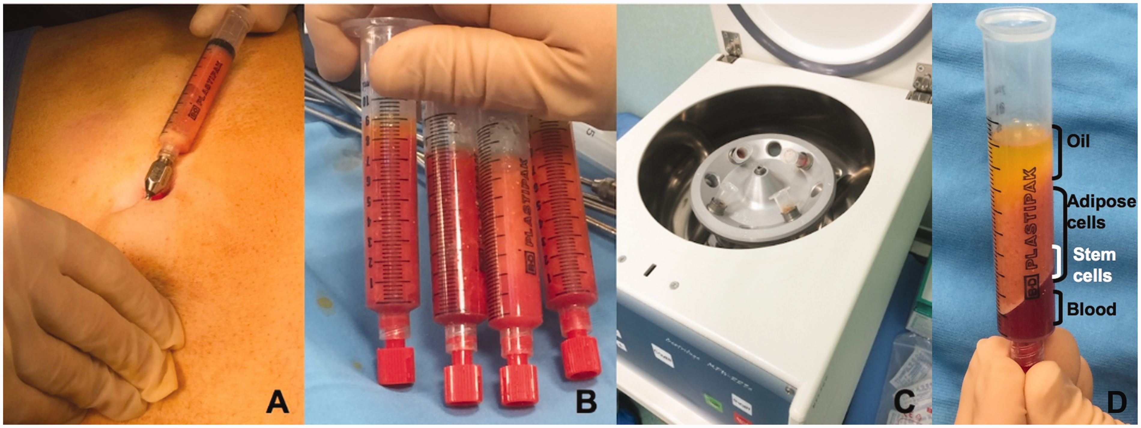
The supernatant oil in the upper layer is discarded by ejection after rotating the syringe. Codman neuropads (Codman Neuro Sciences, Sarl, Switzerland) are used to wick the residual oil component. The lower layer is discarded by opening the plug from the Luer-Lock connection.
Following these procedures, the micro-fat graft is placed into 1 ml syringes for transfer through a blunt Coleman microcannula (1 mm) into the tissue to be grafted (Figure 2).
(a) After fat and blood elimination the ADSCs are aspirated by Luer-Lock syringe technique; (b) Syringes loaded with stem cells.
Small aliquots of fat are injected using a multilayer technique of implantation. Multiple sub dermal and hypodermal tunnels are designed through multiple tissue planes releasing only small amounts of fatty tissue in the recipient area to improve the likelihood of graft implantation. 26 This method reduces fat damage and adipocyte necrosis, improving graft vascularization and three-dimensional fat distribution. Indeed, minimizing the volume grafted with each pass of the cannula will maximize the surface area of contact between the grafted fat and the recipient tissue. The proximity of the newly grafted fat to a blood supply encourages survival and minimizes the potential for fat necrosis and later calcification. 29
Safety of Coleman’s procedure for ADSCs harvesting
Current stem cell harvesting techniques focus on bone marrow, peripheral blood, and umbilical cord blood. In comparison to these techniques, the procedure for harvesting and fat grafting, initially introduced by Coleman, 23 yields a higher number of stem cells that can be used intra-operatively with minor organizational and legal limitations that can prove expensive for clinical use.
Complications for harvesting and fat grafting procedures.

In contrast, data reported on the recovery and safety profiles following bone marrow collections in 9245 donors identified 345 cases (3.7%) with potentially medical complications. In 125 cases (1.35% of the 9245 total), post-harvesting complications were classified as serious. Of these 125 serious cases, 116 were classified as directly related to the collection, being: mechanical injury to tissue, bone or nerve 69 (0.7%), anesthesia 45 (0.5%), infection 1 (0.01%), and grand mal seizure 1 (0.01%). Of the 116 patients with serious complications, 67 (0.7% of the 9245 total) experienced prolonged recovery times related to mechanical injury to tissue from needle aspirations and required interventions ranging from limited physician involvement and/or physical therapy to surgical intervention and ongoing disability (1 to 10 years). Of the remaining 49 patients (0.5% of the 9245 total) with severe reactions, most were due to severe acute reactions related to the anesthesia (complicated post-spinal headaches, cardiac arrhythmia, and pulmonary edema).44−46
Coleman’s procedure for ADSCs harvesting therefore seems advantageous when compared with other techniques for stem cell harvesting. It seems safer as there is less discomfort and risk of complications for the patients.
ADSCs in wound healing
Cutaneous wound healing is a complex biological process involving regenerating dermal and epidermal tissues consisting of four different but partially overlapping steps: hemostasis, inflammation, proliferation (with formation of granulation tissue), and remodeling. 47 Non-healing wounds are the result of an interruption in the progression of this normal sequence of cellular and biochemical events towards the restoration of the skin’s integrity. The characteristics of chronic wounds are prolonged inflammatory phase, lack of appropriate metabolism and clearance of toxic substances from the wound, persistent infections, formation of drug resistant microbial bio-films, and inability of dermal and/or epidermal cells to respond to regenerative stimuli. 48
As conventional treatment strategies for chronic wounds are increasingly reaching their limits and often fail, advanced healing therapies including biological dressings, skin substitutes, growth factor-based therapies and synthetic acellular matrices are being used to correct irregular and dysfunctional cellular pathways present in chronic wounds. 49
In the investigation of new wound healing strategies, increasing angiogenesis and vasculogenesis is the key. 50 Vascular endothelial growth factor (VEGF) and fibroblast growth factor (FGF) are potent angiogenic factors. FGF application to wounds has been shown to increase granulation, decrease contraction, and increase angiogenesis. 51 In addition, keratinocyte growth factor (KGF) and FGF were clarified to activate fibroblast and keratinocyte proliferation and migration, collagen synthesis, and induce angiogenesis.52,53
Vascularization plays a fundamental role in wound healing and therefore is an important parameter for novel therapies. 54 So, innovative and alternative treatment options including stem cell-based therapies have been investigated over the last decade. 55 Indeed, ADSCs have great potential in their ability to release angiogenic factors and have shown increased angiogenesis in wound healing whenever injected or delivered via a scaffold. 56 Increased vascular tissue has been seen in models using endothelial cells and fibroblasts. Furthermore, ADSCs have shown increased healing in both burn models and full-thickness wound models. 55
In addition to their angiogenic potential, ADSCs have shown a positive impact on wound healing in clinical and pre-clinical studies. Recent ADSCs applications in vitro and in vivo showed that they are attracted to the wound site and influence regeneration processes via paracrine mechanisms as well as fusion and differentiation, for example, into keratinocytes or dermal fibroblasts.57−59
Clinical trials are under way using ADSCs for regenerative medical and tissue engineering applications.60,61 Promising applications in wound and ulcer healing have been reported, although thus far from small studies and a total of only 62 patients.62−65
Bartsich and Morrison 66 discussed the long-term treatment of chronic sickle cell ulcers and the possible use of a skin graft and fat grafting to achieve healing. Current treatment using skin grafting and local wound-care is often unsuccessful long-term, as wounds that have healed break down again. In their study, treatment involving permanent alteration of the wound bed with recruitment of a new cell population and subsequent fat grafting was satisfactorily applied. Cervelli et al. 62 demonstrated the ability of the combination of autologous adipose grafts and PRP injected intralesionally or perilesionally to regenerate tissue and to induce epithelialization with wound closure in patients with a loss of substance on the lower limb. They reported a significant healing-time reduction (57% of patients achieved complete healing within three months) and a noteworthy improvement in quality of life, along with cost reduction due to the lower amount of medication required. Marino et al. 63 used purified adipose-derived stem and regenerative cells extracted from autologous fat for the care of chronic ulcers of the lower limbs of arteriopathic patients. In all cases, injections into the edges of the ulcer produced a reduction in both the diameter and depth of the ulcer. In more than half of these cases, there was healing with complete re-epithelization. In the first phase I trial, Bura et al. 64 showed that adipose-derived stroma cell transplantation strongly improves revascularization and tissue perfusion with ulcer evolution and wound healing improvement in non-revascularizable critical limb ischemia patients. Lee et al. 65 demonstrated that multiple intramuscular adipose tissue-derived mesenchymal stem cell injections could be a safe alternative method to achieve therapeutic angiogenesis in patients with critical limb ischemia who are refractory to other treatment modalities. Furthermore, various studies on mouse, rat, and rabbit models have obtained encouraging evidence of ulcer evolution and wound healing improvement.57,58, 67−83
Tissue engineering in wound healing
Tissue engineering and regenerative medicine include “an interdisciplinary field that applies the principles of engineering and life sciences towards the development of biological substitutes that restore, maintain, or improve tissue function or a whole organ”. They have become promising and important fields of research in which the deposition, growth and remodeling of tissue are investigated through three tools: cell-based therapy, biomaterials (or scaffolds) alone, or scaffolds seeded with cells.
Cell-based therapy
Cell-based therapy including mesenchymal stem cells such as ADSCs has a potential to improve wound healing conditions without major surgical procedures and donor-site morbidity. The wound healing would benefit because the ADSCs are multipotent to differentiate into other specialized cells and secrete or suppress the growth hormones and cytokines necessary in the environment and are capable of expanding in number while showing a stable phenotype.
Cell therapy can be applied to both acute and chronic wounds. In the treatment of acute wounds, ADSCs can increase wound healing, reduce scar contracture, and minimize donor-site morbidity. Conversely, in the treatment of chronic wounds, the wound bed should be the environment where maximum wound healing can be achieved by transplanting cells with an excellent wound healing capacity. 84
We emphasize that wound bed preparation needs to be perfect before implantation, according to the TIME protocol (tissue, infection, moisture, and environment protocol). 85
In addition, all techniques which permit the elimination of the effects of reflux and venous stasis have to be also used in order to improve the cell habitat and favor as much as we can the attachment of ADSCs.
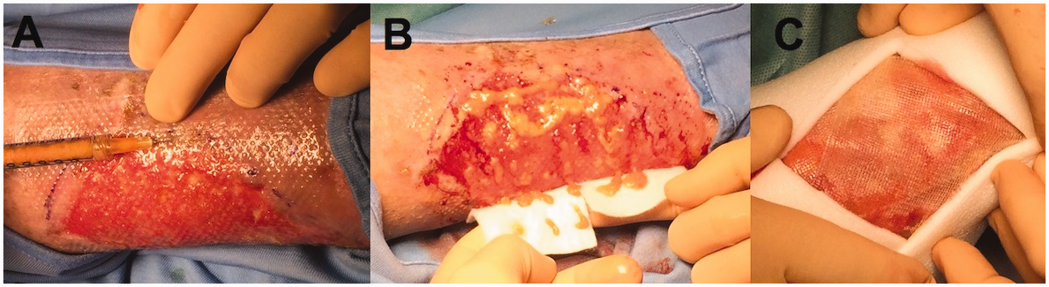
Figure 3 shows a mixed VLU before debridement and the application of stem cells, either by injection into the margin or by using a scaffold with islets of adipose cells (Figure 4).
Mixed VLU before debridement. (a) Margin injection with a suspension of ADSCs. The wound bed preparation is apparent; (b) Islets of adipose cells in the wound bed together with a scaffold of hyaluronic acid; (c) Foam advanced dressing protects the healing area before application of compression.
Scaffolds
The use of scaffolds and cellular matrices is essential to differentiate ADSCs into the required cells and use them to construct the three-dimensional tissues useful in reconstructive medicine.
An essential strategy for tissue engineering is the selection and the construction of a good quality scaffold. Ideally, the scaffold should be a functional and structural platform able to mimic the native extracellular matrix and support the morphogenesis of multiple tissues. Based on the literature, tissue-engineering scaffolds should (1) be biodegradable, (2) not trigger inflammatory responses, (3) have surface properties that enhance the attachment, proliferation and differentiation of cells, (4) mimic the skin in vitro, (5) have suitable mechanical properties, and (6) be suitable for manufacturing into different shapes. 86
Scaffolds seeded with cells
Engineered tissue regeneration uses a biocompatible scaffold which, in combination with living cells and/or bioactive molecules replaces, regenerates, or repairs damaged tissues. This kind of scaffold should be biocompatible, porous and permeable to support cell attachment and proliferation.
Suitable scaffolds available for adipose tissue engineering include: type I collagen sponge, non-woven polyglycolic acid (PGA) and hyaluronic acid (HA) gel. HA is a naturally occurring non-sulfated glycosaminoglycan present in connective tissue, in the synovial fluid of articular joints and in the vitreous humor of the eye. It is one of the natural extracellular matrix components and it is important in tissue hydration, cell differentiation and tissue repair. HA is defined as a “suitable scaffold for adipose tissue engineering”. It is highly biocompatible, does not elicit any adverse reaction and is reabsorbed by the host tissues. 87 In particular, in vivo experiments confirmed the optimal tissue repair and regeneration of full-thickness wounds for the treatment of ulcers in a placebo-controlled study. 88 Moreover, HA and its derivatives are actively angiogenic: preliminary results in in vivo models demonstrate the complete vena cava regeneration inside hyaluronic acid-based prosthesis, opening new perspective in microvascular surgery applications.87,89
In a recent study, Altman at al. 90 elucidated that a new scaffold consisting of silk fibroin-chitosan when combined with ADSCs supports the engraftment of stem cells and their differentiation into epithelial and fibrovascular components increasing the repairing and healing potential of damaged tissues.
The microenvironment for wound regeneration mainly depends on interactions between stem cell progenitors and their niche; 91 therefore, any tissue-engineered reconstruction should provide a suitable microenvironment for the cells to proliferate and differentiate.
Scaffolds and nanotechnology
ADSCs differentiation can also be induced by physical factors and modulation of extracellular matrix nanostructures. Indeed, since the majority of signaling molecules interact with ADSCs at the nanoscale level, scaffolds with surface nanostructures have potential applications for ADSCs in the field of tissue engineering and regenerative medicine. The literature offers several different methods to induce such differentiation through the use of high quality nanoparticles of various chemical compositions. However, the real biological effects of nanoparticles have to be carefully assessed before introducing their use in clinical practice: the release of active peptides may possibly cause interferences with some biological functions and cellular processes. 92
Histology of the wound healing process and potential role of ADSCs
The normal wound healing process in vascular ulcerations is usually aborted for a variety of reasons depending on the single clinical case. Usually, the first phase of wound healing is the inflammatory phase that starts very soon after the wound arises. Immediately after injury, platelets adhere to damaged blood vessels, initiate a release reaction, and begin a hemostatic reaction. Blood platelets release growth factors, cytokines, and other survival or apoptosis-inducing agents. Leukocytes release reactive oxygen species (ROS) with antimicrobial and proteases roles that clear the wound of foreign bodies and bacteria. Resolution of the inflammatory phase is accompanied by gradual inflammatory cell apoptosis a few days after the event.
As the inflammatory phase ends, the proliferative phase of repair begins in order to form the granulation tissue that support re-epithelialization. As dermal and epidermal cells migrate and proliferate within the injury bed, there is a clear requirement for an adequate blood supply for nutrient delivery, gas, and metabolite exchange. Wound healing angiogenesis begins immediately after injury when local hypoxia, secondary to injury-induced blood vessel disruption, occurs. This event stimulates the production of proangiogenic factors such as vascular endothelial growth factor (VEGF), fibroblast growth factor 2 (FGF-2), and PDGF. In response, endothelial cells degrade basal membrane, migrate into the wound site, proliferate, and form new blood vessels (Figure 5(a) and (b)). More recently, it has been revealed that endothelial progenitor cells (EPCs) are also required for wound revascularization.93,94 Endothelial progenitor cell mobilization is mediated by nitric oxide, VEGF, and matrix metalloproteinases (MMP9 particularly);94,95 in this phase, ADSCs could play a main role secreting bFGF, KGF, TGF-β, HGF, and VEGF. Although more research needs to be done to further elucidate the mechanisms of EPC and ADSC recruitment and homing, it is clear that these progenitor cells are necessary for normal wound healing—associated neovasculogenesis and injury repair.21,48,56
Exemplification of the healing process 2 weeks after implantation. (a) Wound site at 4×, H&E. At this magnification, the acanthosis of epidermidis detached by papillary dermis cohabited by granulation tissue rich in vascular structures and remodeling collagen can be seen. In the lower portion of the dermis, the incipient remodeling reaction is evident; (b) Ulcerated site at 10×, H&E. No epidermidis covers the superficial dermis that is repopulated by granulation tissue rich in neovascular structures and collagen; (c) CD34 immunohistochemistry marks neovascular endothelial cells and underlines the typical wound site morphology during repair time characterized by the presence of granulation tissue and scar formation with fibrosis and lost of adnexae structures in the lower dermis.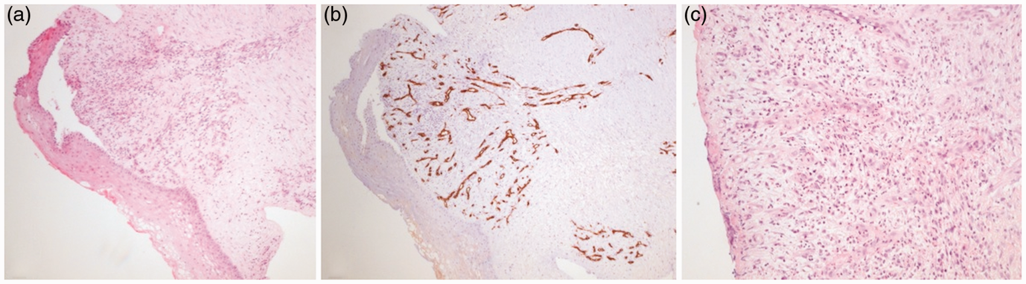
Reestablishment of a normal blood supply provides a favorable microenvironment for epidermal and dermal cell migration and proliferation. Fibroblasts proliferate within the wound and synthesize extra-cellular matrix (ECM) forming granulation tissue perfused with newly formed blood vessels. It seems that ADSCs, when applied after wound bed preparation according to TIME protocol, particularly enhance blood vessel proliferation as well as synthesis of matrix components (unpublished data). In Figure 5(c), one week after ADSCs treatment, blood vessel proliferation together with a provisional matrix can easily be seen.
We know that such a matrix mainly consisting of collagen III, fibrin, fibronectin, and HA is progressively substituted with ECM mainly containing collagen I. Next, wound contraction and matrix remodeling take place. Finally, apoptosis of fibroblastic cells occurs, leading to the formation of a relatively acellular scar tissue, whose tensile strength is comparable with unwounded skin.
Diagnosis of ulcer pathogenesis and synchronous vascular treatments and wound care
Impairment of venous and arterial hemodynamics represents an essential factor in the pathogenesis of CVU. However, other associated medical conditions are reported: obesity, smoking, diabetes, hemolytic anemia and/or iron-deficiency anemia and/or malnutrition, inability to walk, severe cardiac and/or hepatic and/or renal and/or pulmonary insufficiency or chronic administration of cortisones for chronic inflammatory disease and/or auto-immune disease.96,97
ADSCs grafts in successful CVU outcomes need a significant improvement of the venous function by means of associated and customized case-by-case treatments. All of these treatments aim to improve tissue drainage and perfusion and include: compression,8,98−102 intermittent pneumatic compression and other novel pneumatic device,103,104 superficial venous surgery complemented by drugs.105−107
Before starting, it is important to obtain a healthy and clean wound bed without devitalized tissue, an increased drainage and an around-wound tissue that is not macerated or indurated. 85
Moreover, in addition to wound care and infection, the level of proteinases also needs to be corrected before implantation of ADSCs. There is now substantial evidence in animals and humans that proteases in general, human neutrophil elastase (HNE) and matrix metalloproteinases (MMP-9) in particular, are highly elevated in wounds with delayed healing compared to healing wounds.108,109 In healing wounds, there is a rapid initial increase in protease levels. If healing progresses normally, levels peak at about day three and start to reduce by about day five. In non-healing wounds, not only do proteases reach higher levels than in healing wounds but also they persist, resulting in a highly destructive environment.
The neutrophil-derived protease elastase is also important. This is because it has been found to be the biggest contributor to fibronectin degradation in non-healing wounds and fibronectin degradation products stimulate the release of MMPs.110,111 Intact fibronectin (which is necessary for cell adhesion and growth factor signaling) is absent in non-healing wounds but has been shown to reappear in the wound bed as a wound begins to heal. 112
It appears clear from the above how important it is to include treatments improving drainage and perfusion, wound care, and proteases management to prepare the tissue for subsequent ADSCs to achieve a successful implantation.
Conclusion
ADSCs are a promising alternative to embryonic or bone marrow-derived stem cells in the tissue engineering therapy of chronic wounds, because of competing complications rates and simplicity of harvesting.
The development of ADSCs strategies requires the study of all aspects of the tissue engineering process: selection of a cell source, scaffold biomaterial and local microenvironment to provide for cell growth.
In the present review, we have provided evidence that ADSC use by Coleman’s procedure may seem safe, innovative and promising for the treatment of ulcers and wound healing. However, randomized controlled trials demonstrating the clinical effectiveness of ADSC are still poor, but because of promising basic scientific research, ADSC should be considered for patients with recalcitrant VLUs. Moreover, we must underline that the surgical procedure should not be used as a first-line treatment but should be reserved for cases which do not respond to the lines of therapy described above.
Indeed, cell therapies are very costly and can therefore only be justified when traditional treatment has stalled completely.10,103
Cell therapies represent a potential and promising treatment which should be used along with other standard principles of chronic wound management including debridement, infection control, pressure off-loading, and revascularization.
Footnotes
Declaration of Conflicting Interests
The author(s) declared no potential conflicts of interest with respect to the research, authorship, and/or publication of this article.
Funding
The author(s) disclosed receipt of the following financial support for the research, authorship, and/or publication of this article: This work is supported by the Italian Ministry of Education, University and Research (MIUR Programme PRIN 2010-2011), Grant No. 2010XE5L2R.