



Abstract
Objective
Diagnosis of deep vein thrombosis remains a challenging problem. Various clinical prediction rules have been developed in order to improve diagnosis and decision making in relation to deep vein thrombosis. The purpose of this review is to summarise the available clinical scores and describe their applicability and limitations.
Methods
A systematic search of PubMed, MEDLINE and EMBASE databases was conducted in accordance with Preferred Reporting Items for Systematic Reviews and Meta-Analyses guidance using the keywords: clinical score, clinical prediction rule, risk assessment, clinical probability, pretest probability, diagnostic score and medical Subject Heading terms: ‘Venous Thromboembolism/diagnosis’ OR ‘Venous Thrombosis/diagnosis’. Both development and validation studies were eligible for inclusion.
Results
The search strategy returned a total of 2036 articles, of which 102 articles met a priori criteria for inclusion. Eight different diagnostic scores were identified. The development of these scores differs in respect of the population included (hospital inpatients, hospital outpatients or primary care patients), the exclusion criteria, the inclusion of distal deep vein thrombosis and the use of D-dimer. The reliability and applicability of the scores in the context of specific subgroups (inpatients, cancer patients, elderly patients and those with recurrent deep vein thrombosis) remains controversial.
Conclusion
Detailed knowledge of the development of the various clinical prediction scores for deep vein thrombosis is essential in understanding the power, generalisability and limitations of these clinical tools.
Introduction
Deep vein thrombosis (DVT) is a common problem affecting both ambulatory and hospitalised patients. The reported incidence of DVT varies between 48/100,0001 and 160/100,000.2,3 DVT can potentially lead to life-threatening complications, such as pulmonary embolism (PE), and chronic complications, including the post thrombotic syndrome and recurrent DVT, with a significant social and economic impact. Therefore, prompt and accurate diagnosis is essential.
Clinical symptoms are not specific for DVT, making diagnosis challenging. Symptoms of DVT include leg swelling, heaviness, leg pain, redness and tenderness, all of which may be found in a number of other conditions. Initially, all patients with suspected DVT would have duplex ultrasound (DUS) and, if normal, a repeat scan after a week. 4 Only 3% of patients with DVT were identified during the second DUS, raising the question of the cost-effectiveness of the second visit, especially with the introduction at the time of D-dimer testing and clinical decision algorithms. Considering that less than 25% of suspected DVTs will have a confirmed DVT, the policy of repeating DUS has been described as leading to a significant waste of recourses. 5
The use of a clinical pretest probability (PTP) score and a D-dimer test was proven to be more cost effective than undertaking venous imaging on every patient. 6 PTP scores were developed to improve and complement the standard clinical decision making in the referral of patients with suspected DVT for further imaging and the initiation of treatment. These are tools that combine multiple variables (e.g., patient characteristics, history, symptoms, signs from clinical examination and test results) in order to estimate the probability that a condition or disease is present or may occur. These rules are developed after multivariate analysis of a specific population and validated in another sample of the same population or in a different population. 7 The most well-known and validated test is the Wells test, but other clinical tools have been developed and validated in different populations.
Methods
A protocol was established prior to the study and is outlined herein. Using the PubMed, MEDLINE and EMBASE interface, medical Subject Heading terms were used to construct two search domains with each term being linked with an ‘OR’ function. The domains were coupled using the ‘AND’ function. One domain related to the condition (DVT) and one to the desired outcomes (clinical prediction rules): ((“Venous Thromboembolism/diagnosis”[Mesh]) OR “Venous Thrombosis/diagnosis”[Mesh])) AND (((clinical score*) OR risk assessment*) OR clinical probability*) OR pretest probability*) OR clinical prediction rule*) OR diagnostic score*)) Search strategy – PRISMA diagram.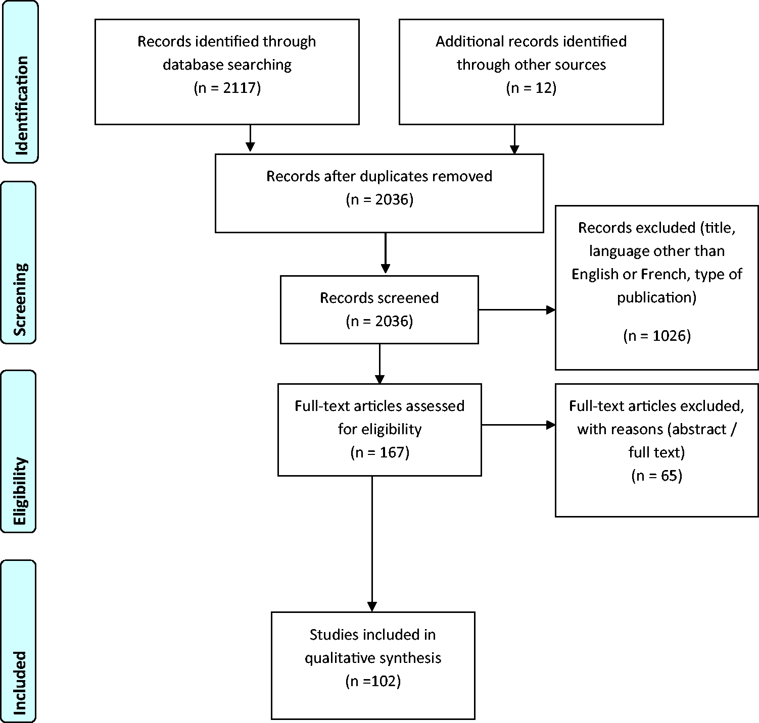
Results
The search strategy identified 2036 articles. Within these articles, 102 were considered relevant to the study. Articles referring to the development studies of clinical scores were included. Validation articles were analysed, especially if they involved specific subgroups.
The search strategy identified eight distinct clinical PTP scores, developed in a number of clinical settings (inpatients or outpatients, including patients with previous thromboembolic events, validated on specific subgroups). The clinical scores are described in the following sections.
Score development studies varied in relation to the following criteria: inclusion and exclusion criteria, imaging modality and strategy of DVT, imaging of distal lower limb deep veins, diagnostic and treatment strategy and type of D-dimer test.
The clinical scores
The Wells score
Available clinical scores for DVT prediction.

DVT: deep vein thrombosis.
Analysing the data from this very first step, a simplified score was developed and validated in a prospective cohort study of 593 outpatients (Table 1). 13 Some of the variables were excluded (hospitalisation within six months, recent trauma, erythema, family history of DVT). This classic Wells score is a 9-point score, giving one point for each clinical presentation (Table 1) and two negative points if an alternative diagnosis is possible. The test categorises patients into three groups of low (<1 points), moderate (1–2) and high suspicion (>3 points). The Wells score allowed patients to be categorised into high, moderate and low risk with a prevalence of DVT of 75%, 17% and 3%, respectively. The Wells score in combination with a low D-dimer level showed a high negative predictive value (NPV) as confirmed by several validation studies.10,14
The score was modified in 200315,16 when another parameter, the history of a previous DVT was added. This modified score was a simplified two-level score 16 with a score of <2 indicating unlikely DVT and ≥2 indicating high possibility of DVT or a three-level score (>3 for high-risk group, 1–2 for moderate risk and <1 for low risk). 15 Adding previous history of DVT in the modified score increases the number of patients in the high-risk group when compared to the classic score. Anderson et al. 15 using the three-level score found an NPV of D-dimer test (SimpliRed or Il-test) of 99.1% for the low-risk group, 94.1% for the moderate-risk group and 76.9% for the high-risk group. Comparing classic Wells score and modified score showed similar accuracy of scores, 17 especially for outpatients with proximal DVT. 18 Geersing et al., 19 in a meta-analysis of 13 studies using classic or modified Wells score and different point of care D-dimer tests, including 10,002 patients concluded that an unlikely Wells score with a negative D-dimer can safely exclude DVT in outpatients or primary care patients with less than 2% cases missed. A low Wells score alone could not exclude DVT. Patients with active malignancy or previous DVT may be more difficult to evaluate.
The specificity of the D-dimer test is lower in higher grades of PTP.
In the 2012 Chest guidelines, 20 both the original (classic) and modified Wells scores are mentioned. Modified Wells score was not considered widely evaluated. In the Institute of Clinical System Improvement guidelines (2013), the original Wells score is recommended. 21
There are several commercial D-dimer assays, which vary in their methodology, sensitivity and specificity. 22 Enzyme-linked immunosorbent assays (ELISA) and enzyme-linked immunofluorescence assays (ELFAs) are more sensitive but less specific. Qualitative or semi-quantitative and immunoturbimetric latex agglutination assays and whole blood agglutination assays are now widely used. Whole blood assays and latex qualitative and semi-quantitative are rapid assays with lower sensitivity and better specificity.23,24 Stein et al. 25 found higher sensitivity of ELISAs and ELFAs comparing to other tests including quantitative immunoturbimetric latex assays, while a review by Di Nisio et al. 23 showed that ELISAs, ELFAs and quantitative latex assays have similar sensitivity (94%, 97% and 93%, respectively) and specificity (53%, 46% and 53%, respectively). Qualitative, whole blood test are point of care tests. The use of a qualitative test for D-dimer (SimpliRED) in combination with classic Wells score results in high sensitivity and NPV.26–28 A systematic review of the point of care available tests (SimpliRED, Clearview Simplify, Cardiac and Triage D-dimer) showed that the sensibility of these tests was 0.85, 0.78, 0.96, 0.93, respectively, and the specificity was 0.74, 0.62, 0.57 and 0.48, respectively, making these tests accurate in excluding DVT in low-risk patients. 29 Other studies have reiterated that a negative D-dimer can exclude DVT in the low clinical probability group30–34 even when a point of care D-dimer test is used. 33
HemosIL D-dimer test (a turbimetric latex test) had sensitivity 100% and a specificity of 42.1% when used in combination with an original Wells clinical score. 35 The specificity is higher for lower PTP (32.4% for high PTP group, 42.1% for moderate and 45.5% for low). A low or moderate modified Wells score in combination with a turbimetric latex test (HemosIL D-dimer Test) can exclude DVT in outpatients. 36 Funfsinn et al., 37 comparing different D-dimer tests (ELISA or turbimetric latex or ELFA), also concluded that a negative test in combination with a low clinical score can exclude DVT.
Using a latex quantitative D-dimer test, Bates et al. 38 and Schutgens et al. 39 showed that DVT can be safely excluded in the low-risk group as well as in the moderate-risk group when D-dimer levels are low. This was confirmed using different turbimetric D-dimer tests.35,40 Yamaki et al. 41 suggested that a different cut-off point for a quantitative latex D-dimer test should be used for the moderate clinical risk group of patients. Similarly, Fancher et al. 42 in a review of studies using a rapid, point of care D-dimer test or a sensitive D-dimer test, showed that the three-months incidence of DVT was 0.5% in the rapid D-dimer group with low clinical Wells score and 0.4% in the sensitive D-dimer group with a low or moderate clinical score. They suggested using a rapid test for the low clinical probability group and a more sensitive test for the moderate clinical probability groups.
Based on CHEST 2012 guidelines, 20 a negative moderate sensitivity D-dimer test or a high sensitivity D-dimer test in combination with a low pretest probability can exclude DVT. A high sensitivity D-dimer test and a moderate PTP can also exclude DVT. Patients with high PTP should undergo imaging.
The Wells score has been shown to be equally reliable when applied by the emergency department nurses or doctors in training, thus increasing the applicability of the test.43–45
Both the two-level Wells score and the three-level score apply equally in men and women, although men were found to have a higher prevalence of confirmed DVT than women.46,47
The development and validation studies excluded patients receiving anticoagulation treatment in order to avoid unreliability of the D-dimer test, as shown by Aguilar and del Villar. 48
Janes and Ashford 49 suggested a simplified form of the Wells score in order to be easily applied by junior doctors. The points included were: history of previous DVT, malignancy, paralysis, bedridden, pregnancy, strong family history, thigh and calf swelling, calf swelling >3 cm, localised tenderness along the deep system, D-dimer test. Only if all the points of the clinical score were negative in conjugation with a negative D-dimer (SimplyRed test), DVT was excluded. Junior doctors applied the score, and only 1 patient of the 98 patients with negative score and D-dimer presented venous thromboembolism (VTE) during follow-up.
Another variation of the modified Wells score was published by Grune et al. 12 and included the first nine points of the modified Wells, complimented by the presence of alternative diagnosis (−2 points), muscle hardening (1 point) and Meyer’s calf pressure sign (1 point) and the absence of concomitant existence of muscle hardening, Meyer’s sign and calf swelling (−2 points) (Table 1). The probability of having DVT is 88% if nine signs are present and 78% if three signs are present. The NPV is 91%–95%, but the specificity is 100%.
Michiels et al. 50 suggested that the component ‘alternative diagnosis’ of Wells score is subjective and only useful and accurate to experienced physicians. Therefore, they proposed a slightly different score, named Rotterdam, consisting of eight points, excluding the ‘alternative diagnosis’ component (Table 1). The strategy suggested involved a DUS and a D-dimer test at presentation, followed by the PTP if D-dimer is positive.
The Oudega score
The Wells score was developed in outpatients, and therefore, its accuracy in other subgroups has been questioned.51,52 The Oudega score was suggested as an alternative score able to include primary care patients. The score was developed after analysing the medical history of 1295 consecutive patients presenting with suspected DVT and included patients with a past history of confirmed DVT.53,54
The D-dimer status is added as a variable as part of the Oudega score. The following points were included in the assessment: male sex (1 point), use of oestrogens (1 point), malignancy (1 point), recent surgery (1 point), absence of trauma (four weeks) (1 point), vein distension (1 point), calf difference >3 cm (2 points) and positive D-dimer test (6 points) (Table 1). D-dimer was measured with two commercial quantitative assays, and with two different cut-off points (for the VIDAS assay, the cut-off point was 500 ng/mL, and while for the Tinaquant assay, it was 400 ng/mL). This resulted in a 13-point (maximum) score, separating patients into four risk groups for DVT probability (very low, low, moderate and high, with prevalence rates of 0.7%, 4.5%, 78.3% and 48.7%, respectively). A negative D-dimer result would miss 1.2% of DVTs, while a negative D-dimer and a score less than 2 would miss only 0.7% of DVTs.
Some differences relating to the defining of risk factors included the definition for bedridden (immobilisation for more than three days), recent surgery (within four weeks) and leg trauma (within four weeks). Venous DUS was limited to the proximal leg. The score was validated by the same team in 532 primary care patients confirming the accuracy of the test. External validation by Buller et al. 55 using a point of care D-dimer also confirmed the findings, missing 1.4% of cases with DVT in the low-risk group.
Geersing et al. 56 compared the efficiency of Oudega score with clinical judgment of family doctors in a group of primary care patients. Both scores had a similar utility in diagnosing DVT, with less than 2% of missing cases; family doctors may refer a larger number of patients for DUS.
The Hamilton score
The Hamilton score 57 was developed in 214 consecutive ambulatory patients presenting to the emergency department and validated in 312 patients in the same department (temporal validation). The Hamilton score includes six objective components (immobilisation of the lower limb (2 points), active malignancy (2 points), bed rest or recent surgery (<4 weeks) (1 point), male sex (1 point), calf circumference difference greater than 3 cm (1 point), erythema (1 point)) and a subjective component (strong clinical suspicion of DVT without other diagnostic possibilities by the emergency physicians (2 points)). A score of 3 or greater indicates a likely probability, and a score of 2 or less represents an unlikely probability for DVT. A combined diagnostic strategy of unlikely probability Hamilton score and a negative D-dimer (Simplify) had an NPV of 99% (95% confidence interval (CI): 94.7%–100%).
The Hamilton score was compared to the modified Wells score, and the scores performed similarly. 58 The sensitivity for the Hamilton score was 66.67% and the specificity 71.14%, positive predictive value 39% and NPV 88%. When the score was combined with a bedside D-dimer test, these values were 99%, 42%, 32% and 99%, respectively. If a modified Wells score was applied to the same population, the values were 99%, 33%, 29% and 99%, respectively. In the Hamilton study, the whole leg was scanned using DUS, and patients with distal DVT were treated with anticoagulation; in the Wells study only the proximal leg underwent DUS. The Hamilton score was found to be able to identify 27% more patients with in the unlikely probability score with low D-dimer compared with the modified Wells score. 58 The combination of a low Hamilton score and a negative immunochromatographic point of care D-dimer test Simplify could exclude proximal or distal DVT. 59
The Kahn score
In 1999, another PTP was developed by Kahn for outpatients and inpatients. The Kahn score 60 is a 4-point score including the following variables: sex, history of orthopaedic surgery during the last six months, superficial vein dilatation and local warmth. For the development study, 324 patients with suspected DVT were included; DVT was confirmed with impedance plethysmography or venogram or both. Positive plethysmographic tests were usually confirmed with a venogram. D-dimers are not reported. The score categorises patients into three groups of probability. The prevalence of DVT within these three categories was 76%, 26% and 9% for the high, moderate and low probability groups, respectively. In a study comparing Wells, Constans (described below), Kahn and St Andre (described below) scores in Chinese hospitalised population, the area under the receiver-operator characteristic curve (AUC-ROC) was 0.79 for the Kahn score, lower than Constans and Wells scores, indicating that Constans and Wells scores performed better in that specific population. 61
The Constans score
The Constans score is an outpatient score combining six variables: male sex (+1 point), paralysis or immobilisation of lower limb (+1 point), confinement to bed for three days (+1 point), lower limb enlargement (+1 point), unilateral lower limb pain (+1 point) and other diagnosis at least as plausible (−1 point) (Table 1). The component added is the unilateral leg pain. 62 The score was developed in 444 outpatients and validated in 282 patients. Patients had a whole leg DUS. The AUC-ROC was slightly higher for the Constans score (0.79, 95% CI: 0.74–0.84) compared to Wells (0.76), Kahn (0.57) and St Andre scores (0.67).
St Andre score
St Andre test was developed for inpatients but evaluated in outpatients by Constans et al. 62 The score was developed as a simpler and accurate alternative to Wells score. 63 The St Andre score includes the following parameters: cancer, unilateral pitting oedema, local warmth, superficial venous dilatation, lower limb immobility and other diagnosis as plausible (−1). The results were compared to Kahn score and Wells score for inpatients. Both proximal and distal DVT were evaluated. The prevalence of DVT was 59% with a Wells score ≥3 and 13% with a Wells score of 0, 81% with a St Andre score of 3 and 13% with a St Andre score of 0. Amongst the 17 patients classified as having a high St Andre score, 13 (76%) had a confirmed DVT. When Wells score was applied 33 of the 65 patients (3%) with a score ≥3 had DVT. A total of 141 and 73 patients were classified as low St Andre score or Wells score, respectively, and 15 or 9 of them had a DVT. The test classifies a higher number of patients in the lower risk group with a slight increase in DVT prevalence. Kahn score was less accurate in this inpatient population compared to the Wells and St Andre scores.
The Gagne score
The Gagne score 11 was developed in order to identify patients with DVT in primary care. In contrast to the Oudega score that includes D-dimer value as a variable, the Gagne score does not require D-dimer measurements. Primary care doctors recruited 276 non-consecutive patients. Patients with signs of PE and patients receiving anticoagulation for more than five days were excluded. The score included four variables of medical history (history of VTE (+1), active cancer (+3), immobilisation in previous month (+1), use of contraceptives (+2)), one clinical variable (reduced ankle mobility (+1)) and the absence of alternative diagnosis (−3). The score classifies patients into a two- or three-grade system. Within the high-risk group, 63% of patients had confirmed DVT, while 26% of patients with a low risk had DVT. The DVT was confirmed if proximal or distal thrombus was present. Thrombosis of calf muscular veins was not considered as confirmed DVT.
The score was validated with 164 patients recruited in a similar way, within the same population. 64 In this study, the clinical variable is replaced by the ankle mobility estimation instead of calf swelling. Amongst the patients with a high score (≥2) 43% were found to have DVT, while 29% of patients with a low score (<2) had confirmed DVT. The results were less discriminating than the development study, and therefore, the authors suggested to modify the level between the two probability groups (a score of <1 rather than <2 for the low-risk group). If the level of score is reduced and patients with proximal DVT are only considered as affected, then the percentages of DVT are 23% and 6.5% in the high- and low-risk groups, respectively. The score was validated on 3523 patients of the OPTIMEV study, 65 including ambulatory or hospitalised patients referred to a vascular specialist.
Other scores
The LEFT (Left, Oedema, First Trimester) clinical prediction score is a diagnostic score developed in 194 pregnant women with suspected DVT recruited over eight years 66 and validated later.67,68 The analysis of known risk factors revealed that three characteristics were related to DVT: the left side, diameter >2 cm and first trimester of pregnancy. The sensitivity and specificity for a LEFT score of zero was 100% and 50%, respectively; the NPV was 100%.
Accuracy in different subgroups
Inpatients, outpatients and primary care patients
Although the Wells score was developed on outpatients, a study from the same team validated the test on inpatients, 69 finding an NPV for a low D-dimer and low clinical score of 96.2%. Aschwanden et al. 70 found an NPV of 98.6% for outpatients and 97.2% for inpatients when applying the classic Wells score in combination with the SimpliRed D-dimer test. The D-dimer value among inpatients is very often elevated. Silveira et al. 71 conducted a study of 1135 inpatients with suspected DVT, including medical and surgical inpatients; 137 (12.1%) had confirmed proximal DVT. The AUC-ROC of the Wells score accuracy (not combined with D-dimer test) was 0.6, showing that the Wells score was only slightly better than chance in diagnosing DVT amongst inpatients. Of the D-dimer tests requested, 93.5% were positive (the modified Wells score was used, but patients were classified into three grades of DVT risk). Similar findings have been published by other groups. 18
Sermsathanasawadi et al. 72 applied and compared five different clinical scores (Wells, modified Wells, Kahn, St Andre and Constans scores) on 500 consecutive outpatients and inpatients. They showed that modified Wells score and Constans score were more accurate for unselected outpatients and inpatients (based upon AUC-ROC), but they had poor prediction values for inpatients (AUC-ROC: 0.59–0.60). A retrospective analysis of data showed that the original Wells score was accurate when applied to outpatients with musculoskeletal disorders reported. 73
van der Velde et al. 74 used the data from the AMUSE study to compare Wells and Oudega score in primary care patients, finding similar results for both scores, with and without consideration of the D-dimer levels. A low modified Wells score or Oudega score and a normal rapid bedside D-dimer test (Clearview, Simplify) could avoid referring 50% of patients for DUS. That resulted in 1.6% missed VTE for Wells score and 1.4% for Oudega score. Geersing et al., 19 in their meta-analysis of 13 studies, showed that the Wells score could be safely applied to primary care patients. The explanation that the team suggested for these conflicting findings 53 is that the Oudega study had included many patients with recurrent DVT (24%) and the original Wells score was used which is not developed to include recurrent DVT, while Van der Velde included substantially fewer patients with recurrent DVT (15%). The application of modified Wells score in combination with a point of care D-dimer test or selective DUS in primary care patients has an NPV of 99%. 75 If clinical judgement is used instead of Wells score, there is an overestimation of patients likely to have DVT and more tests are requested.
Martin et al. 36 in a prospective study, examining the feasibility and safety of out of hours DUS, included inpatients and outpatients. The NPV of the strategy followed was similar in the two groups, and the protocol followed could safely rule out DVT. A negative D-dimer (HemosIL) in combination with a modified Wells score ≤2 could exclude DVT safely. Patients with high D-dimer or Wells score were given an interim dose of anticoagulation and had a DUS performed the following day. Only 1 inpatient of the 345 inpatients had a moderate Wells score and negative D-dimer test and subsequently had a confirmed DVT. For the inpatients with confirmed DVT, the Wells score was ≤0 in 9.8% of patients, 1–2 in 15.4% and ≥3 in 35.7%. For outpatients, these values were 3.3%, 16.8% and 69%, respectively.
Previous DVT
The difference between the original Wells and the modified Wells score is the addition of the history of previous DVT. Engelberger et al., 18 however, found similar results when applying either modified or original Wells score in an outpatient population including participants with previous DVT. Similarly, Cornuz et al. 76 showed that the original Wells score can be applied to patients with previous DVT. A meta-analysis of 29 studies by Goodacre et al., 77 including several available scores, showed that Wells score performs worse on patients with previous DVT or older patients.
The majority of the studies in this field excluded patients with previous DVT. Within the development studies, Oudega et al. had included patients with previous DVT history; 24% of the recruited patients had a history of DVT. In the primary care setting, Buller also identified that 15% had a history of DVT. Silveira et al. 71 found that amongst 1135 inpatients in different departments presenting with suspected DVT, 14.5% had a history of DVT. A previous DVT event was more common in medical units than surgical units. Anderson et al. 15 included patients with previous thromboembolic events, and the NPV of low prediction risk in combination with a low D-dimer was still 99%.
Isolated Distal DVT
The original and modified Wells scores were developed and validated in outpatients that underwent DUS of only the proximal leg, and a venogram was performed when necessary.9,13 Only 5.6% of patients required venography; of the 17 patients with a negative DUS in the high-risk group, 4 had confirmed DVT (2 proximal, 1 proximal and calf and 1 distal DVT). 13 When the modified Wells score is applied, of the 94 patients with high clinical risk and normal DUS, 4 were found to have DVT on venography (2 proximal and 2 distal). 15 The accuracy of the Wells score in predicting distal DVT has been questioned. Engelberger et al. 18 compared the classic and modified Wells score in 298 outpatients and inpatients and concluded that both scores were more accurate in predicting proximal DVT than distal, and in predicting DVT in outpatients than inpatients. The participants were examined by a vascular specialist and had a complete lower limb DUS. Sartori et al., 78 examining outpatients with distal DVT, found an 8.3% DVT rate in the low pretest risk subgroup, 13.5% in the moderate risk group and 22.2% in the high-risk group. Although the modified Wells score was not as accurate when applied to patients with isolated distal DVT compared to proximal DVT, the combination of a low clinical pretest with a low level of D-dimer had an NPV >95%. Nonetheless, D-dimer levels tend to be higher in patients with proximal DVT 79 than in patients with distal DVT.
Anderson et al. 15 included venography when necessary, but not regularly, in their study, with DUS confined to proximal veins. Patients with a high suspicion of DVT, a normal DUS and positive D-dimer would undergo venography. They found two additional cases of distal DVT in the subgroup of 94 patients with high clinical suspicion and one additional case in the moderate risk subgroup of 80 patients. These patients received anticoagulation, although they were not considered as part of the positive DVT results. In the low clinical risk group (n = 448), two patients were found with distal DVT. In total, five distal DVTs were identified.
In the low-risk clinical groups, DVT cases can be restricted to distal veins more often than in moderate- to high-risk groups. 70 This was found to be the case when the Gagne score was applied to a large population. 65
Cornuz et al., 76 in an external Wells score validation, had two patients with positive DUS within the group of 49 patients with negative D-dimers measured with a rapid ELISA test. One of these scans concerned distal veins. D-dimer is probably less sensitive in distal DVT.10,34,80 Luxemburg et al. 81 showed that some D-dimer tests are less accurate for distal DVT than others.
Kahn et al. 60 performed venography or plethysmography in their derivation study; 30.8% of the venograms detected isolated DVTs. Almost all the positive plethysmographs were confirmed with venography. No difference in the clinical score’s performance in relation to proximal and distal DVT is mentioned. All patients with DVT (proximal or distal) received treatment and no new events during the follow-up period were reported. Repeated negative plethysmography was not confirmed with venography; considering the inferior sensitivity of plethysmography for distal DVT, 82 cases may have been missed.
Furthermore, Ambid-Lacombet et al. 17 did not find any difference when applying the Wells score in patients with proximal or distal DVT. Walsh et al. 83 found an NPV of the original Wells score combined with a low D-dimer test to be 100%, including patients with distal DVT.
Elderly patients
The accuracy of clinical scores and D-dimer test in elderly patients has been questioned, as clinical signs are felt to be less reliable.84,85 A meta-analysis of studies using the original or modified Wells score and different D-dimer assays showed that a low D-dimer level in combination with a low clinical probability could exclude DVT in elderly individuals, 86 while other studies have questioned the accuracy of the clinical scores. Schouten et al., 87 studying the elderly population in the primary care setting, found a high number of misdiagnosed cases: 5.8% of DVT cases were missed in the low-risk groups if the Oudega score was applied, and 4.9% if the Wells score in combination with a qualitative D-dimer test was used. They suggested a simplified Oudega score which included three variables (calf circumference difference, gender and D-dimer levels); the missed DVT rate decreased to 2.3%. According to this suggestion, patients with negative D-dimer test should be send for additional imaging only if the leg circumference is >3 cm. If D-dimer is positive, then additional imaging is justified. In the case of males, or females with leg circumference >3 cm, it was recommended that treatment should be prior to obtaining imaging.
Another option would be to adapt the D-dimer diagnostic level. As D-dimer levels increase with age, 88 an alternative cut-off point for the test has been suggested for older patients.
Douma et al. 89 suggested an age-adjusted value of D-dimer ((patient’s age × 10) µg/L in patients aged > 50). The validation of the findings was done using VIDAS or Tinaquant (latex quantitative) test on patients with suspicion of PE. Haas et al. suggested a different cut-off value of 750 µg/L for patients older than 60 with suspected DVT. Haas et al. used three different latex quantitative D-dimer tests (STA Latest, Tinaquant and Innovance). The cut-off value of 750 µg/L for patients >60 years old gives an NPV of 100% and a specificity of 48.5% for Liatest, 60.6% for Tinaquant and 49.2% for innovance, resulting in a decrease of the number of scans needed.
In primary care patients, the modified Wells score and either a cut-off D-dimer value of 750 µg/L for patients older than 60, 90 or the use of an age-dependent D-dimer cut-off value could safely exclude DVT. Schouten et al. 91 showed that the use of a fixed cut-off value of 750 µg/L after the age of 60 years increases the number of patients that can be excluded by 5.4%, while the number of false-negative cases remains the same. The use of the age-dependent algorithm for D-dimer cut-off point in the same population increased the number of patients excluded by 5.7%, with a slight increase in false-negative cases from 0.3% to 0.5%. The age-dependent D-dimer cut-off value is calculated as age (years) × 10 µg/L, in patients older than 50 years. The age-adjusted D-dimer cut-off algorithm increases the specificity of the test in the elderly people without affecting sensitivity. 92 Douma et al., 93 in a meta-analysis of five trials, showed that if the age-adjusted D-dimer value is applied to quantitative latex tests or ELFA D-dimer test, another 8% to 8.6% of patients with non-high clinical score can be excluded, while the number of false-negative results remains the same.
Cini et al. 94 evaluated the performance of two different age-adjusted values of a latex quantitative D-dimer test (HemosIL, Instrumentation Laboratory) in 326 outpatients, applying the modified Wells score. The use of a higher cut-off value (376 ng/mL) compared to the conventional value (320 ng/mL) for patients >60 years, or the use of an age-adjusted cut-off point (age × 5 ng/mL) for patients >50 years in combination with a low modified Wells score could safely exclude more patients than the conventional cut-off value. There was an absolute increase of the number of patients in the non-high clinical risk group that could be excluded using the higher cut-off value (20.8%) or the age-adjusted value (24.1%), and this was more evident in older groups.
Cancer
The accuracy of prediction rules in cancer patients may be lower than for the general population. A meta-analysis of 13 studies 19 applying the Wells score in outpatients and primary care patients showed that the combination of a low D-dimer and a low Wells score could exclude DVT in both sexes, outpatients or primary care patients, but not in patients with DVT recurrence or cancer.
Carrier et al. 86 retrospectively compared the results of three studies conducted in emergency and outpatients departments using either the original or the modified Wells score. They concluded that although a low Wells score with a negative D-dimer test can safely exclude DVT, the specificity of the prediction rule is low as many patients with cancer have a positive D-dimer.
It has been noted that D-dimer levels are elevated in cancer patients and older individuals, and therefore, it has been suggested that D-dimer cut-off points for these patients should be higher. Chen et al. 95 suggested that a cut-off point of 981 ng/dL could increase the sensitivity of the test.
Other studies have shown that in cancer patients, a low D-dimer and a low clinical score is able to exclude DVT. 96 The modified Wells score in combination with a low D-dimer test was used to exclude pre-operative asymptomatic DVT in a group of patients with colorectal cancer with an NPV of 99%. 97
Dermatology patients
The Wells score and the D-dimer also have limited value in dermatology patients, as the D-dimer is often found to be positive. 98 Still, the NPV for D-dimer is 100%. Haenssle et al. 98 analysed the data of 102 patients hospitalised in a dermatology unit with different primary diagnoses (erysipelas, chronic leg ulcers, venous stasis, inflammatory skin lesions, skin cancer or other skin disorders). The Wells score could not differentiate DVT from non-DVT patients. The NPV for D-dimer remains high for patients with no inflammatory skin disorders.
Paediatric patients
VTE is a rising problem in children, and the diagnosis is often challenging. Wells score alone cannot exclude DVT in children. 99 Kerlin et al. 100 retrospectively analysing data from 389 children with a suspicion of VTE found the following variables to be related to VTE: male gender, asymmetric extremity (swelling, oedema, discoloration) central line catheter utilisation or dysfunction, active cancer and absence of alternative diagnosis. The ability of this score to discriminate was significant in the development study (AUC 0.73; p < 0.0010) but moderate in a validation study (AUC 0.64; p < 0.011).
Other
Clinical diagnosis of DVT can be challenging in postoperative orthopaedic patients. Baud et al. 101 developed two different scoring systems in order to select patients after hip or knee replacement that would benefit from DUS. The following eight points were included in the first score: spontaneous pain, provoked pain, high temperature, oedema, ecchymosis, calf circumference >3 cm, female gender and previous VTE. Each parameter attributes one point. For the second score, created after multivariate analysis, temperature, gender, previous VTE, provoked pain and circumference >3 cm give 2 points, and total maximum score is 13. For the first score, a value >4 is related to a risk of DVT of 34% and a value >6 to a risk 56%. When the second score is used, a value <6 is related to low DVT probability and a score >9 to high DVT probability.
Discussion
Clinical prediction rules are developed to support clinical decision making; however, we must be aware of their limitations. Comparing the different scores, one can appreciate that development studies had different target populations (outpatients, inpatients or other specific subgroups) and different exclusion criteria. Patients with a history of DVT were often excluded, which invalidates the test in cases of recurrent DVT. Patients with signs of PE were also often excluded from these studies, excluding a subgroup of patients with respiratory symptoms of other cause, for example, individuals with heart failure. The validation studies have shown that specific algorithms might be more sensitive for specific subgroups of patients. Retrieving generalised conclusions and suggesting algorithms from these studies would be inaccurate as they are very heterogeneous in design.
The application of the scores can also be influenced by the experience and expertise of the person using them. The inter-observer agreement between emergency physicians and vascular physicians in applying the classic Wells score was excellent; however, the accuracy of the empirical assessment performed by specialised physicians was higher 102 or equal 103 compared with that of the Wells score. In another study, the sensitivity was higher when the Wells score was applied by a specialist nurse than when applied by a physician. 76 When the Wells score was used by physicians in training, the accuracy was satisfactory with the physicians in training slightly overestimating the pretest probability when compared to the supervising physicians’ assessments. 43 Miron et al. 104 also evaluated the empirical assessment compared to Wells clinical score, calculated retrospectively and found a similar accuracy. The likelihood ratio of DVT in the low empirically estimated risk group was 0.05 and 0.12 when Wells score was applied. The empirical assessment could slightly more accurately categorise patients in the high-risk group. More patients were also categorised in the low or high probability group by Wells score and less in the moderate risk group comparing to empirical assessment. Although both scores had similar accuracy, the agreement between them was low, and the accuracy depended also on the strategy applied.
Regarding the definition of the variables, localised tenderness is probably the most difficult variable to standardise. 62 The term immobility could also pertain to various degrees of severity and aetiology. Beam et al. 105 underlined the importance of clearly stating the cause of immobility when calculating DVT risk. They showed that different levels and underlying causes of immobility are correlated to different risks of DVT. In a retrospective study, 7940 self-presenting emergency department patients were categorised according to their degree of immobility in six grades: no immobility, general or whole-body immobility greater than 48 h, limb (orthopaedic) immobility, travel greater than 8 h causing immobility within the previous seven days, neurologic paralysis or other immobility not listed above. Limb immobility, or whole body, or neurological immobility was related to higher risk.
Amongst the clinical scores, only the Oudega score includes D-dimer levels as a variable. In addition, the D-dimer tests used in the various studies have different accuracy. When comparing different D-dimer tests and different cut-off values, in combination with the described clinical prediction scores, variation in NPVs is reported, suggesting that each test may be adequate for a specific strategy. 106 The use of different D-dimer cut-off points for different grades of clinical risk has also been proposed. Linkins et al. 107 suggested that a D-dimer of less 1000 µg/dL could exclude DVT in patients with low clinical score, while the cut-off value should be 500 µg/dL when patients are at moderate or high risk. Specific subgroups might benefit from different D-dimer tests and cut-offs. 108 As D-dimer increases progressively during pregnancy, 109 cut-off values can be adapted or D-dimer test with higher specificity could be used. 110
Conclusion
We describe the available clinical diagnostic scores for DVT with a focus on development studies and validation studies in different populations. The different scores have been developed within different populations, and this should be considered when employing the scores in clinical practice. Specific groups like cancer patients or patients with recurrent DVT may be more difficult to diagnose clinically, and scores may apply less readily to these subgroups. The clinical presentation alone often cannot diagnose DVT owing to the low specificity of symptoms. The detailed knowledge of the methodology and development of the various clinical prediction rules results in being able to understand the power and the limitations of the clinical PTP scores.
Footnotes
Declaration of Conflicting Interests
The author(s) declared no potential conflicts of interest with respect to the research, authorship, and/or publication of this article.
Funding
The author(s) received no financial support for the research, authorship, and/or publication of this article.