



Abstract
Background
Cyanoacrylate adhesive closure is a technically simple alternative to endothermal ablation of peripheral veins. N-butyl cyanoacrylate is delivered via catheters or by percutaneous injection resulting in occlusion of target veins. The local tissue reaction or the systemic immune response that may follow have not been characterised.
Aim
To characterise the late local tissue reaction to N-butyl cyanoacrylate glue injected in peripheral vessels.
Methods
Biopsies were obtained from two patients. In patient one, distal tributaries of the great saphenous vein were injected with VenaBlock™ glue under ultrasound guidance. Ultrasound-guided incisional biopsies were performed at one week, six weeks and 12 months. In patient two, a peripheral arterio-venous malformation was injected with Venablock™ and biopsy was performed 12 months later. Histological analysis was performed using haematoxylin and eosin and immunofixation with CD-4, CD-31, CD-34, CD-68 and D2-40.
Results
Echogenic material with a strong shadow artefact consistent with the injected N-butyl cyanoacrylate was observed on ultrasound on all follow-up occasions. Biopsies taken at one week showed intravascular glue without histiocytes. Biopsies at six weeks showed isolated foreign body histiocytes coating intravascular fibrillary glue spicules but no granuloma formation. The one-year biopsies showed extravascular changes including fibrosis, lymphoid aggregates and multiple extravascular foreign body cavitated granulomas. Some vessel lumens contained residual spicules of glue but no intravascular granulomas. The extravascular granulomas were deeply located, asymptomatic and not complicated by clinical ulceration. Histologically, there was no evidence of transepidermal elimination.
Conclusion
Extravascular foreign body cavitated granulomas containing spicules of glue with fibrosis and lymphoid aggregates occur as a delayed finding following the use of N-butyl cyanoacrylate.
Keywords
Introduction
Cyanoacrylate adhesive closure (CAC) is a technically simple alternative to endothermal ablation (ETA) or surgical stripping of saphenous veins. The procedure can be completed in less than 30 min and is marketed as a non-thermal non-tumescent (NTNT) technique not requiring compression or tumescent anaesthesia. 1 The cyanoacrylate (CA) glue is delivered into target veins via specialised catheters or by direct percutaneous injections under ultrasound guidance. On exposure to intra-vascular blood, CA monomers polymerise to form a solid cast which adjusts to the shape of the target vessel causing occlusion.
N-butyl CA (n-BCA) is used as an embolising agent to treat vascular anomalies and gastro-oesophageal varices. There are currently four products marketed internationally for CAC treatment of peripheral veins. These are VenaSeal™ (Medtronic, Minnesota, USA), Venablock™ and Veinoff™ (Invamed, Ankara, Turkey) and Variclose™ (Biolas, Ankara, Turkey). Similar products are being manufactured and will enter the market in various countries in future.
The late pathological reaction that follows CAC treatmet of peripheral veins has not been investigated in humans. Histopathological studies in pig arteries and veins, as well as arteriovenous malformations (AVMs) in humans have consistently demonstrated chronic granulomatous reactions.2–7 A worrying recent clinical case report described suppurative granulomas with extrusion of the n-BCA four months after bilateral treatment of great saphenous veins (GSVs) with VenaSeal™ system. 8 The complication occurring in both legs after CAC treatment in both legs indicates a probable immunological tissue response rather than a local technical complication. 8
The only available histological study of a human saphenous vein after catheter-delivery of VenaSeal™ glue is by Shaidakov et al. 9 The GSV was removed 10 min after the procedure and sent for histology (as confirmed through personal communication with Prof. Shaidakov) giving inadequate time for a tissue response. However, perivascular mast cell degranulation was noted. The aim of the present study was to characterise the late tissue reaction to n-BCA injection in peripheral veins and AVMs and whether this involves granuloma formation and immune activation.
Materials and methods
Materials
The materials included: VenaBlock™ (Invamed, Ankara, Turkey); 25G 1.5 inch (0.5mm × 38mm) needle (BD PrecisionGlide™, Franklin Lakes, USA); 19G 1.5 inch TW (1.1mm × 38mm) needle (BD PrecisionGlide™, Franklin Lakes, USA) and 1mL SOFT-JECT® Luer Lock no dead-space syringe (Henke-Sass Wolf GmbH, Tuttlingen, Germany); TOSHIBA Aplio™ 500 Platinum Series (Canon, Tokyo, Japan).
Methods
Informed consent was obtained for the procedure and the subsequent biopsies.
Duplex ultrasound
A complete mapping of the venous system of the affected leg was conducted prior to interventions and on each follow-up occasion at one week, six weeks and one year following the completion of all procedures. This included interrogation of deep and superficial venous systems and perforating veins. Patency, competence, flow characteristics, evidence of previous thrombosis, arteriovenous anomalies were all looked for and if present, documented. Mapping of the venous system included both competent and incompetent veins. Reflux was defined as retrograde flow (superficial veins >0.5 s, deep veins >1 s).
Injection technique
The procedures were performed under aseptic conditions. VenaBlock™ glue was drawn up in a no dead-space 1 mL syringe via the 19G needle. The glue was then percutaneously delivered using a 25G needle in alliquots of 0.2 mL under ultrasound-guidance. Care was taken to remove the needle from the injected vessel within a few seconds. The injected target vessel was compressed with the ultrasound probe for 10 s after each injection. Glue was observed to occlude the target vessel on ultrasound. The target vessel was non-compressible at the end of the procedure and the echogenic appearance of glue was observed and recorded.
Patient 1 – peripheral venous glue ablation
A 28-year-old male with no history of autoimmune, inflammatory, granulomatous disease or allergies taking no medications, underwent treatment for incompetent peripheral veins. Two procedures were performed a week apart. On the first occasion, a right medial calf tributary of the GSV was injected with VenaBlock™ glue. On the second occasion, a right lateral calf tributary of the GSV was treated.
Incisional biospies to include skin, subcutaneous fat and the target vessels were obtained at one week, six weeks and one year post-glue injection. On each occasion, the injected veins were identified on ultrasound and an incisional biopsy was performed for histology. At one week post-glue injection, the injected medial calf vein was biopsied. At six weeks post-glue injection, samples were obtained from the injected lateral calf vein. At one year post-glue injection, samples were obtained from the lateral calf vein avoiding the six-week biopsy site.
Patient 2 – peripheral arteriovenous malformation glue ablation
This patient was a 27-year-old male with no history of autoimmune, inflammatory, granulomatous disease or allergies taking no medications, undergoing treatment for a peripheral AVM of the right anterior shin. Ultrasound identified the AVM and two feeding arteries, one communicating with the posterior tibial artery and the other with the anterior tibial artery. Draining veins of the AVM had been previously treated with endovenous laser ablation and foam ultrasound-guided sclerotherapy. Venablock™ was delivered percutaneously into the lateral shin feeding artery that communicated with the anterior tibial artery. Ultrasound-guided incisional biopsy was performed 12 months later.
Histopathology and immunohistochemistry
The biopsy specimens were stained with hematoxylin and eosin (H&E). Immunohistochemistry markers included the T-cell marker CD-4 used to help identify the lymphocytic infiltrate; CD-31 (PECAM-1), a vascular marker used to detect vascular endothelium and angiogenesis; CD-34, a surrogate marker for hematopoietic stem and progenitor cells but widely regarded as a marker for vascular endothelial progenitor cells; CD-68, marker for histiocytes and histiocytic granulomas and D2-40, a lymphatic marker used to detect lympahtic vessels.
Samples were tested for light polarisation, a technique used in light microscopy to identify foreign bodies found in cutaneous granulomas such as those found in sarcoidosis and identification of deposits such as amyloid, oxalate and gout.
Results
Clinical follow-up
Clinically, there were no systemic or local adverse events on all follow-up occasions (one week, six weeks and one year). No necrosis, dermatitis, phlebitis or other clinical complications occurred in either patient.
Ultrasound appearance
The injected vessels were identified on B-mode ultrasound on all follow-up occasions (one week, six weeks and one year) post-glue injection. The injected glue appeared echogenic and produced a shadow artefact on all occasions (Figure 1).
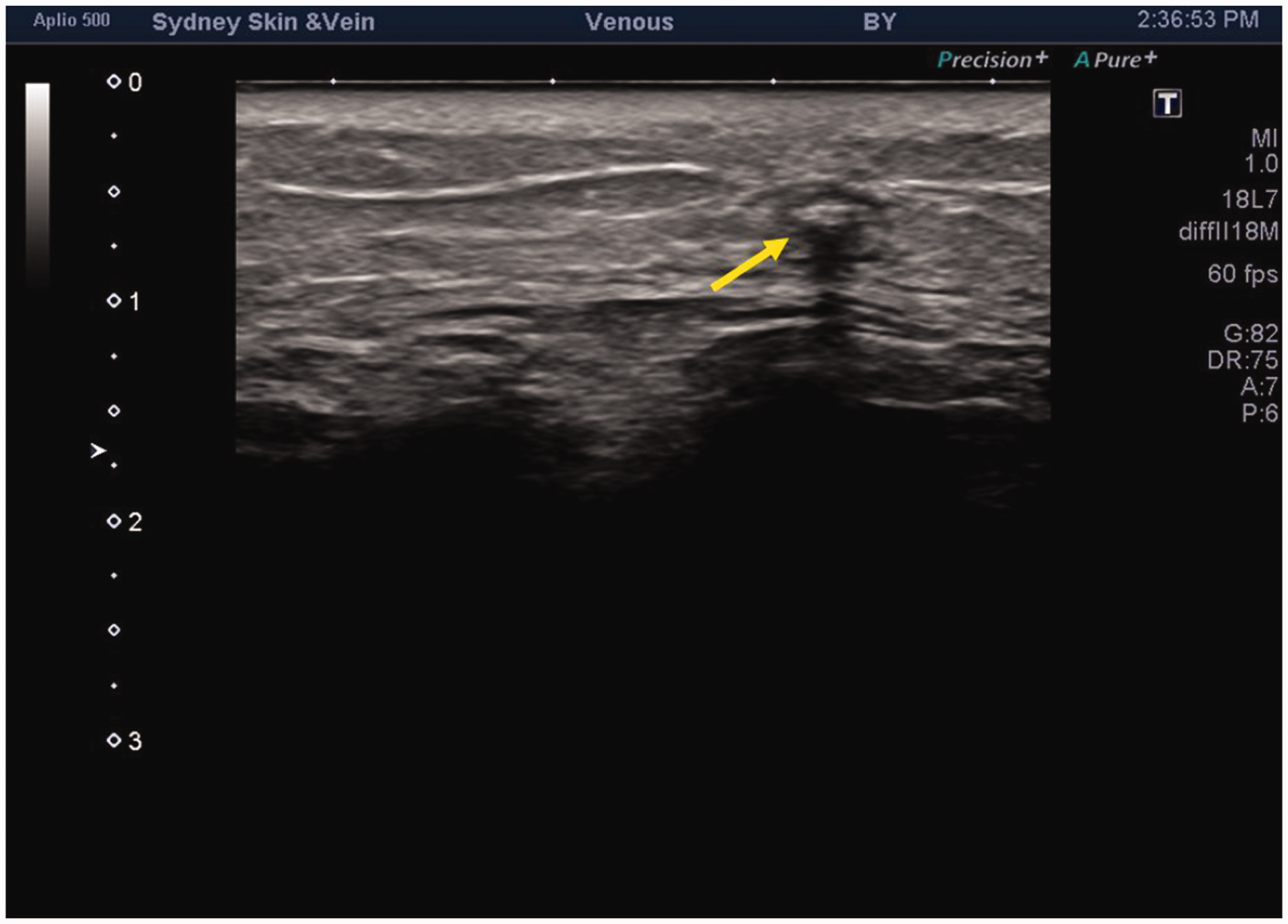
One year: Echogenic glue. B-mode ultrasound of a venous tributary injected with Venablock™. N-BCA is visible 12 months after the injection. Note the echogenic appearance of the foreign body material (yellow arrow) generating a shadow artefact.
Histopathology
1. Patient 1, peripheral vein – one week
The epidermis and dermis appeared normal. The glue was seen in the vein lumen presenting as a homogenous eosinophilic material that was crystallised to produce a snowflake-like appearance merging with more diffuse deposits coated with erythrocytes (Figure 2). No histiocytes or granulomas were evident around the glue. The glue did not polarise.

One week: Glue within the vein lumen. Injected vein lumen at one week demonstrating homogeneous, fibrillar and crystallised glue (arrows) without inflammatory response (hematoxylin and eosin, original magnification ×50).
2. Patient 1, peripheral vein – six weeks
The epidermis and dermis appeared normal. Glue and fibrillary spicules were observed in the sampled vein lumen. Fibrinous material and erythrocytes were adherent to the glue. No granulomatous reaction was seen. A small area in the lumen showed isolated foreign body histiocytes but no granulomas.
3. Patient 1, peripheral vein – one year
The epidermis and dermis appeared normal. In the deep tissue, there were multiple granulomas with multinucleated foreign body giant cells lining empty spaces with matted fibrillary borders of spicules of glue and surrounding fibrosis (Figure 3). The observed granulomas had typical features of foreign body granulomas and contained foreign body-type giant cells. All granulomas were extra-vascular and none were observed in the vessel walls or within the lumen of the treated vessels. Lymphoid hyperplasia with angioplasia was also evident (Figure 4). Multiple venules devoid of glue or a granulomatous reaction were present adjacent to the granulomatous cavities (Figures 5 and 6). A larger vein section in the deep subcutaneous tissue had residual glue spicules but no intravascular granulomas.

One year: Empty vein and extensive peri-vascular fibrosis. Biopsy taken at one year showing deep tissue with extensive fibrosis, large empty vascular lumen (arrow A) and focal lymphoid aggregates (arrow B) (hematoxylin and eosin, original magnification ×12.5).

One year: Fibrosis and lymphoid hyperplasia. Details of gross fibrosis and lymphoid hyperplasia at one year (arrows) (hematoxylin and eosin, original magnification ×50).

One year: Empty veins and extruded glue within cavitated granulomas. Details of empty vein (arrow A) and cavitated extravascular granuloma (arrow B) containing glue spicules (arrow C) at one year (hematoxylin and eosin, original magnification ×50).
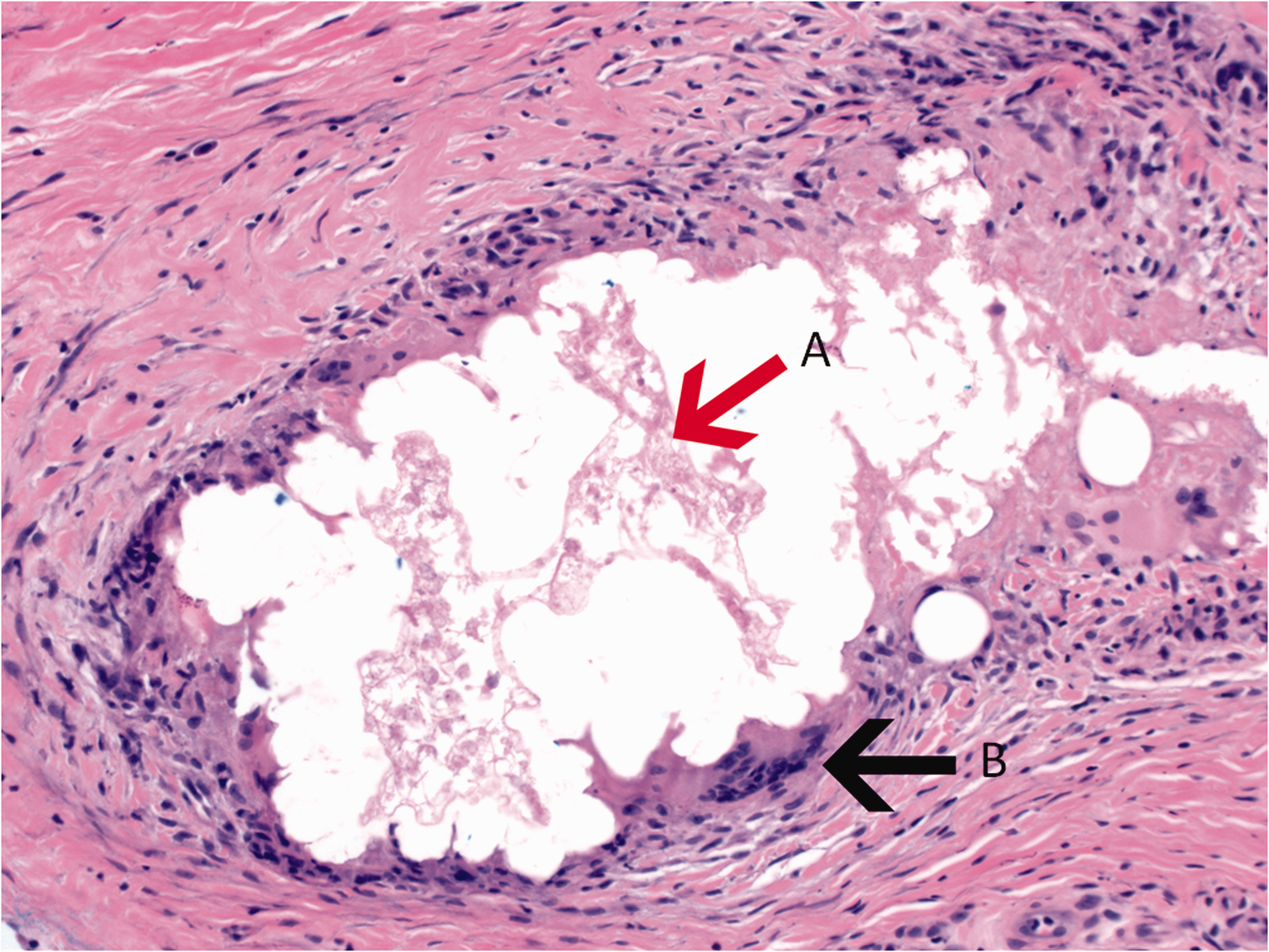
One year: Glue contained in extravascular cavitated granulomas. Details of cavitated extravascular granuloma at one year with spiked glue (arrow A) centrally outlined by foreign body granuloma with multi-nucleated giant cells (arrow B) and surrounding fibrosis (hematoxylin and eosin, original magnification ×100).
Vascular marker CD-31 and CD-34 (Figure 7) showed no evident vascular remnants within the glue-based cavitated granulomas. CD-68 for histiocytes highlighted the cavitated granulomas (Figure 8).

One year: Glue contained in cavitated granulomas. CD-34 positive vessels (staining brown) demonstrating prominent angioplasia in the fibrosis (arrows A) surrounding the cavitated granuloma (asterix), which has no endothelial cells or a vessel associated with the central glue (arrow B) (CD-34 immunoperoxidase stain, original magnification ×50).

One year: Histiocytes coating the residual glue within the granulomas. CD-68 stain for histiocytes (staining brown) showing aggregates forming granulomas, which surround cavities and outline glue coated with histiocytes (CD-68 immunoperoxidase stain, original magnification ×50).
4. Patient 2, peripheral arteriovenous malformation – one year
The epidermis and dermis appeared normal. The dermis showed no evident fibrosis or granulomatous inflammation. There was fibrosis and lymphoid aggregates in the subctaneous fat and its septa. The fibrosis with lymphoid aggregates extended into the deep tissue and surrounded a nerve. Focal empty cavities were associated with a fibrillar outline surrounded by foreign body histiocytes with surrounding fibrosis and lymphocytic inflammation. Immunoperoxidase stains for CD-34 was positive for the vascular endothelium and interstitial cells within the dermis. The cavities and the glue spicules did not stain with CD-34 indicating a lack of endothelial lining.
Discussion
We present the first longitudinal histological study in humans following the use of n-BCA in treatment of peripheral vessels. The first patient underwent percutaneous glue ablation of peripheral tributaries of the GSV, while the second patient underwent glue ablation of the feeding artery of a peripheral AVM. There were no systemic or local adverse events such as necrosis, dermatitis, phlebitis or other clinical complications. The treated vessels in both cases showed similar ultrasonic and histological features on follow-up. Ultrasound imaging demonstrated the glue to be persistently present on all follow-up occasions, appearing highly echogenic and creating a shadow artefact. The one-year follow-up biopsies demonstrated lymphoid hyperplasia and surrounding tissue fibrosis. The glue appeared to be mostly extruded from the treated vessels and was contained within extra-vascular granulomas.
To our knowledge, this is the first report of the late tissue reaction to the injection of n-BCA in peripheral vessels. The only other histological report of a human peripheral vein injected with n-BCA was limited by the fact that the vein was removed just 10 min after treatment not allowing adequate time for a tissue response. 9 In the present study, biopsies were taken at one week, six weeks and one year after the glue injection. The one- and six-week samples did not demonstrate a granulomatous tissue reaction although there were small foci of foreign body histiocytes observed at six weeks. The one-year biopsies revealed striking features that included extra-vascular foreign body granulomas, glue extrusion, lack of an endothelial lining in the treated vessels, lymphoid hyperplasia and fibrosis of the surrounding tissue.
The observed granulomas had typical features of foreign body granulomas and contained foreign body-type giant cells. All granulomas were extra-vascular and none were observed in the vessel walls or within the lumen of the treated vessels and hence there was no evidence of a granulomatous phlebitis that would suggest a vasculitic immune response. The granulomas appeared to contain the released extra-vascular glue, which histologically appeared as fibrillary material. Some of the granulomas appeared cavitated, but these cavities may be artefactual as central dry glue may have been shattered during the biopsy or tissue sectioning.
Granulomas detected in this study were a late finding observed in samples obtained 12 months following the procedure. The extra-vascular location of the granulomas containing glue implies extrusion of some of the glue material. Only small amounts of glue appearing as fibrillary material were observed to remain within treated vessels. The delayed extra-vascular extrusion of glue from the treated vessels in our study is consistent with previous histological studies on n-BCA where complete extrusion of glue and formation of glue casts within the lumen of oesophagus were observed on endoscopy following n-BCA treatment of oesophageal varices. 10
At one year, the vascular marker CD-34 showed no evident endothelial cells in the treated vessel although other layers of the vessel wall were histologically still present. Similarly, the treated vessels could still be seen on ultrasound creating a strong shadow artefact. This indicates that n-BCA does not result in vessel degradation, but vessel occlusion followed by intimal necrosis.
In one-year biopsies, we observed lymphoid hyperplasia indicating a possible reactive immunological response to the injected glue. The morphology and non-sheet like distribution of the observed lymphoid hyperplasia indicates a reactive nature that is very different to that observed in malignancies or those seen in relation to breast implants. An immune response to n-BCA is not surprising as there are multiple case reports of delayed-type hypersensitivity reactions and in particular, contact dermatitis to CA.11–22 A recent case report described contact dermatitis to Venaseal™ glue, another n-BCA used to treat peripheral veins. 23
Our histological findings are consistent with the published literature demonstrating foreign body granuloma formation as a late tissue reaction to n-BCA.2–4 Studies on cerebral AVMs embolised with n-BCA showed acute inflammation at 24 h followed by foreign body giant cells surrounding the glue at three weeks and consistently observed for 52–60 months.2,3 Similarly, studies on the internal carotid arteries of pigs have shown acute inflammation in the injection site in the first week, but granuloma formation and fibrosis appearing at around two months. 5
Foreign body granuloma formation may progress to necrosis, ulceration, foreign body extrusion or immunological reaction to the foreign material with sarcoidal features or lymphoid hyperplasia. Whether granuloma formation can progress to ulceration and abscess formation may depend on the size and depth of the vessels treated and glue extrusion to the surrounding tissue. Consistently, clinical complications of n-BCA as an embolic agent have included ulceration, abscess and fistula formation. Abscesses were reported in association with the stomach, brain, and peritoneal cavity. 10 A case report by Zernovicky 8 highlighted the possible devastating effects of a granulomatous reaction following CAC treatment of saphenous veins. A 54-year-old female treated with VenaSeal™ developed spontaneous skin perforations with extrusion of glue pieces from the treated sites bilaterally four months after a bilateral CAC procedure on GSVs. The microbiology was repeatedly negative. Progressive erythema, pain and swelling was present on both extremities and worsened despite treatment with steroids. The patient eventually required bilateral saphenectomies, resulting in chronic lymphoedema.
To our knowledge, the report by Zernovicky 8 has been the only recorded and published case of a complication of this nature to date. The scarcity of published reports may be due to the overall safety of the procedure but may also be due to under-reporting of complications. Both Venaseal™ and Venablock™ gained regulatory approval for use in peripheral veins only in the past four years and complications may evolve long term and gradually appear in the medical literature. National and international registers of complications of CAC should be encouraged. International guidelines are required to recommend evidence-based criteria for patient selection and to define contraindications. We strongly recommend against the use of CAC in patients with chronic granulomatous disorders. The granulomatous reaction that follows n-BCA injection may be marked and dominated by epithelioid histiocytes forming sarcoidal granulomas particularly in patients who have underlying sarcoidosis.
This study had several limitations. We used only one n-BCA agent (VenaBlock™). We expect a similar tissue reaction to other n-BCA agents but this needs to be demonstrated in studies using those agents. While to our knowledge, the present study is the only available long-term histological follow-up of CAC, longer-term studies (5–10 years) are required to assess the potential for longer-term complications especially when these agents are injected dermally, sub-dermally or in superficially located venous tributaries.
In conclusion, our serial histological findings demonstrate a delayed extra-vascular granuloma formation, glue extrusion and lymphoid hyperplasia detected one year following injection into peripheral vessels. The clinical safety of n-BCA glue especially when injected superficially needs to be monitored given the risk of ulcerating granulomas and glue extrusion.
Footnotes
Declaration of Conflicting Interests
The author(s) declared no potential conflicts of interest with respect to the research, authorship, and/or publication of this article.
Funding
The author(s) received no financial support for the research, authorship, and/or publication of this article.
Ethical approval
Not applicable.
Gurantor
Not applicable.
Contributorship
Mina Kang: design and execution of the methodology described in this article, sourcing of figures, literature research, article writing and final editing of the paper. Steve Kossard: results analysis and interpretation, sourcing of figures, article writing and final editing of the paper. Kurosh Parsi: design and execution of the methodology described in this article, sourcing of figures, literature research, article writing and final editing of the paper.