



Abstract
Objective
We aim to report on the Laser-Sclerosing Foam Hybrid Treatment (LSFHT) and its outcomes when used on patients with great saphenous vein (GSV) insufficiency.
Methods
This was a single center retrospective cohort study on patients with GSV insufficiency that were treated with the LSFHT technique, a surgical procedure that comprises the use of both sclerosing foam and endovenous ablation and avoids the use of tumescent anesthesia. Occlusion rates and complications were reported.
Results
139 legs from 106 patients were operated, achieving a 100% occlusion rate, while only a small burn and 2 popliteal vein thrombosis cases occurred.
Conclusion
The study suggests that the LSFHT is a feasible fast procedure that proved both effective and safe for the treatment of GSV insufficiency.
Keywords
Introduction
Chronic venous insufficiency (CVI) is a disease estimated to affect around 64% of the population 1 that can cause, in the most severe cases, complications like activity limiting pain that requires regular use of analgesics, severe inferior extremity edema and venous ulcers that may be impossible to heal without surgical or endovascular intervention. Thus, chronic venous insufficiency is a very common disease that significantly impacts quality of life, increases healthcare costs and disability. 2
While surgical ligation and stripping of saphenous veins is still regarded as the gold standard for the treatment of CVI, it can be a painful and traumatic procedure associated with slow recovery. Recent developments, however, have provided new and revolutionary alternatives to the standard surgical treatment, such as endovenous laser ablation (EVLA) and ultrasound-guided foam sclerotherapy (UGFS).
Ultrasound-guided foam sclerotherapy has proved to be a safe way to achieve good occlusion rates of both great and small saphenous veins, as well as perforators, tributaries, and smaller reticular branches, with minimal discomfort for the patient, low complication rate and little significant morbidity.3,4 The introduction of EVLA offered an even more effective solution for the treatment of great and small saphenous veins insufficiency, with greater reported occlusion rates than UGFS, minimal need of reintervention, lower morbidity, and faster recovery than conventional surgical techniques.3–5
Both EVLA and UGFS, however, still have important limitations. Excessive amounts of foam can cause visual disturbances, dysesthesia, headaches, migraine, and increase the risk of stroke and both deep and superficial vein foam-induced thrombosis; plus, the sclerosants commonly used degrade in contact with blood. Therefore, the effectiveness of UGFS reduces with larger veins, and consequently the European Society for Vascular Surgery recommends it only for saphenous trunks that are less than 6 mm in diameter. 6
Endovenous laser ablation, on the other hand, requires the use of high volumes of tumescent anesthesia, which can result in higher levels of pain and extensive hematomas, hyperpigmentation, skin burns, and paresthesias due to skin and nerve damage, thrombophlebitis, vessel perforation, and heat-induced thrombosis, that can lead to life-threatening outcomes.6–8
Techniques that combine EVLA and UGFS have recently been investigated, starting with Tessari et al, 9 with contributions from Frullini et al, 10 Ulloa et al, 7 Boné, 11 and Carmelino et al, 12 with the aim to achieve better occlusion rates even in severely dilated venous trunks, decreasing the sclerosing foam (SF) volume and the power needed to achieve a successful ablation with the laser and eliminating the need for tumescent anesthesia, thus decreasing complications, improving patient satisfaction and allowing for better outcomes.
Objective
In this article we aim to report our experience in the first cases performed with the use of Laser-Sclerosing Foam Hybrid Treatment (LSFHT) of the great saphenous vein (GSV) while using sedation only, without the need for tumescent anesthesia nor general anesthesia, and the reduction in adverse effects that can be achieved while maintaining a high rate of success.
Method
Venous Doppler ultrasound (US) examination was performed on 139 legs from 106 patients (22 males and 84 females) with symptomatic GSV incompetence, while they were standing on their feet and a reflux time greater than 0.5 s in duration after a Valsalva or compression/relaxation maneuver was found in all patients. The distribution of the patients according to the CEAP classification was: 5 C1, 12 C2, 67 C3, 21 C4, 1 C5.
The patients were premedicated with diphenhydramine before the procedure. During the procedure, the patients were given about 25 to 50 mcg of fentanyl, and approximately 20% of patients were also sedated with sevoflurane, if they were deemed too anxious or agitated. Neither antibiotics nor thromboprophylaxis were needed.
A second US evaluation was repeated immediately before the procedure, to mark on the skin the course of the saphenous vein as well as the access point for the endovenous laser fiber. To effectively treat the whole incompetent segment of the GSV, we tried to mark the access point for the laser fiber as distal as possible, usually just above the malleolus.
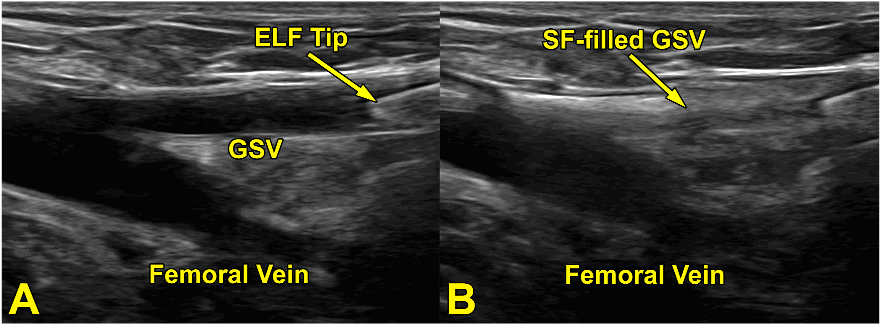
The procedure started with a percutaneous GSV access under local anesthesia, in the previously marked access point for the laser fiber, followed by the insertion of a guidewire, through which a 6Fr Glidesheath Slender Introducer [Terumo, Tokio, Japan] or a 5Fr conventional introducer was placed. Then, the 360°-radial-tipped fiber of a 1470-nm laser with automatic retractor was inserted, under US (ultrasound) guidance, up to 2 to 2.5 cm distal to the saphenofemoral junction (SFJ), as recommended by the American Venous Forum and the Society for Vascular Surgery, 13 as seen in Figure 2(a).
Meanwhile, 10 mL of SF were prepared using 1% Polidocanol [Aethoxysklerol] and room air at a 1:4 drug/air ratio, shaking the foam 40 times between two 10 mL syringes, as per the Tessari method.
14
Then, through the introducer side tube, the GSV was flushed with 15 mL of saline solution, as seen in Figure 1, quickly followed by the injection of a bolus of 10 mL of SF, also through the introducer. We did not use the US transducer to trap or restrict the flow of SF. Instead, we let the SF flow freely, from the introducer in the distal leg or thigh through the GSV up to the SFJ, while the Trendelenburg position was used to improve the venous drainage of the leg. US imaging was used to check for adequate spasm of the treated GSV tract up to the SFJ, as seen in Figure 2(b). The procedure setup: the introducer, placed just above the ankle, connected through a 3-way valve to syringes with saline, to flush the vein, and the sclerosing foam, that has just been prepared; the endovenous laser fiber, inserted through the same introducer up to 2-2,5 cm from the saphenofemoral junction, is ready to be activated as soon as the saline and the foam are injected. Endovenous laser fiber (ELF) inside the great saphenous vein (GSV), at 2.5 cm from the saphenofemoral junction, before (a) and immediately after (b) the sclerosing foam injection (in the distal leg). In figure b we can appreciate the foam filling the GSV up to the saphenofemoral junction.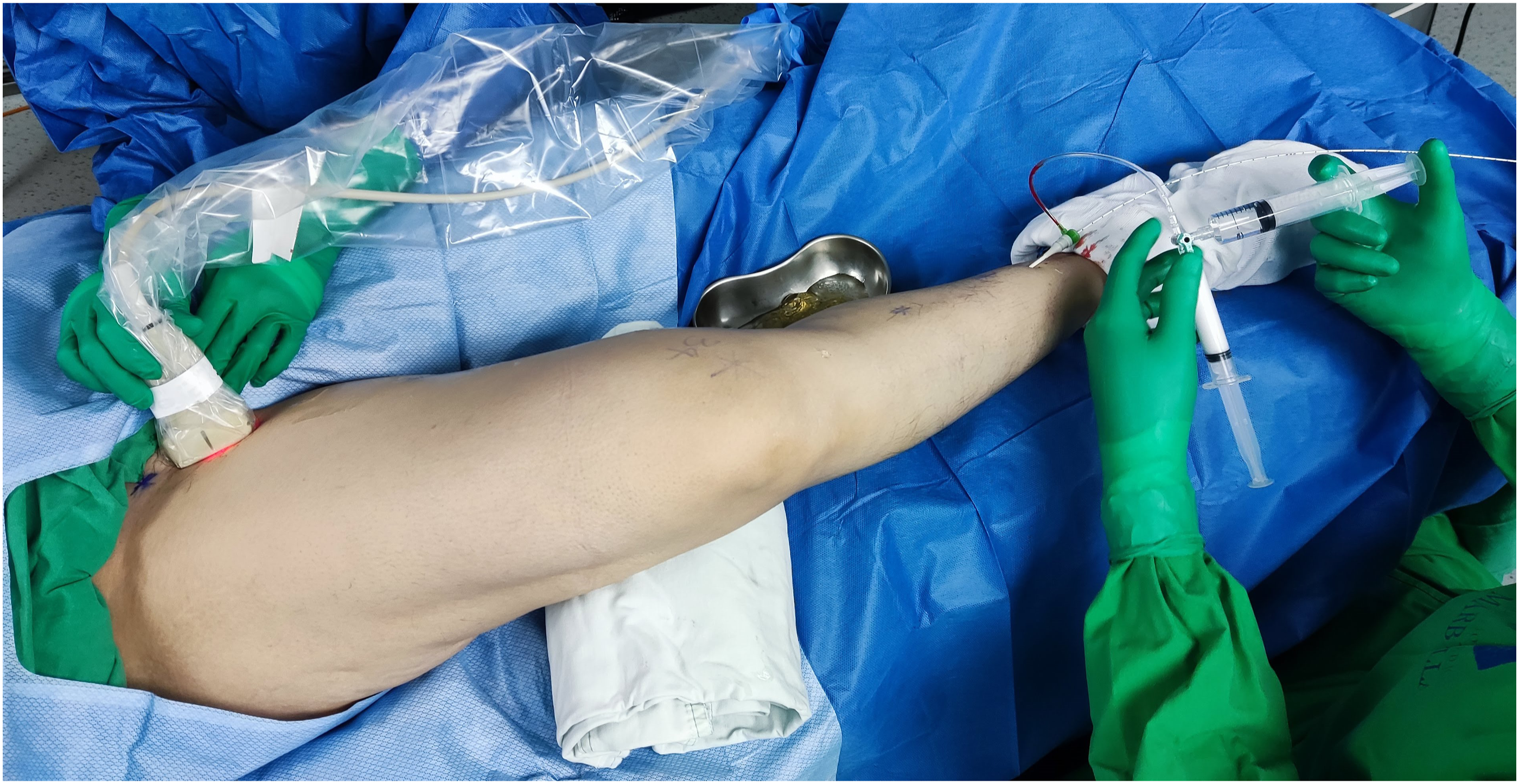
Next, the patients were sedated with a bolus of propofol and the endovenous laser fiber was activated. Propofol dose was generally around 100-150 mg, enough to deeply sedate the patient while maintaining spontaneous respiration.
Laser fiber power and retraction speed parameters varied between patients, depending on characteristics such as vein diameter and patient’s tolerance to pain, but in general power settings were set higher for the proximal segment of the GSV (usually starting at 10–8 W, with a retraction speed of 2 mm/s (with a calculated laser fluence of 50–40 J/cm), and were then gradually reduced; an example of a commonly used set of parameters would be 8 W, 2 mm/s (40 J/cm) for the high-thigh segment of the GSV, then 8 W, 3 mm/s (26.7 J/cm) for the mid-to-low-thigh segment, 7 W, 4 mm/s (17.5 J/cm) for the patellar segment, and 6 W, 4 mm/s (15 J/cm) for the infrapatellar segment. Laser tip retraction was always performed under US guidance.
After the laser was removed, a continuous compression/relaxation maneuver was performed on the patient’s leg to spread the SF thoroughly and deeply along the patent tributaries.
Patients were prescribed paracetamol and tramadol prn for analgesia for the first 3 days post-op and were instructed to apply Eucerin Aquaphor Ointment Body Spray to the affected leg twice a day.
30–40 mmHg graduated compression stockings were prescribed to all patients for 24 continuous hours, then only during daytime for an additional 48 hours, and they were thereafter switched to 20–30 mmHg compression stockings. All patients were encouraged to walk and to perform bending exercises in the days following the procedure.
All patients were recommended clinical follow-ups at 3- and 7-days, 1-, 3-, and 6-months post-op. The follow-up visits always included US evaluation to assess the ablated GSV and its tributaries, and to search for complications such as deep vein thrombosis. Out of all the patients that underwent this procedure, however, many were lost, especially after the 3-month follow-up. Therefore, we decided to include in this article only the 106 patients that attended at least up to the 3-month follow-up visit, in the 4 years prior to the writing of this paper.
Results
Satisfactory ablation was achieved in all patients, and at the follow-up visits all showed complete occlusion and absence of blood flow in the treated GSV tract.
All patients reported that, in the first 3 days, post-operative pain was low enough that it was effectively treated with the prescribed oral analgesics; no need for oral analgesics was reported afterward. The few patients that reported pain after the first 3 days post-op were effectively treated with topical analgesics only.
Some patients presented small bruises in the site of punction, but no major hematomas or ecchymoses were detected, a clear difference compared to the patients subjected to tumescent anesthesia. Two patients showed a thrombosis at the 7-days follow-up: the first had a popliteal vein thrombosis that required anticoagulant treatment (rivaroxaban) for the following 4 months; the second refused to use the recommended compression stockings and developed a popliteal and superficial femoral vein thrombosis that required anticoagulant treatment (rivaroxaban) for 2 months; neither thrombosis case lead to further complications.
One patient showed a small first degree burn at follow-up, and another showed an allergic rash that may have been related to the procedure. No patient has reported long-lasting neurologic symptoms that may indicate nerve injury.
In one case, the introducer decannulated after the laser fiber was inserted, but before the SF could be injected. It was decided to continue the ablation procedure with the laser only, which resulted in a satisfactory occlusion at follow-up.
Discussion
Endovenous laser ablation is one of the most recommended treatments of saphenous vein insufficiency, 6 thanks to it being a scar-free procedure, aesthetically pleasing, minimally invasive procedure with a high rate of success.
Endovenous laser ablation, however, has always been strictly related to the tumescent anesthesia, as this has been considered necessary to protect the perivascular tissue from heating and burns thanks to the cooling effect, to control pain, to guarantee a sufficient vein depth from the skin and to reduce the GSV diameter, eliminating blood and improving surface contact.15–17
Tumescent anesthesia, however, is a long and relatively invasive procedure that can lead to both pain and unpleasant aesthetic consequences, such as perivenous ecchymosis and hematoma, leading to patient dissatisfaction. Deleterious effects can present using too little or too much tumescent anesthesia, 17 and complications, albeit rare, can be very serious, including fracture of the laser fiber by the anesthesia needle and necrotizing fasciitis. 9
Nowadays, thanks to new, higher-wavelength laser devices with higher affinity for water that seem to better target the vein wall, rather than the hemoglobin that was primarily targeted by older lasers, and radial laser tips, that better distribute the laser light along the vein wall, the same rate of occlusion can be achieved with lower power delivery settings.
The addition of SF to the technique induces a vasospasm that gets the vein wall closer to the laser tip and directly contributes to the vein wall damage that will ultimately occlude it, thus allowing us to use power delivery settings so low that no tumescent anesthesia is needed, as we have demonstrated in this paper.
The interaction between the SF and the laser beam has already been evaluated from the molecular point of view 18 and in vivo, initially with transdermal lasers19,20 and more recently with endovascular lasers9,11 and a synergistic effect has since been hypothesized, while neither of the studies we found reported SF inactivation or laser beam attenuation. Even in the case that the SF in the GSV was indeed inactivated by the laser, it would still partially damage the vein endothelium in the few seconds or minutes between the SF injection and the laser activation, and it would still provoke the vasospasm needed to successfully ablate the vein with low laser power settings and no tumescent anesthesia.
While with the traditional technique, using tumescent anesthesia, the infrapatellar GSV is usually left untreated, our technique allows the surgeon to treat larger portions of the GSV or even the whole vein down to the malleolus, if possible. Furthermore, the absence of tumescent anesthesia makes for a shorter and simpler procedure that only requires sedation for a short amount of time, and this, together with the low levels of pain, the minimal reported need for analgesics and the absence of large ecchymosis, leads to greater patient satisfaction.
Interestingly, since we let the SF flow freely through the GSV, if the GSV being treated also has insufficient tributary branches, some amount of SF could flow backwards into them and incidentally occlude these insufficient veins that would otherwise require a separate treatment if the classic EVLA was performed.
In the method first proposed by Tessari et al, 9 a short vein segment is treated with a traditional EVLA at a high power delivery setting to create a “plug,” then the SF is injected and the rest of the vein is treated by the laser at a lower power delivery setting; by contrast, in our technique, the SF is injected before laser activation, in the distal GSV and the Trendelenburg position is used to improve blood drainage and spread the SF to the patent tributaries (rather than the “plug” in the Tessari technique), so that the entire length of the vein can then be treated with the laser at a lower power setting.
Even with this low-power combined technique there is still a risk of complications such as endovenous heat-induced thrombosis, nerve injury, and burns; however, we hope that by reducing power delivery, the risk of these complications can also be reduced, while the relatively low amounts of SF used should decrease the risk of endovenous foam-induced thrombosis and other side effects.
While we observed 2 cases of deep vein thrombosis out of 139 legs, which may seem quite a high incidence, it must be noted that one of the patients refused the recommended thromboprophylaxis and therefore may not have developed any complications if he had adhered with the post-op recommendations; nevertheless, further studies are needed to determine if the combination of these two techniques does indeed increase the overall risk of deep vein thrombosis. Although we did not observe any neurological symptoms, saphenous nerve injury is still a concern due to the absence of space and cooling around the GSV provided by the tumescent anesthesia and should also be further investigated.
Passing the endovenous laser fiber through a very tortuous vein can sometimes be particularly challenging, thus making the treatment of these patients with EVLA more difficult. We found that this problem can also be overcome using LSFHT: if, for example, the distal tract of the GSV is too tortuous, the laser fiber can be inserted just below the knee or even above the knee, while the SF is injected more distally, to both treat the lower tract of the vein and assist the laser in the treatment of the higher tract.
Further investigations about the non-inferiority of this technique in terms of effectiveness and adverse effects are ongoing, including quantifying post-operative pain and ecchymoses more precisely, and the details of the procedure are still being improved. Long-term outcomes, at least up to 2 years post-op, and patient-reported improvement of symptoms and quality of life should also be investigated. The exact power delivery parameters for each situation are currently at the discretion of the surgeon and very much unstandardized, thus more experience is needed in this regard, as well as in the application of this strategy with other techniques, such as with radiofrequency devices. Finally, new devices that facilitate the use of this combined technique, such as the endovenous laser fiber proposed by Boné, 11 should be developed and more thoroughly investigated.
Conclusion
This study reveals that the LSFHT is a feasible and effective option for the treatment of the insufficient GSV. This fast and minimally invasive technique can achieve high occlusion rates, and looks like a promising option to avoid some of the complications that can occur with EVLA and SF only procedures.
Further studies are needed, however, to better assess the technique, its occlusion rates and the possible complications, to demonstrate its non-inferiority to other techniques and whether it significantly improves patient satisfaction and quality of life compared to other techniques.
Footnotes
Acknowledgements
We would like to express our sincere gratitude to the following personnel from Clínica Vascular de Panamá for their contributions to this research, including aiding with initial patient selection and follow-ups, venous Doppler ultrasound examinations and data collection: Dr Ana Caballero, Dr Jaime Vásquez, Dr Estefanía Ulloa, Dr Ian Bustamante, Johanna Cárdenas, Dr María Adames, Dr María Fernández, Dr Yarubis Vásquez.
Authors’ contribution
GV and AR researched literature, conceived the surgical technique and the study, were involved in protocol development and patient recruitment. GZ was involved in data analysis and wrote the first draft of the manuscript. All authors reviewed and edited the manuscript and approved the final version of the manuscript.
Declaration of conflicting interests
The author(s) declared no potential conflicts of interest with respect to the research, authorship, and/or publication of this article.
Funding
The author(s) received no financial support for the research, authorship, and/or publication of this article.
Guarantor
GV.
