



Abstract
Introduction
Depending on the severity of chronic venous disease (CVD), it has been estimated to affect between 38% to 90% of the adult population 1 and can be accompanied by a significantly decreased quality of life. 2 With a wide range of clinical manifestations, CVD is widely underdiagnosed and because of this many patients may not be obtaining timely and appropriate referral and treatment. 3
Clinically, CVD patients may be identified on physical examination by the presence of ankle edema, a common sign and symptom. A semi-quantitative method for detecting ankle edema is the pitting test wherein a finger presses firmly along the tibia bone above the medial malleolus and the resultant dermal pitting is subjectively graded on a scale of 0 to 4+ based on the depth of indentation produced. 4 Beyond the inherent observer variability with this subjective methodology, the presence of ankle edema is significantly underrecognized even in patients presenting for evaluation of superficial venous disease. 5 Alternative objective methods for measuring lower extremity edema have been proposed but none have been extensively studied or been accepted into clinical practice.4,6–13 We have developed a 3D-printed Edema RulerTM as an inexpensive and objective method to easily quantify ankle edema. 14
Duplex ultrasound (DUS) analysis is another method to evaluate ankle edema severity due to its wide availability and non-invasiveness . 15 Suehiro et al. proposed a method for characterizing ankle edema on DUS though a grading system based on subcutaneous echogenicity (SE) and subcutaneous echo free space (SES).16,17 Inherently, this method is largely qualitative relying on an observer’s interpretation of image echogenicity and echogenic lines.16,17 In our study we modified the Suehiro scale by adding a quantitative metric.
Our objective was to prospectively determine the prevalence of ankle edema in an outpatient vein center and measure its severity objectively with both the Edema RulerTM and DUS analysis with the Modified Suehiro Grade.
Methods
Patients and edema measurements
All adult patients with initial appointments for suspected superficial venous disease who were consecutively referred to Vein911 Vein Treatment Centers outpatient offices in Tampa, FL, between May 2023 to August 2023, were included in this study. As part of the standard initial examination, each patient was evaluated for lower extremity edema both by clinical examination and DUS. Clinical assessments including CEAP, Total rVCSS, and Edema rVCSS were assigned to each patient by a physician or physician’s assistant (PA) independently of the Edema RulerTM and DUS analysis. A pitting test using the Edema RulerTM (Figure 1) was utilized for the clinical exam performed by a PA or medical assistant (MA). The patient was kept in a sitting position with the feet flat on the floor. The index finger of the examiner applied firm pressure for 20 seconds at a location 5 cm above the medial malleolus on the tibia. The finger was then removed and the Edema RulerTM was used to measure the depth of the indentation on the skin. The process was performed on both legs after which the patient had an DUS performed and underwent an office clinical examination. Before discharge from the office, a different PA or MA repeated the clinical pitting test, blinded to the results from the first examination. The PAs and MAs were instructed on how to perform the pitting test and utilize the Edema RulerTM. Medical assistants were required to show competence in the test under the supervision of a PA on a minimum of three patients before the study began. For the Edema RulerTM this study had seven evaluators across three Vein911 Vein Treatment Centers locations. (a) The Edema RulerTM inner tube with millimeter scale markings at its tip is used to measure the depth of indentation on the skin. (b) Moveable locator ring is placed on the inner tube (c) and advanced to the level where it touches the skin (d) thereby measuring the depth of indentation.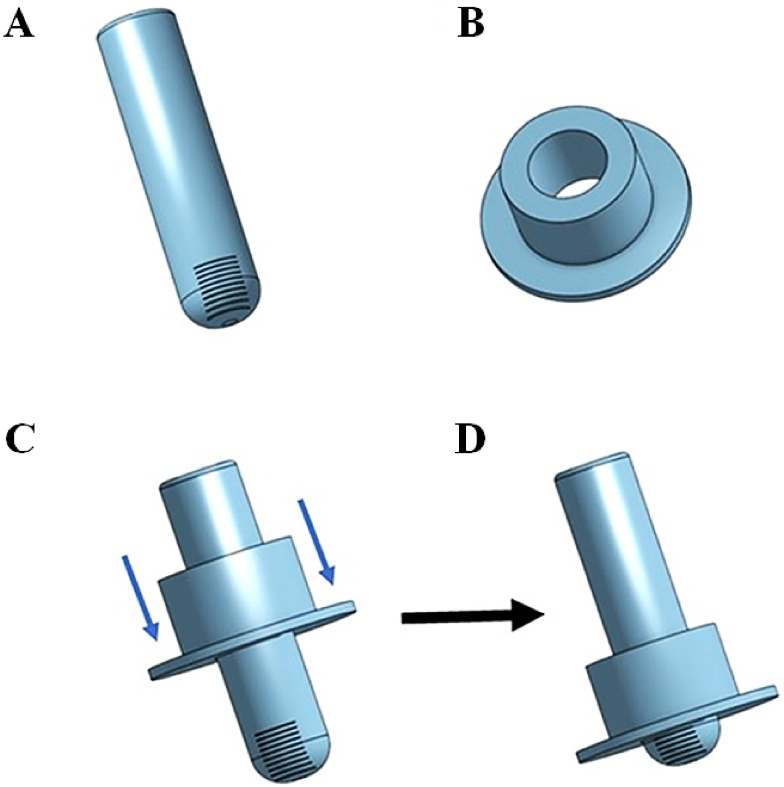
The 3D-printed Edema RulerTM is composed of two parts (Figure 1). 14 The inner tube with a length of 6 cm has a curved tip to fit the indentation on the skin caused by the examiner’s index finger. The tip has integral measurement markings in one-millimeter increments. The second component of the ruler is the locator ring which slides up and down the inner tube to mark the depth of pitting. The entire device is used by placing the inner tube onto the center of the indentation and then, while holding the inner tube steady, the examiner slides the locator ring down until the disc is flush with the skin on the sides of the pit. The device is then removed and the measurement is taken by seeing where the lip of the locator ring lines up on the measurement lines.
Ultrasound
Modified suehiro 16 ultrasound scale for edema.
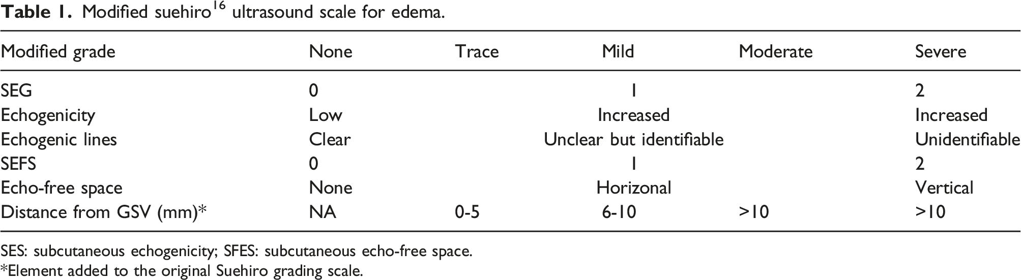
SES: subcutaneous echogenicity; SFES: subcutaneous echo-free space.
Element added to the original Suehiro grading scale.
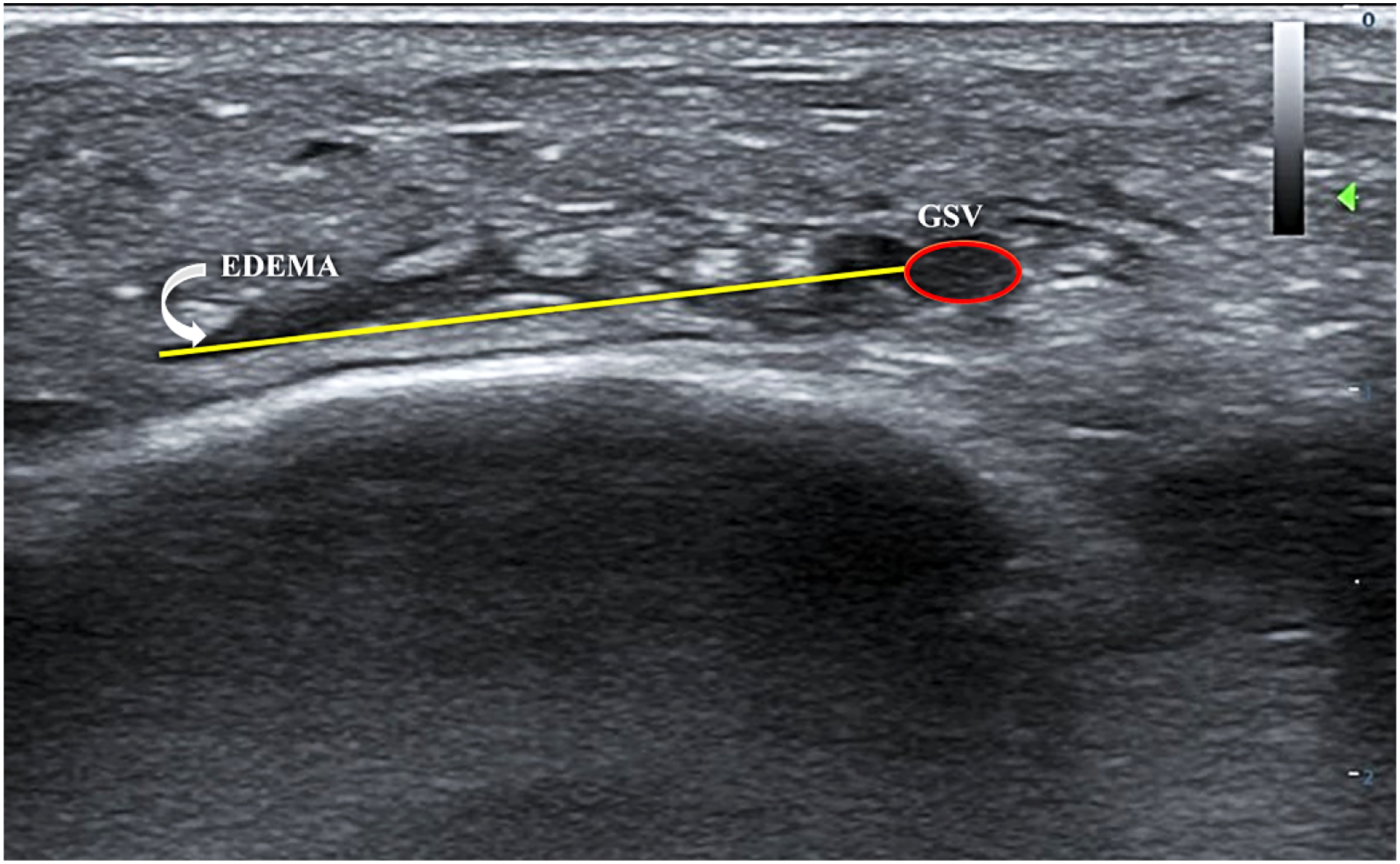
Duplex ultrasound measurement of edema from the wall of the GSV (ellipse) to furthest lacunae of edema (arrow).
This study was granted Institutional Review Board exemption by the University of South Florida IRB (Study #5470) as all patients underwent a non-invasive, minimal-risk examinations which was standard practice at Vein911 Vein Treatment Centers for which the patients signed consent-to-treat forms.
Statistical analysis
Statistical analysis was performed using IBM SSPS Statistics 29.0 (IBM; Armonk, NY). Continuous data was reported as mean
Results
Of the 47 patients (94 legs), 37 were females (79%) and 10 were males (21%) with a mean age of 57 ± 14 years and mean body mass index (BMI) of 27 ± 5. The racial distribution reflected 41 patients who were Caucasian (87%), 1 was African-American (2%), 1 was Asian-American (2%), and the remaining 4 patients classified their race as Other (9%). In addition to their race, 14 participants classified their ethnicity as Hispanic or Latino (30%).
Leg CEAP and rVCSS Scores.

rVCSS - revised Venous Clinical Severity Score.
The mean Total rVCSS of the participants was 4.84 ± 2.46. Most of the participants (67%) were classified with an Edema rVCSS score of 1 and a Varicose Vein rVCSS (VV rVCSS) of 1 or below (59%) (Table 2).
Edema measurements.
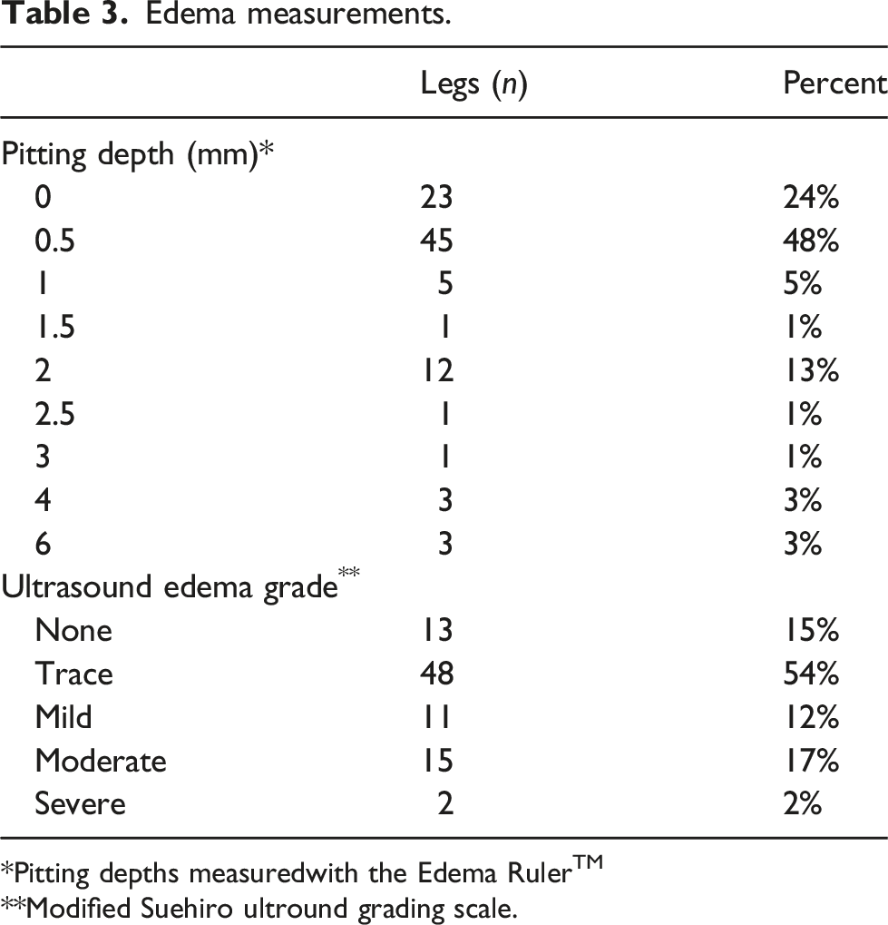
Pitting depths measuredwith the Edema RulerTM
Modified Suehiro ultround grading scale.
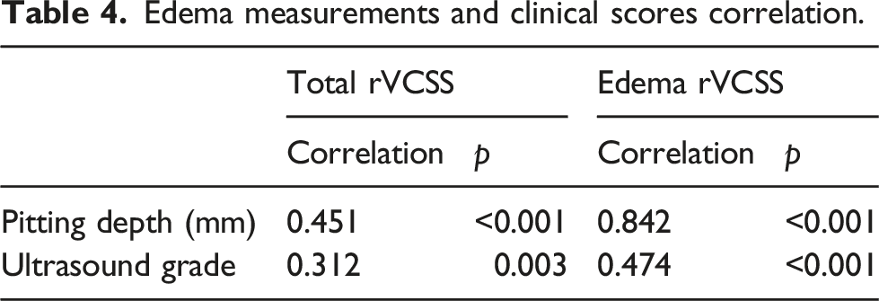
Edema measurements and clinical scores correlation.
The interobserver reliability with the Edema RulerTM was excellent with an intra-class correlation coefficient of 0.985 (p < .001). Similarly, the DUS analysis had an interobserver reliability of 0.883 (p < .001).
The correlations between the measured edema pitting depth and DUS Modified Suehiro Grade with the Total and Edema rVCSS scores were highly significant but more so with the Edema RulerTM measurements as compared to the DUS (Table 4). The highest association, with a correlation coefficient of 0.842, was between pitting depth and the Edema rVCSS score. The correlation between the Edema RulerTM and the DUS Modified Suehiro Grade was 0.441 (p < .001).
Edema measurements and CEAP comparison.
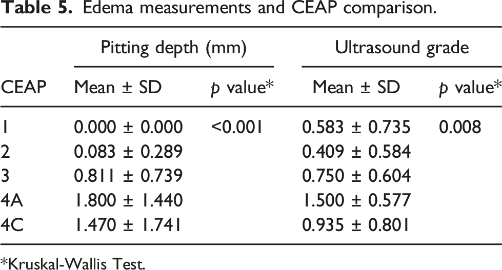
*Kruskal-Wallis Test.
Discussion
Lower extremity edema is a common symptom and sign of CVD and the defining characteristic of the C3 clinical category of the CEAP classification. 18 This study adheres to the 2020 update of the CEAP classification system. 18 Leg edema, through early detection, can lead to the proper diagnosis of venous disease and more appropriate referral and treatment of patients.3,5 It is typically detected during the clinical examination visually and with palpation and the pitting test. However, previous research has demonstrated the unreliability of the classical pitting exam. Brodovicz et al. showed clinical pitting assessment had poor inter-examiner reliability, with interclass correlation coefficients ranging from −0.01 to 0.53 depending on its location. 4 A recognized limitation of the pitting examination is its non-quantified visual estimate of pitting depth and time of its resolution.
Applying more quantitative methods to diagnose and measure edema severity can improve diagnosis and clinical documentation. In addition to assessing the classical clinical assessment, Brodovicz et al. examined six other objective methods for evaluating lower extremity edema. 4 These methods included (1) water displacement, (2) ankle circumference, (3) figure-of-eight, (4) an edema tester, (5) a modified edema tester, and (6) indirect leg volume. Water displacement involved the patient placing their foot and ankle into a container of water and measuring the water displaced. 6 The ankle circumference method measured the ankle circumference 5 cm above the medial malleolus. 7 The figure-of-eight method involved marking the foot and ankle based on 5 anatomical landmarks and placing a tape measure on these landmarks to obtain a measurement of ankle circumference. 8 The Edema Tester is a plastic card with a series of holes placed under a blood pressure cuff under varying pressures with the resulting markings evaluated by time of resolution. 9 The Modified Edema tester involved the same method as the Edema Tester but instead of holes on the plastic card there were protrusions. 9 The indirect leg volume is assessed using the disc method in which multiple ankle and leg circumferences are recorded to estimate total leg volume. 10
Water displacement and ankle circumference measurements had the best interobserver reliability of >0.90. 4 However, water displacement measurement is cumbersome and impractical in the usual clinical setting. 4 Ankle circumference measurements, performed by marking with a tape measure precluded successive observers to be blinded and may have resulted in an overestimation of interobserver reliability. 4 Our own clinical experience with serial ankle circumference measurements has also found them to have greater interobserver variability (unpublished observations). Other attempts at quantitative methods of edema measurement, using a plastic card with either holes or protuberances pressed into the skin, had poor interobserver reliability and patients reported discomfort. 4 Kogo et al. devised an edema gauge with excellent interobserver reliability but its use on the dorsum of the foot instead of above the medial malleolus has less clinical applicability.11,12
The Edema RulerTM was developed to obviate some of the limitations of prior devices while making it inexpensive and easy to use. Our prior technical study confirmed its ease of use in providing an objective and quantifiably measure of ankle edema with excellent interobserver reliability. 14 This has allowed it to be integrated into a busy multi-location outpatient vein practice with different providers. Our results in the present study found the Edema RulerTM to have an excellent interobserver reliability with an ICC of 0.985 (p < .001). This exceeded the interobserver reliability of other quantitative methods previously reported and was similar to that of the water displacement methodology.4,11,12 The Edema RulerTM is limited to detecting pitting edema in patients who have complaint skin and would not be useful in patients with chronic non-pitting edema or C4 disease with lipodermatosclerosis that prevents adequate pitting.
Duplex ultrasound imaging has also been proposed as a method to both detect and quantify lower extremity edema.15,16 This technology is non-invasive and widely available in venous centers. Suehiro et al. proposed using DUS to measure subcutaneous edema with a grading system based on subcutaneous echogenicity and subcutaneous echo free space.15–17 They found a correlation with different degrees of leg edema, but it was inadequate in differentiating edema severity because their evaluation was qualitative and subjective. Our clinical observation that early ultrasonographic evidence of edema in CVD patients typically presents as wispy, low echogenicity stranding around the great saphenous vein (GSV) wall, just above the medial malleolus, prompted us to modify Suehiro’s scale by adding the objectively- measured distance of edema from the wall of the GSV. This showed a very good interobserver reliability of 0.883 (p < .001). As would be expected, DUS imaging was slightly more sensitive than pitting examination using the ruler for the detection of ankle edema. In 85% of the patients, DUS found at least trace edema as compared with 76% of patients showing measurable pitting depth (Table 3). Overall, more than 75% of our study patients were found to have detectable edema both clinically or on DUS imaging.
However, the interobserver reliability of the DUS grading was not as high as that of the Edema RulerTM. The ruler demonstrated better interobserver reliability (0.985) and was well accepted by clinical users who stated that the device was easy to use. A limitation with the DUS analysis was the use of a single saved image for the assessment of edema. This occasionally made it difficult for observers to later distinguish anechoic areas around the GSV to be edema or a vein branch. Examining more than one image would be more useful. Another limitation of the DUS analysis in our study was potential compression of the GSV by the ultrasound probe during image collection, which can interfere with the measured distance of edema from the wall of the GSV.
Both DUS and the Edema RulerTM provide quantifiable methods of detecting edema. The Edema RulerTM had stronger correlations with Total rVCSS and Edema rVCSS (Table 4). As would be expected, the strongest clinical correlation was between the Edema rVCSS and the Edema RulerTM (0.842, p < .001), reflecting its specific use for the measurement of leg edema. Overall, there was a weak correlation between the Edema RulerTM and DUS analysis (0.441, p < .001) suggesting the potential complementary use of these two modalities. A potential reason for this weaker correlation is that the Edema RulerTM was used to detect gross clinical edema, while the Modified Suehiro Grade includes edema that is not yet clinically detectable. Ultrasound may be most useful in identifying, rather than necessarily quantifying, early stages of edema which have not yet become clinically evident. 5
Potential limitations of our study include the lack of measurement for intra-observer variability in the clinical assessment with the Edema RulerTM. However, as all measurements were collected at a patient’s initial appointment, it would have been difficult for the investigator to be blinded to their earlier measurement. In addition, the degree of finger pressure applied during the pitting test could not be uniformly controlled.
Conclusion
Chronic venous disease is a widely prevalent and underdiagnosed condition. A quantitative and reproducible method of measuring lower extremity edema would be clinically useful. Our study found that DUS can identify lower extremity edema, particularly less severe degrees that are not readily detected on physical examination. The Edema RulerTM provides a quantitative measurement of lower extremity edema with high reliability which can be easily integrated into clinical practice.
Footnotes
Acknowledgements
MC was the recipient of a summer research grant from USF Health Morsani College of Medicine, Research, Innovation & Scholarly Endeavors (USF MCOM RISE)
Author contributors
MC, JB, and CP were involved in research conception/design, research analysis/interpretation, and writing the manuscript. MC was involved in data collection and statistical analysis. JB and CP were also involved in critical revision of the manuscript.
Declaration of conflict of interest
The author(s) declared the following potential conflicts of interest with respect to the research, authorship, and/or publication of this article: MC has a U.S. Provisional Coversheet Application for Patent for the Edema RulerTM. JB and CP declare that there is no conflict of interest.
Funding
The author(s) received no financial support for the research, authorship, and/or publication of this article.
Guarantor
MC.
Ethical statement

Data availability statement
For data relating to this research, please contact the corresponding author.